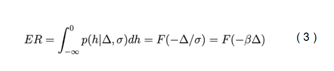
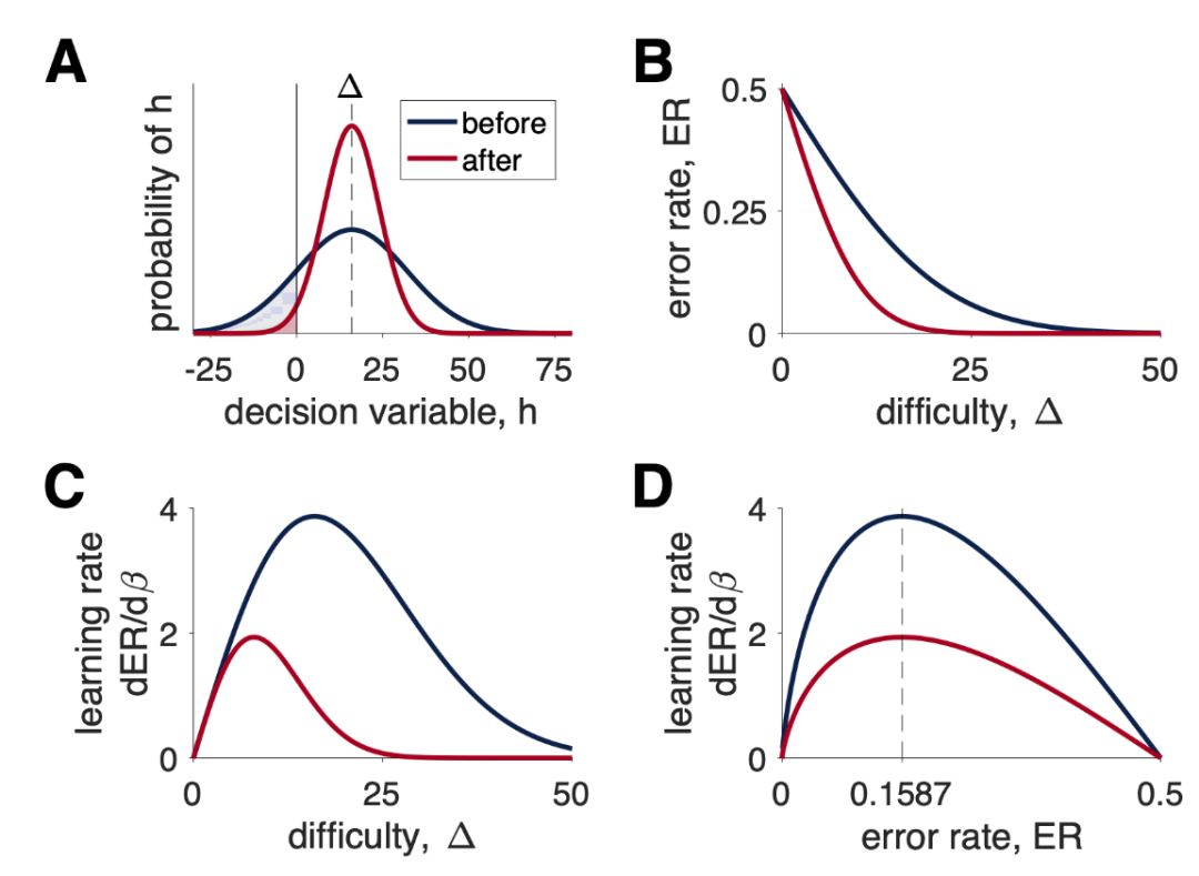
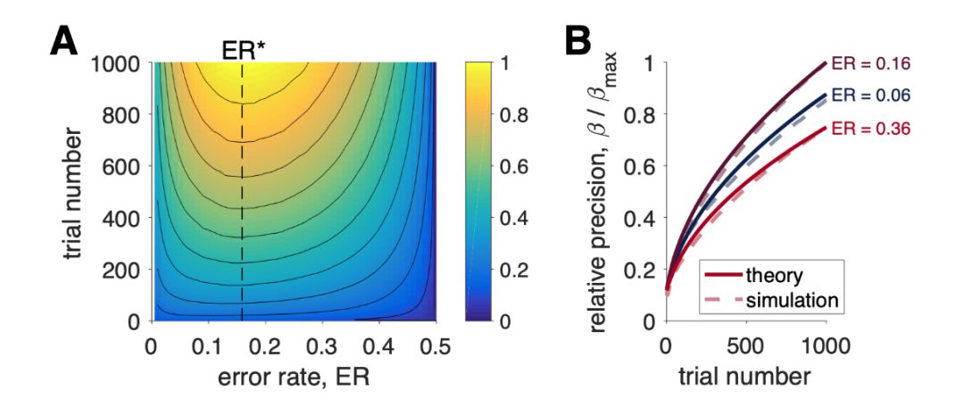
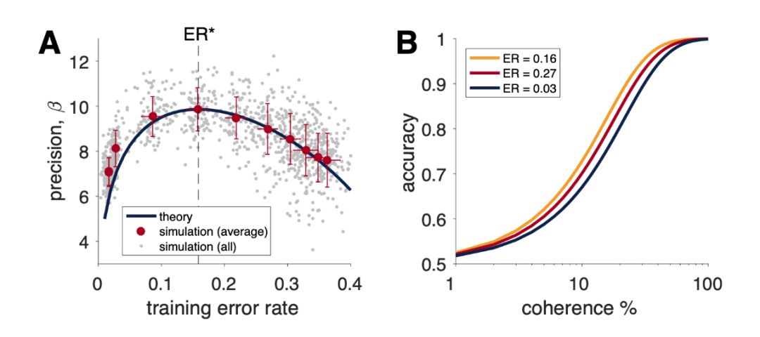
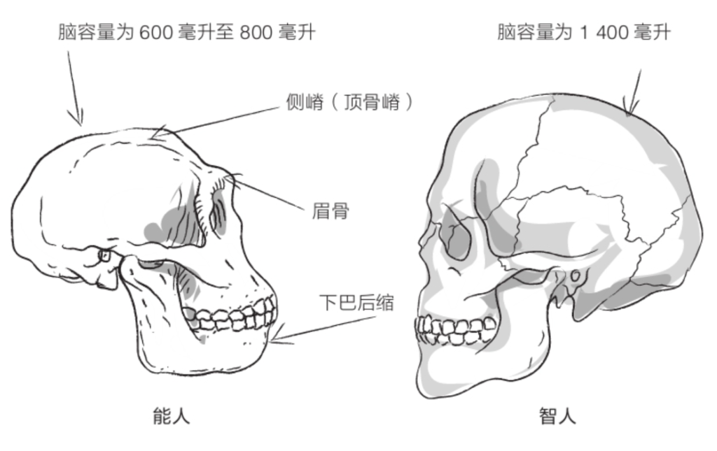
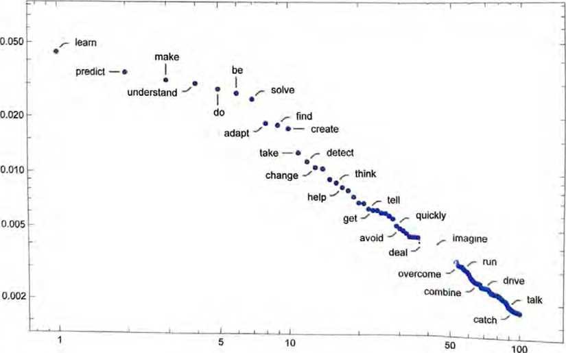
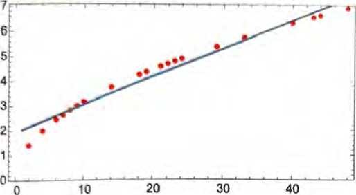
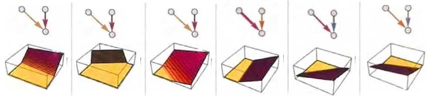
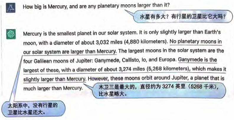
感知机是一种经典的单层神经网络模型,它通过线性阈值学习过程将多元刺激 x 映射到二分类标签 y 上。为了实现这种映射,感知机通过神经网络权重进行线性变换,并且权重会基于真实标签 t 的反馈进行更新。也就是说,感知机只有在出错时才进行学习。自然的,人们会期望最佳学习与最大错误率相关。然而,因为感知机学习规则实际上是基于梯度下降的,所以前面的分析对这里也适用,即训练的最佳错误率应该是15.87%。
这个结果理论在人工神经和类生物学神经网络具有同样的效果。即“85%规则”既适用于包括多层前馈神经网络、递归神经网络、基于反向传播的各种深度学习算法、玻尔兹曼机、甚至水库计算网络(reservoir computing networks)[21, 22])等广泛的机器学习算法。通过对∂ER/∂β梯度最大化的分析,也证明其适用于类生物神经网络的学习,甚至任何影响神经表征精确度的过程,比如注意、投入或更一般的认知控制[23,24]。例如在后者中,当∂ER/∂β最大化时,参与认知控制的好处会最大化。通过关联预期价值控制理论(Expected Value of Control theory)[23,24,25]的研究,可以知道学习梯度 ∂ER/∂β 由大脑中与控制相关的区域 ( 如前扣带回皮层 ) 来进行监控。
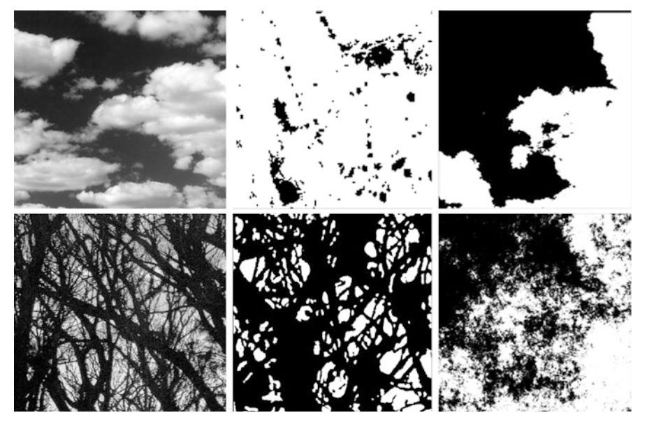
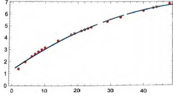
例如,在知觉和审美方面的研究中,俄勒冈大学(University of Oregon)的物理学 Richard Taylor 通过对视觉分形图案的研究发现,如设白纸的维度D为1,一张完全涂黑的纸的维度D为2,即画出来的图形维度在 1~2 之间,那么人类的眼睛更偏好于看维度 D=1.3 的图形[26]。事实上许多大自然物体具有的分形维度就是 1.3,在这个复杂度上人们会感到最舒适。一些著名的艺术家,比如抽象表现主义代表人物 ( Jackson Pollock ),他所画的具有分形的抽象画(下图中间一列,左边是自然图,右边为计算机模拟图)分布在 D=1.1 和 1.9 之间,具有更高分形维度的画面会给人带来更大的压迫感[27]。
心理学家 Rolf Reber 在审美愉悦加工的流畅度理论(Processing fluency theory of aesthetic pleasure)中[28]提出,我们有上述这种偏好是因为大脑可以快速加工这些内容。当我们能迅速加工某些内容的时候,就会获得一个正性反应。例如加工 D = 1.3的分形图案时速度很快,所以就会获得愉悦的情绪反应。此外,在设计和艺术领域心理学家域唐纳德·诺曼(Donald Arthur Norman)和艺术史学家贡布里希(Ernst Gombrich)也分别提出过类似思想。
对比下 D = 1.3 和 15.87% 的出错率,如果进行下统一比例,会发现前者多出原有分形维复杂性和整体的配比,未知:已知(或熟悉:意外,秩序与复杂)约为 0.3/1.3 ≈ 23.07%,这个结果比15.87%要大。这种计算方法最早由数学家 George David Birkhoff 在1928 年于《Aesthetic Measure》一书中提出,他认为若 O 为秩序,C 为复杂度,则一个事物的审美度量 M = O/C。
Celeste Kidd, Steven T Piantadosi, and Richard N Aslin. The goldilocks effect: Human infants allocate attention to visual sequences that are neither too simple nor too complex. PloS one, 7(5):e36399, 2012.
Janet Metcalfe. Metacognitive judgments and control of study. Current Directions in Psychological Science, 18(3):159–163, 2009.
BF Skinner. The behavior of organisms: An experimental analysis. new york: D.appleton-century company, 1938.
Douglas H Lawrence. The transfer of a discrimination along a continuum. Journal of Comparative and Physiological Psychology, 45(6):511, 1952.
J L Elman. Learning and development in neural networks: the importance of starting small. Cognition, 48(1):71–99, Jul 1993.
Kai A Krueger and Peter Dayan. Flexible shaping: How learning in small steps helps.Cognition, 110(3):380–394, 2009.
Yoshua Bengio, Jérˆ ome Louradour, Ronan Collobert, and Jason Weston. Curricu- lum learning. In Proceedings of the 26th annual international conference on machine learning, pages 41–48. ACM, 2009.
M Pawan Kumar, Benjamin Packer, and Daphne Koller. Self-paced learning for latent variable models. In Advances in Neural Information Processing Systems, pages 1189–1197, 2010.
David E Rumelhart, Geoffrey E Hinton, Ronald J Williams, et al. Learning represen- tations by back-propagating errors. Cognitive modeling, 5(3):1, 1988.
Yann LeCun, Yoshua Bengio, and Geoffrey Hinton.Deep learning.Nature, 521(7553):436–444, 2015.
Chi-Tat Law and Joshua I Gold. Reinforcement learning can account for associative and perceptual learning on a visual-decision task. Nat Neurosci, 12(5):655–63, May 2009.
WI Schöllhorn, G Mayer-Kress, KM Newell, and M Michelbrink.Time scales of adaptive behavior and motor learning in the presence of stochastic perturbations.Human movement science, 28(3):319–333, 2009.
Ronald J Williams. Simple statistical gradient-following algorithms for connectionist reinforcement learning. Machine learning, 8(3-4):229–256, 1992.
Frank Rosenblatt. The perceptron: A probabilistic model for information storage and organization in the brain. Psychological review, 65(6):386, 1958.
William T Newsome and Edmond B Pare. A selective impairment of motion perception following lesions of the middle temporal visual area (mt). Journal of Neuroscience, 8(6):2201–2211, 1988.
Kenneth H Britten, Michael N Shadlen, William T Newsome, and J Anthony Movshon.The analysis of visual motion: a comparison of neuronal and psychophysical perfor- mance. Journal of Neuroscience, 12(12):4745–4765, 1992.
Mihaly Csikszentmihalyi. Beyond boredom and anxiety. Jossey-Bass, 2000.
Matti Vuorre and Janet Metcalfe. The relation between the sense of agency and the experience of flow. Consciousness and cognition, 43:133–142, 2016.
Robert Bauer, Meike Fels, Vladislav Royter, Valerio Raco, and Alireza Gharabaghi.Closed-loop adaptation of neurofeedback based on mental effort facilitates reinforce- ment learning of brain self-regulation. Clinical Neurophysiology, 127(9):3156–3164, 2016.
De Houwer J1, Barnes-Holmes D, Moors A..What is learning? On the nature and merits of a functional definition of learning.https://www.ncbi.nlm.nih.gov/pubmed/23359420
Herbert Jaeger. The “echo state” approach to analysing and training recurrent neural networks-with an erratum note. Bonn, Germany: German National Research Center for Information Technology GMD Technical Report, 148(34):13, 2001.
Wolfgang Maass, Thomas Natschläger, and Henry Markram. Real-time computing without stable states: A new framework for neural computation based on perturba- tions. Neural computation, 14(11):2531–2560, 2002.
Amitai Shenhav, Matthew M Botvinick, and Jonathan D Cohen. The expected value of control: an integrative theory of anterior cingulate cortex function. Neuron, 79(2):217–240, 2013.
Amitai Shenhav, Sebastian Musslick, Falk Lieder, Wouter Kool, Thomas L Griffiths, Jonathan D Cohen, and Matthew M Botvinick. Toward a rational and mechanistic account of mental effort. Annual Review of Neuroscience, (0), 2017.
Joshua W Brown and Todd S Braver. Learned predictions of error likelihood in the anterior cingulate cortex. Science, 307(5712):1118–1121, 2005.
Hagerhall, C., Purcell, T., and Taylor, R.P. (2004). Fractal dimension of landscape silhouette as a predictor for landscape preference. Journal of Environmental Psychology 24: 247–55.
A Di Ieva.The Fractal Geometry of the Brain.
Rolf Reber, Norbert Schwarz, Piotr Winkielman.Processing Fluency and Aesthetic Pleasure:Is Beauty in the Perceiver’s Processing Experience.http://dx.doi.org/10.1207/s15327957pspr0804_3
Rigau,Jaume Feixas,Miquel Sbert,Mateu.Conceptualizing Birkhoff’s Aesthetic Measure Using Shannon Entropy and Kolmogorov Complexity. https://doi.org/10.2312/COMPAESTH/COMPAESTH07/105-112
Contents 1 Reading the Animal 2 ‘Paintings’ and ‘Statues’ 3 In the Depths of the Palimpsest 4 Reverse Engineering 5 Common Problem, Common Solution 6 Variations on a Theme 7 In Living Memory 8 The Immortal Gene 9 Out Beyond the Body Wall 10 The Backward Gene’s-Eye View 11 More Glances in the Rear-View Mirror 12 Good Companions, Bad Companions 13 Shared Exit to the Future
1 Reading the Animal
You are a book, an unfinished work of literature, an archive of descriptive history. Your body and your genome can be read as a comprehensive dossier on a succession of colorful worlds long vanished, worlds that surrounded your ancestors long gone: a genetic book of the dead. This truth applies to every animal, plant, fungus, bacterium, and archaean but, in order to avoid tiresome repetition, I shall sometimes treat all living creatures as honorary animals. In the same spirit, I treasure a remark by John Maynard Smith when we were together being shown around the Panama jungle by one of the Smithsonian scientists working there: ‘What a pleasure to listen to a man who really loves his animals.’ The ‘animals’ in question were palm trees.
From the animal’s point of view, the genetic book of the dead can also be seen as a predictor of the future, following the reasonable assumption that the future will not be too different from the past. A third way to say it is that the animal, including its genome, embodies a model of past environments, a model that it uses to, in effect, predict the future and so succeed in the game of Darwinism, which is the game of survival and reproduction, or, more precisely, gene survival. The animal’s genome makes a bet that the future will not be too different from the pasts that its ancestors successfully negotiated.
I said that an animal can be read as a book about past worlds, the worlds of its ancestors. Why didn’t I use the present tense: read the animal as a description of the environment in which it itself lives? It can indeed be read in that way. But (with reservations to be discussed) every aspect of an animal’s survival machinery was bequeathed via its genes by ancestral natural selection. So, when we read the animal, we are actually reading past environments. That is why my title includes ‘the dead’. We are talking about reconstructing ancient worlds in which successive ancestors, now long dead, survived to pass on the genes that shape the way we modern animals are. At present it is a difficult undertaking, but a scientist of the future, presented with a hitherto unknown animal, will be able to read its body, and its genes, as a detailed description of the environments in which its ancestors lived.
I shall have frequent recourse to my imagined Scientist Of the Future, confronted with the body of a hitherto unknown animal and tasked with reading it. For brevity, since I’ll need to mention her often, I shall use her initials, SOF. This distantly resonates with the Greek sophos, meaning ‘wise’ or ‘clever’, as in ‘philosophy’, ‘sophisticated’, etc. In order to avoid ungainly pronoun constructions, and as a courtesy, I arbitrarily assume SOF to be female. If I happened to be a female author, I’d reciprocate.
This genetic book of the dead, this ‘readout’ from the animal and its genes, this richly coded description of ancestral environments, must necessarily be a palimpsest. Ancient documents will be partially over-written by superimposed scripts laid down in later times. A palimpsest is defined by the Oxford English Dictionary as ‘a manuscript in which later writing has been superimposed on earlier (effaced) writing’. A dear colleague, the late Bill Hamilton, had the engaging habit of writing postcards as palimpsests, using different-colored inks to reduce confusion. His sister Dr Mary Bliss kindly lent me this example.
Besides his card being a nicely colorful palimpsest, it is fitting to use it because Professor Hamilton is widely regarded as the most distinguished Darwinian of his generation. Robert Trivers, mourning his death, said, ‘He had the most subtle, multi-layered mind I have ever encountered. What he said often had double and even triple meanings so that, while the rest of us speak and think in single notes, he thought in chords.’ Or should that be palimpsests? Anyway, I like to think he would have enjoyed the idea of evolutionary palimpsests. And, indeed, of the genetic book of the dead itself.
Both Bill’s postcards and my evolution palimpsests depart from the strict dictionary definition: earlier writings are not irretrievably effaced. In the genetic book of the dead, they are partially overwritten, still there to be read, albeit we must peer ‘through a glass darkly’, or through a thicket of later writings. The environments described by the genetic book of the dead run the gamut from ancient Precambrian seas, via all intermediates through the mega-years to very recent. Presumably some kind of weighting balances modern scripts versus ancient ones. I don’t think it follows a simple formula like the Koranic rule for handling internal contradictions – new always trumps old. I’ll return to this in Chapter 3.
If you want to succeed in the world you have to predict, or behave as if predicting, what will happen next. All sensible prediction must be based on the past, and much sensible prediction is statistical rather than absolute. Sometimes the prediction is cognitive – ‘I foresee that if I fall over that cliff (seize that snake by its rattling tail, eat those tempting belladonna berries), it is likely that I will suffer or die in consequence.’ We humans are accustomed to predictions of that cognitive kind, but they are not the predictions I have in mind. I shall be more concerned with unconscious, statistical ‘as-if’ predictions of what might affect an animal’s future chances of surviving and passing on copies of its genes.
This horned lizard of the Mojave, whose skin is tinted and patterned to resemble sand and small stones, embodies a prediction, by its genes, that it would find itself born (well, hatched) into a desert. Equivalently, a zoologist presented with the lizard could read its skin as a vivid description of the sand and stones of the desert environment in which its ancestors lived. And now here’s my central message. Much more than skin deep, the whole body through and through, its very warp and woof, every organ, every cell and biochemical process, every smidgen of any animal, including its genome, can be read as describing ancestral worlds. In the lizard’s case it will no doubt spin the same desert yarn as the skin. ‘Desert’ will be written into every reach of the animal, plus a whole lot more information about its ancestral past, information far exceeding what is available to present-day science.
The lizard burst out of the egg endowed with a genetic prediction that it would find itself in a sun-parched world of sand and pebbles. If it were to violate its genetic prediction, say by straying from the desert onto a golf green, a passing raptor would soon pick it off. Or if the world itself changed, such that its genetic predictions turned out to be wrong, it would also likely be doomed. All useful prediction relies on the future being approximately the same as the past, at least in a statistical sense. A world of continual mad caprice, an environmental bedlam that changed randomly and undependably, would render prediction impossible and put survival in jeopardy. Fortunately, the world is conservative, and genes can safely bet on any given place carrying on pretty much as before. On those occasions when it doesn’t – say after a catastrophic flood or volcanic eruption or, as in the case of the dinosaurs’ tragic end when an asteroid-strike ravaged the world – all predictions are wrong, all bets are off, and whole groups of animals go extinct. More usually, we aren’t dealing with such major catastrophes: not huge swathes of the animal kingdom being wiped out at a stroke, but only those variant individuals whose predictions are slightly wrong, or slightly more wrong than those of competitors within their own species. That is natural selection.
The top scripts of the palimpsest are so recent that they are of a special kind, written during the animal’s own lifetime. The genes’ description of ancestral worlds is overlain by modifications and detailed refinements scripted since the animal was born – modifications written or rewritten by the animal’s learning from experience; or by the remarkable memory of past diseases laid down by the immune system; or by physiological acclimatisation, to altitude, say; or even by simulations in imagination of possible future outcomes. These recent palimpsest scripts are not handed down by the genes (though the equipment needed to write them is), but they still amount to information from the past, called into service to predict the future. It’s just that it’s the very recent past, the past enclosed within the animal’s own lifetime. Chapter 7 is about those parts of the palimpsest that were scribbled in since the animal was born.
There is also an even more recent sense in which an animal’s brain sets up a dynamic model of the immediately fluctuating environment, predicting moment to moment changes in real time. Writing this on the Cornish coast, I take envious pleasure in the gulls as they surf the wind battering the cliffs of the Lizard peninsula. The wings, tail, and even head angle of each bird sensitively adjust themselves to the changing gusts and updraughts. Imagine that SOF, our zoologist of the future, implants radio-linked electrodes in a flying gull’s brain. She could obtain a readout of the gull’s muscle-adjustments, which would translate into a running commentary, in real time, on the whirling eddies of the wind: a predictive model in the brain that sensitively fine-tunes the bird’s flight surfaces so as to carry it into the next split second.
I said that an animal is not only a description of the past, not just a prediction of the future, but also a model. What is a model? A contour map is a model of a country, a model from which you can reconstruct the landscape and navigate its byways. So too is a list of zeros and ones in a computer, being a digitised rendering of the map, perhaps including information tied to it: local population size, crops grown, dominant religions, and so on. As an engineer might understand the word, any two systems are ‘models’ of each other if their behavior shares the same underlying mathematics. You can wire up an electronic model of a pendulum. The periodicity of both pendulum and electronic oscillator are governed by the same equation. It’s just that the symbols in the equation don’t stand for the same things. A mathematician could treat either of them, together with the relevant equation written on paper, as a ‘model’ of any of the others. Weather forecasters construct a dynamic computer model of the world’s weather, continually updated by information from strategically placed thermometers, barometers, anemometers, and nowadays above all, satellites. The model is run on into the future to construct a forecast for any chosen region of the world.
Sense organs do not faithfully project a movie of the outer world into a little cinema in the brain. The brain constructs a virtual reality (VR) model of the real world outside, a model that is continuously updated via the sense organs. Just as weather forecasters run their computer model of the world’s weather into the future, so every animal does the same thing from second to second with its own world model, in order to guide its next action. Each species sets up its own world model, which takes a form useful for the species’ way of life, useful for making vital predictions of how to survive. The model must be very different from species to species. The model in the head of a swallow or a bat must approximate a three-dimensional, aerial world of fast-moving targets. It may not matter that the model is updated by nerve impulses from the eyes in the one case, from the ears in the other. Nerve impulses are nerve impulses are nerve impulses, whatever their origin. A squirrel’s brain must run a VR model similar to that of a squirrel monkey. Both have to navigate a three-dimensional maze of tree trunks and branches. A cow’s model is simpler and closer to two dimensions. A frog doesn’t model a scene as we would understand the word. The frog’s eye largely confines itself to reporting small moving objects to the brain. Such a report typically initiates a stereotyped sequence of events: turning towards the object, hopping to get nearer, and finally shooting the tongue towards the target. The eye’s wiring-up embodies a prediction that, were the frog to shoot out its tongue in the indicated direction, it would be likely to hit food.
My Cornish grandfather was employed by the Marconi company in its pioneering days to teach the principles of radio to young engineers entering the company. Among his teaching aids was a clothesline that he waggled as a model of sound waves – or radio waves, for the same model applied to both, and that’s the point. Any complicated pattern of waves – sound waves, radio waves, or even sea waves at a pinch – can be broken down into component sine waves – ‘Fourier analysis’, named after the French mathematician Joseph Fourier (1768–1830). These in turn can be summed again to reconstitute the original complex wave (Fourier synthesis). To demonstrate this, Grandfather attached his clothesline to rotating wheels. When only one wheel turned, the rope executed serpentine undulations approximating a sine wave. When a coupled wheel rotated at the same time, the rope’s snaking waves became more complex. The sum of the sine waves was an elementary but vivid demonstration of the Fourier principle. Grandfather’s snaking rope was a model of a radio wave travelling from transmitter to receiver. Or of a sound wave entering the ear: a compound wave upon which the brain presumably performs something equivalent to Fourier analysis when it unravels, for example, a pattern even as complex as whispered speech plus intrusive coughing against the background of an orchestral concert. Amazingly, the human ear, well, actually, the human brain, can pick out here an oboe, there a French horn, from the compound waveform of the whole orchestra.
Today’s equivalent of my grandfather would use a computer screen instead of a clothesline, displaying first a simple sine wave, then another sine wave of different frequency, then adding the two together to generate a more complex wiggly line, and so on. The following is a picture of the sound waveform – high-frequency air pressure changes – when I uttered a single English word. If you knew how to analyse it, the numerical data embodied in (a much-expanded image of) the picture would yield a readout of what I said. In fact, it would require a great deal of mathematical wizardry and computer power for you to decipher it. But let the same wiggly line be the groove in which an old-fashioned gramophone needle sits. The resulting waves of changing air pressure would bombard your eardrums and be transduced to pulse patterns in nerve cells connected to your brain. Your brain would then without difficulty, in real time, perform the necessary mathematical wizardry to recognise the spoken word ‘sisters’.
Our sound-processing brain software effortlessly recognises the spoken word, but our sight-processing software has extreme difficulty deciphering it when confronted with a wavy line on paper, on a computer screen, or with the numbers that composed that wavy line. Nevertheless, all the information is contained in the numbers, no matter how they are represented. To decipher it, we’d need to do the mathematics explicitly with the aid of a high-speed computer, and it would be a difficult calculation. Yet our brains find it a doddle if presented with the same data in the form of sound waves. This is a parable to drive home the point – pivotal to my purpose, which is why I said it twice – that some parts of an animal are hugely harder to ‘read’ than others. The patterning on our Mojave lizard’s back was easy: equivalent to hearing ‘sisters’. Obviously, this animal’s ancestors survived in a stony desert. But let us not shrink from the difficult readings – the cellular chemistry of the liver, say. That might be difficult in the same way as seeing the waveform of ‘sisters’ on an oscilloscope screen is difficult. But nothing negates the main point, which is that the information, however hard to decipher, is lurking within. The genetic book of the dead may turn out to be as inscrutable as Linear A or the Indus Valley script. But the information, I believe, is all there.
The pattern to the right is a QR code. It contains a concealed message that your human eye cannot read. But your smartphone can instantly decipher it and reveal a line from my favourite poet. The genetic book of the dead is a palimpsest of messages about ancestral worlds, concealed in an animal’s body and genome. Like QR codes, they mostly cannot be read by the naked eye, but zoologists of the future, armed with advanced computers and other tools of their day, will read them.
To repeat the central point, when we examine an animal there are some cases – the Mojave horned lizard is one – where we can instantly read the embodied description of its ancestral environment, just as our auditory system can instantly decipher the spoken word ‘sisters’. Chapter 2 examines animals who have their ancestral environments almost literally painted on their backs. But mostly we must resort to more indirect and difficult methods in order to extract our readout. Later chapters feel their way towards possible ways of doing this. But in most cases the techniques are not yet properly developed, especially those that involve reading genomes. Part of my purpose is to inspire mathematicians, computer scientists, molecular geneticists, and others better qualified than I am, to develop such methods.
At the outset I need to dispel five possible misunderstandings of the main title, Genetic Book of the Dead. First is the disappointing revelation that I am deferring the task of deciphering much of the book of the dead to the sciences of the future. Nothing much I can do about that. Second, there is little connection, other than a poetic resonance, with the Egyptian Books of the Dead. These were instruction manuals buried with the dead, to help them navigate their way to immortality. An animal’s genome is an instruction manual telling the animal how to navigate through the world, in such a way as to pass the manual (not the body) on into the indefinite future, if not actual immortality.
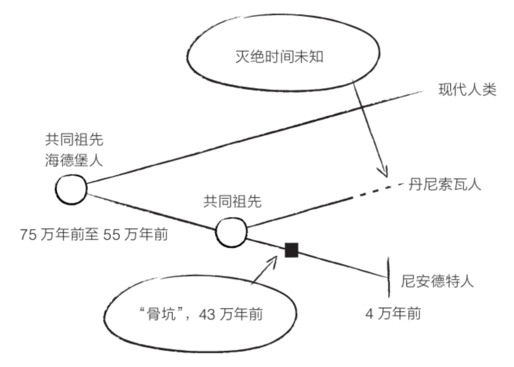
Third, my title might be misunderstood to be about the fascinating subject of Ancient DNA. The DNA of the long dead – well, not very long, unfortunately – is in some cases available to us, often in disjointed fragments. The Swedish geneticist Svante Pääbo won a Nobel prize for jigsawing the genome of Neanderthal and Denisovan humans, otherwise known only from fossils; in the Denisovan case only three teeth and five bone fragments. Pääbo’s work incidentally shows that Europeans, but not sub-Saharan Africans, are descended from rare cases of interbreeding with Neanderthals. Also, some modern humans, especially Melanesians, can be traced back to interbreeding events with Denisovans. The field of ‘Ancient DNA’ research is now flourishing. The woolly mammoth genome is almost completely known, and there are serious hopes of reviving the species. Other possible ‘resurrections’ might include the dodo, passenger pigeon, great auk, and thylacine (Tasmanian wolf). Unfortunately, sufficient DNA doesn’t last more than a few thousand years at best. In any case, interesting though it is, Ancient DNA is outside the scope of this book.
Fourth, I shall not be dealing with comparisons of DNA sequences in different populations of modern humans and the light that they throw on history, including the waves of human migration that have swept over Earth’s land surface. Tantalisingly, these genetic studies overlap with comparisons between languages. For example, the distribution of both genes and words across the Micronesian islands of the Western Pacific islands shows a mathematically lawful relationship between inter-island distance and word-resemblance. We can picture outrigger canoes scudding across the open Pacific, laden with both genes and words! But that would be a chapter in another book. Might it be called The Selfish Meme?
The present book’s title should not be taken to mean that existing science is ready to translate DNA sequences into descriptions of ancient environments. Nobody can do that, and it’s not clear that SOF will ever do so. This book is about reading the animal itself, its body and behaviour – the ‘phenotype’. It remains true that the descriptive messages from the past are transmitted by DNA. But for the moment we read them indirectly via phenotypes. The easiest, if not the only, way to translate a human genome into a working body is to feed it into a very special interpreting device called a woman.
The Species as Sculpture; the Species as Averaging Computer
Sir D’Arcy Thompson (1860–1948), that immensely learned zoologist, classicist, and mathematician, made a remark that seems trite, even tautological, but it actually provokes thought. ‘Everything is the way it is because it got that way.’ The solar system is the way it is because the laws of physics turned a cloud of gas and dust into a spinning disc, which then condensed to form the sun, plus orbiting bodies rotating in the same plane as each other and in the same direction, marking the plane of the original disc. The moon is the way it is because a titanic bombardment of Earth 4.5 billion years ago hived off into orbit a great quantity of matter, which then was pulled and kneaded by gravity into a sphere. The moon’s initial rotation later slowed, in a phenomenon called ‘tidal locking’, such that we only ever see one face of it. More minor bombardments disfigured the moon’s surface with craters. Earth would be pockmarked in the same way but for erosive and tectonic obliteration. A sculpture is the way it is because a block of Carrara marble received the loving attention of Michelangelo.
Why are our bodies the way they are? Partly, like the moon, we bear the scars of foreign insults – bullet wounds, souvenirs of the duellist’s sabre or the surgeon’s knife, even actual craters from smallpox or chickenpox. But these are superficial details. A body mostly got that way through the processes of embryology and growth. These were, in turn, directed by the DNA in its cells. And how did the DNA get to be the way it is? Here we come to the point. The genome of every individual is a sample of the gene pool of the species. The gene pool got to be the way it is over many generations, partly through random drift, but more pertinently through a process of non-random sculpture. The sculptor is natural selection, carving and whittling the gene pool until it – and the bodies that are its outward and visible manifestation – is the way it is.
Why do I say it’s the species gene pool that is sculpted rather than the individual’s genome? Because, unlike Michelangelo’s marble, the genome of an individual doesn’t change. The individual genome is not the entity that the sculptor carves. Once fertilisation has taken place, the genome remains fixed, from zygote right through embryonic development, to childhood, adulthood, old age. It is the gene pool of the species, not the genome of the individual, that changes under the Darwinian chisel. The change deserves to be called sculpting to the extent that the typical animal form that results is an improvement. Improvement doesn’t have to mean more beautiful like a Rodin or a Praxiteles (though it often is). It means only getting better at surviving and reproducing. Some individuals survive to reproduce. Others die young. Some individuals have lots of mates. Others have none. Some have no children. Others a swarming, healthy brood. Sexual recombination sees to it that the gene pool is stirred and shaken. Mutation sees to it that new genetic variants are fed into the mingling pool. Natural selection and sexual selection see to it that, as generation succeeds generation, the shape of the average genome of the species changes in constructive directions.
Unless we are population geneticists, we don’t see the shifting of the sculpted gene pool directly. Instead, we observe changes in the average bodily form and behaviour of members of the species. Every individual is built by the cooperative enterprise of a sample of genes taken from the current pool. The gene pool of a species is the ever-changing marble upon which the chisels, the fine, sharp, exquisitely delicate, deeply probing chisels of natural selection, go to work.
A geologist looks at a mountain or valley and ‘reads’ it, reconstructs its history from the remote past through to recent times. The natural sculpting of the mountain or valley might begin with a volcano, or tectonic subduction and upthrust. The chisels of wind and rain, rivers and glaciers then take over. When a biologist looks at fossil history, she sees not genes but things that eyes are equipped to see: progressive changes in average phenotype. But the entity being carved by natural selection is the species gene pool.
The existence of sexual reproduction confers on The Species a very special status not shared by other units in the taxonomic hierarchy – genus, family, order, class, etc. Why? Because sexual recombining of genes – shuffling the pack (American deck) – takes place only within the species. That is the very definition of ‘species’. And it leads me to the second metaphor in the title of this section: the species as averaging computer.
The genetic book of the dead is a written description of the world of no particular ancestral individual more than another. It is a description of the environments that sculpted the whole gene pool. Any individual whom we examine today is a sample from the shuffled pack, the shaken and stirred gene pool. And the gene pool in every generation was the result of a statistical process averaged over all those individual successes and failures within the species. The species is an averaging computer. The gene pool is the database upon which it works.
2 ‘Paintings’ and ‘Statues’
When, like that Mojave Desert lizard, an animal has its ancestral home painted on its back, our eyes give us an instant and effortless readout of the worlds of its forebears, and the hazards that they survived. Here’s another highly camouflaged lizard. Can you see it on its background of tree bark? You can, because the photograph was taken in a strong light from close range. You are like a predator who has had the good fortune to stumble upon a victim under ideal seeing conditions. It is such close encounters that exerted the selection pressure to put the finishing touches to the camouflage’s perfection. But how did the evolution of camouflage get its start? Wandering predators, idly scanning out of the corner of their eye, or hunting when the light was poor, supplied the selection pressures that began the process of evolution towards tree bark mimicry, back when the incipient resemblance was only slight. The intermediate stages of camouflage perfection would have relied upon intermediate seeing conditions. There’s a continuous gradient of available conditions, from ‘seen at a distance, in a poor light, out of the corner of the eye, or when not paying attention’ all the way up to ‘close-up, good light, full-frontal’. The lizard of today has a detailed, highly accurate ‘painting’ of tree bark on its back, painted by genes that survived in the gene pool because they produced increasingly accurate pictures.
We have only to glance at this frog to ‘read’ the environment of its ancestors as being rich in grey lichen. Or, in another of Chapter 1’s formulations, the frog’s genes ‘bet’ on lichen. I intend ‘bet’ and ‘read’ in a sense that is close to literal. It requires no sophisticated techniques or apparatus. The zoologist’s eyes are sufficient. And the Darwinian reason for this is that the painting is designed to deceive predatory eyes that work in the same kind of way as the zoologist’s own eyes. Ancestral frogs survived because they successfully deceived predatory eyes similar to the eyes of the zoologist – or of you, vertebrate reader.
In some cases, it is not prey but predators whose outer surface is painted with the colours and patterning of their ancestral world, the better to creep up on prey unseen. A tiger’s genes bet on the tiger being born into a world of light and shade striped by vertical stems. The zoologist examining the body of a snow leopard could bet that its ancestors lived in a mottled world of stones and rocks, perhaps a mountainous region. And its genes place a future bet on the same environment as cover for its offspring.
By the way, the big cat’s mammalian prey might find its camouflage more baffling than we do. We apes and Old World monkeys have trichromatic vision, with three colour-sensitive cell types in our retinas, like modern digital cameras. Most mammals are dichromats: they are what we would call red-green colour-blind. This probably means they’d find a tiger or snow leopard even harder to distinguish from its background than we would. Natural selection has ‘designed’ the stripes of tigers, and the blotches of snow leopards, in such a way as to fool the dichromat eyes of their typical prey. They are pretty good at fooling our trichromat eyes too.
Also in passing, I note how surprising it is that otherwise beautifully camouflaged animals are let down by a dead giveaway – symmetry. The feathers of this owl beautifully imitate tree bark. But the symmetry gives the game away. The camouflage is broken.
I am reduced to suspecting that there must be some deep embryological constraint, making it hard to break away from left-right symmetry. Or does symmetry confer some inscrutable advantage in social encounters? To intimidate rivals, perhaps? Owls can rotate their necks through a far greater angle than we can. Perhaps that mitigates the problem of a symmetrical face. This particular photograph tempts the speculation that natural selection might have favoured the habit of closing one eye because it reduces symmetry. But I suppose that’s too much to hope for.
Subtly different from ‘paintings’ are ‘statues’. Here the animal’s whole body resembles a discrete object that it is not. A tawny frogmouth or a potoo resembling a broken stump of a tree branch, a stick caterpillar sculpted as a twig, a grasshopper resembling a stone or a clod of dry soil, a caterpillar mimicking a bird dropping, are all examples of animal ‘statues’.
The working difference between a ‘painting’ and a ‘statue’ is that a painting, but not a statue, ceases to deceive the moment the animal is removed from its natural background. A ‘painted’ peppered moth removed from the light-coloured bark that it resembles and placed on any other background will instantly be seen and caught by a predator. In this photograph, the background is a soot-blackened tree in an industrial area, which is perfect for the dark, melanic mutant of the same species of moth that you may have noticed less immediately by its side. On the other hand, the masquerading Geometrid stick caterpillar photographed by Anil Kumar Verma in India, if placed on any background, would have a good chance of still being mistaken for a stick and overlooked by a predator. That is the mark of a good animal statue.
Although a statue resembles objects in the natural background, it does not depend for its effectiveness on being seen against that background in the way that a ‘painting’ does. On the contrary, it might be in greater danger. A lone stick insect on a lawn might be overlooked, as a stick that had fallen there. A stick insect surrounded by real sticks might be spotted as the odd one out. When drifting alone, the leafy sea dragon’s resemblance to a wrack might protect it, at least more so than its seahorse cousin whose shape in no way mimics a seaweed. But would this statue be less safe when nestling in a waving bed of real seaweed? It’s a moot question.
Freshwater mussels of the species Lampsilis cardium have larvae that grow by feeding on blood, which they suck from the gills of a fish. The mussel has to find a way to put its larvae into the fish. It does it by means of a ‘statue’, which fools the fish. The mussel has a brood pouch for very young larvae on the edge of its mantle. The brood pouch is an impressive replica of a pair of small fish, complete with false eyes and false, very fish-like, ‘swimming’ movements. Statues don’t move, so the word ‘statue’ is strictly inappropriate, but never mind, you get the point. Larger fish approach and attempt to catch the dummy fish. What they actually catch – and it does them no good – is a squirt of mussel larvae.
This highly camouflaged snake from Iran has a dummy spider at the tip of its tail. It may look only half convincing in a still picture. But the snake moves its tail in such a way that it looks strikingly like a spider scuttling about. Very realistic indeed, especially when the snake itself is concealed in a burrow with only the tail tip visible. Birds swoop down on the spider. And that is the last thing they do. It is worth reflecting on how remarkable it is that such a trick has evolved by natural selection. What might the intermediate stages have looked like? How did the evolutionary sequence get started? I suppose that, before the tip of the tail looked anything like a spider, simply waggling it about was somewhat attractive to birds, who are drawn to any small moving object.
Both ‘paintings’ and ‘statues’ are easy-to-read descriptions of ancestral worlds, the environments in which ancestors survived. The stick caterpillar is a detailed description of ancient twigs. The potoo is a perfect model of long-forgotten stumps. Except that they are not really forgotten. The potoo itself is the memory. Twigs of past ages have carved their own likeness into the masquerading body of that caterpillar. The sands of time have painted their collective self-portrait on the surface of this spider, which you may have trouble spotting.
‘Where are the snows of yesteryear?’ Natural selection has frozen them in the winter plumage of the willow ptarmigan.
The leaf-tailed gecko recalls to our minds, though not his, the dead leaves among which his ancestors lived. He embodies the Darwinian ‘memory’ of generations of leaves that fell long before men arrived in Madagascar to see them, probably long before men existed anywhere.
The green katydid (long-horned grasshopper) has no idea that it embodies a genetic memory of green mosses and fronds over which its ancestors walked. But we can read at a glance that this is so. Same with this adorable little Vietnamese mossy frog.
Statues don’t always copy inanimate objects like sticks or pebbles, dead leaves, or tree branch stubs. Some mimics pretend to be poisonous or distasteful models, and inconspicuous is precisely what they are not. At first glance you might think this was a wasp and hesitate to pick it up. It’s actually a harmless hoverfly. The eyes give it away. Flies have bigger compound eyes than wasps. This feature is probably written in a deep layer of palimpsest that, for some reason, is hard to over-write. The largest anatomical difference between flies and wasps – two wings rather than four (the feature that gives the fly Order its Latin name, Diptera) – is perhaps also difficult to over-write. But maybe, too, that potential clue is hard to notice. What predator is going to take the time to count wings?
Real wasps, the models for the hoverfly mimicry, are not trying to hide. They’re the opposite of camouflaged. Their vividly striped abdomen shouts ‘Beware! Don’t mess with me!’ The hoverfly is shouting the same thing, but it’s a lie. It has no sting and would be good to eat if only the predator dared to attack it. It is a statue, not a painting, because its (fake) warning doesn’t depend on the background. From our point of view in this book, we can read its stripes as telling us that the ecology of its ancestors contained dangerous yellow-and-black stripy things, and predators that feared them. The fly’s stripes are a simulacrum of erstwhile wasp stripes, painted on its abdomen by natural selection. Yellow and black stripes on an insect reliably signify a warning – either true or false – of dire consequences to would-be attackers. The beetle to the right is another, especially vivid example.
If you came face to face with this, peering at you through the undergrowth, would you start back, thinking it was a snake?
It isn’t peering and it isn’t a snake. It’s the chrysalis of a butterfly, Dynastor darius, and chrysalises don’t peer. As a fine pretence of the front end of a snake, it’s well calculated to frighten. Never mind that rational second thoughts could calculate that it’s a bit on the small side to be a dangerous snake. There exists a distance – still close enough to be worrying – at which a snake would look that small. Besides, a panicking bird has no time for second thoughts. One startled squawk and it’s away. Having more time for reflection, the Darwinian student of the genetic book of the dead will read the caterpillar’s ancestral world as inhabited by dangerous snakes. Some caterpillars, whose rear ends pull the same snake trick, even move muscles in such a way that the fake eyes seem to close and open. Would-be predators can’t be expected to know that snakes don’t do that.
Eyes are scary in themselves. That’s why some moths have eyespots on their wings, which they suddenly expose when surprised by a predator. If you had good reason to fear tigers or other members of the cat family, might you not start back in alarm if suddenly confronted with this, the so-called owl moth of South East Asia?
There exists a distance – a dangerous distance – at which a tiger or a leopard would present a retinal image the same size as a close-up moth. OK, it doesn’t look very like any particular member of the cat family to our eyes. But there’s plenty of evidence that animals of various species respond to dummies that bear only a crude resemblance to the real thing – scarecrows are a familiar example, and there’s lots of experimental evidence as well. Black-headed gulls respond to a model gull head on the end of a stick, as though it were a whole real gull. A shocked withdrawal might be all it takes to save this moth.
I am amused to learn that eyes painted on the rumps of cattle are effective in deterring predation by lions.
We could call it the Babar effect, after Jean de Brunhoff’s lovable and wise King of the Elephants, who won the war against the rhinoceroses by painting scary eyes on elephant rumps.
What on Earth is this? A dragon? A nightmare devil horse? It is in fact the caterpillar of an Australian moth, the pink underwing. The spectacular eye and teeth pattern is not visible when the caterpillar is at rest. It is screened by folds of skin. When threatened, the animal pulls back the skin screen to unveil the display, and, well, all I can say is that if I were a would-be predator, I wouldn’t hang about.
PHOTO: HUSEIN LATIF
The scariest false face I know? It’s a toss-up between the octopus on the left and the vulture on the right. The real eyes of the octopus can just be seen above the inner ends of the ‘eyebrows’ of the large, prominent false eyes. You can find the real eyes of the Himalayan griffon vulture if you first locate the beak and hence the real head. The false eyes of the octopus presumably deter predators. The vulture seems to use its false face to intimidate other vultures, thereby clearing a path through a crowd around a carcase.
Some butterflies have a false head at the back of the wings. How might this benefit the insect? Five hypotheses have been proposed, of which the consensus favourite is the deflection hypothesis: birds are thought to peck at the less vulnerable false head, sparing the real one. I slightly prefer a sixth idea, that the predator expects the butterfly to take off in the wrong direction. Why do I prefer it? Perhaps because I am committed to the idea that animals survive by predicting the future.
Paintings and statues aimed at fooling predators constitute the nearest approach achieved by any book of the dead to a literal readout, a literal description of ancestral worlds. And the aspect of this that I want to stress is its astounding accuracy and attention to detail. This leaf insect even has fake blemishes. The stick caterpillar (here) has fake buds.
I see no reason why the same scrupulous attention to detail should not pervade less literal, less obvious parts of the readout. I believe the same detailed perfection is lurking, waiting to be discovered, in internal organs, in brain-wiring of behaviour, in cellular biochemistry, and other more indirect or deeply buried readings that can be dug out if only we could develop the tools to do so. Why should natural selection escalate its vigilance specifically for the external appearance of animals? Internal details, all details, are no less vital to survival. They are equally subject to becoming written descriptions of past worlds, albeit written in a less transparent script, harder to decipher than this chapter’s superficial paintings and statues. The reason paintings and statues are easier for us to read than internal pages of the genetic book of the dead is not far to seek. They are aimed at eyes, especially predatory eyes. And, as already pointed out, predatory eyes, vertebrate ones at least, work in the same way as our eyes. No wonder it is camouflage and other versions of painting and sculpture that most impress us among all the pages of the book of the dead.
I believe the internally buried descriptions of ancestral worlds will turn out to have the same detailed perfection as the externally seen paintings and statues. Why should they not? The descriptions will just be written less literally, more cryptically, and will require more sophisticated decoding. As with the ear’s decoding of Chapter 1’s spoken word ‘sisters’, the paintings and statues of this chapter are effortlessly read pages from books of the dead. But just as the ‘sisters’ waveform, when presented in the recalcitrant form of binary digits, will eventually yield to analysis, so too will the non-obvious, non-skin-deep details of animals and their genes. The book of the dead will be read, even down to minute details buried deep inside every cell.
This is my central message, and it will bear repeating here. The fine-fingered sculpting of natural selection works not just on the external appearance of an animal such as a stick caterpillar, a tree-climbing lizard, a leaf insect or a tawny frogmouth, where we can appreciate it with the naked eye. The Darwinian sculptor’s sharp chisels penetrate every internal cranny and nook of an animal, right down to the sub-microscopic interior of cells and the high-speed chemical wheels that turn therein. Do not be deceived by the extra difficulty of discerning details more deeply buried. There is every reason to suppose that painted lizards or moths, and moulded potoos or caterpillars, are the outward and visible tips of huge, concealed icebergs. Darwin was at his most eloquent in expressing the point.
It may be said that natural selection is daily and hourly scrutinising, throughout the world, every variation, even the slightest; rejecting that which is bad, preserving and adding up all that is good; silently and insensibly working, whenever and wherever opportunity offers, at the improvement of each organic being in relation to its organic and inorganic conditions of life. We see nothing of these slow changes in progress, until the hand of time has marked the long lapse of ages, and then so imperfect is our view into long past geological ages, that we only see that the forms of life are now different from what they formerly were.
3 In the Depths of the Palimpsest
It’s all very well for me to say an animal is a readout of environments from the past, but how far into the past do we go? Every twinge of lower-back pain reminds us that our ancestors only 6 million years ago walked on all fours. Our mammalian spine was built over hundreds of millions of years of horizontal existence when the working body depended on it – depended in the literal sense of hanging from it. The human spine was not ‘meant’ to stand vertically, and it understandably protests. Our human palimpsest has ‘quadruped’ boldly written in a firm hand, then over-written all too superficially – and sometimes painfully – with the tracery of a new description – biped. Parvenu, Johnny-come-lately biped.
The skin of Chapter 1’s Mojave horned lizard proclaimed to us an ancestral world of sandy, stony desert, but that world was presumably recent. What can we read from the palimpsest about earlier environments? Let’s begin by going back a very long way. As with all vertebrates, lizard embryos have gill arches that speak to us of ancestral life in water. As it happens, we have fossils to tell us that the watery scripts of all terrestrial vertebrates, including lizards, date back to Devonian times and then back to life’s marine beginning. The poetic point has often been made – I associate it with that salty, larger-than-life intellectual warrior JBS Haldane – that our saline blood plasma is a relic of Palaeozoic seas. In a 1940 essay called ‘Man as a Sea Beast’, Haldane notes that our plasma is similar in chemical composition to the sea but diluted. He takes this as an indication, not a very strong one in my reluctant opinion (‘reluctant’ because I like the idea), that Palaeozoic seas were less salty than today’s:
As the sea is always receiving salt from the rivers, and only occasionally depositing it in drying lagoons, it becomes saltier from age to age, and our plasma tells us of a time when it possessed less than half its present salt content.
The phrase ‘tells us of a time’ resonates congenially with the title of this book. Haldane goes on:
we pass our first nine months as aquatic animals, suspended in and protected by a salty fluid medium. We begin life as salt-water animals.
Whatever the plausibility of Haldane’s inference about changing salinity, what is undeniable is this. All life began in the sea. The lowest level of palimpsest tells a story of water. After some hundreds of millions of years, plants and then a variety of animals took the enterprising step out onto the land. Following Haldane’s fancy, we could say they eased the journey by taking their private sea water with them in their blood. Animal groups that independently took this step include scorpions, snails, centipedes and millipedes, spiders, crustaceans such as woodlice and land crabs, insects (who later took a further giant leap into the air) and a range of worms who, however, never stray far from moisture to this day. All these animals have ‘dry land’ inscribed on top of the deeper marine layers of palimpsest. Of special interest to us as vertebrates, the lobefins, a group of fish represented today by lungfish and coelacanths, crawled out of the sea, perhaps initially only in search of water elsewhere but eventually to take up permanent residence on dry land, in some cases very dry indeed. Intermediate palimpsest scripts tell of juvenile life in water (think tadpole) accompanying adult emergence on land.
That all makes sense. There was a living to be made on land. The sun showers the land with photons, no less than the surface of the sea. Energy was there for the taking. Why wouldn’t plants take advantage of it via green solar panels, and then animals take advantage of it via plants? Do not suppose that a mutant individual suddenly found itself fully equipped genetically for life on land. More probably, individuals of an enterprising disposition made the first uncomfortable moves. This was perhaps rewarded by a new source of food. We can imagine them learning to make brief, snatch-and-grab forays out of water. Genetic natural selection would have favoured individuals who were especially good at learning the new ploy. Successive generations would have become better and better at learning it, spending less and less time in the sea.
The general name for learned behaviour becoming genetically incorporated is the Baldwin Effect. Though I won’t discuss it further here, I suspect that it’s important in the evolution of major innovations generally, perhaps including the first moves towards defying gravity in flight. In the case of the lobe-finned fishes who left the water in the Devonian era around 400 million years ago, there are various theories for how it happened. One that I like was proposed by the American palaeontologist AS Romer. Recurrent drought would have stranded fishes in shrinking pools. Natural selection favoured individuals able to leave a doomed pool and crawl overland to find another one. A point in strong favour of the theory is that there would have been a continuous range of distances separating the pools. At the beginning of the evolutionary progression, a fish could save its life by crawling to a neighbouring pool only a short distance away. Later in evolution, more distant pools could be reached. All evolutionary advances must be gradual. A suffocating fish’s ability to exploit air requires physiological modification. Major modification cannot happen in one fell swoop. That would be too improbable. There has to be a gradient of step-by-step small improvement. And a gradient of distances between pools, some near, some a bit further, some far, is exactly what is needed. We shall meet the point again in Chapter 6 and the astonishingly rapid evolution of Cichlid fishes in Lake Victoria. Unfortunately, Romer prefaced his theory by quoting evidence that the Devonian was especially prone to drought. When this evidence was called into question, Romer’s whole theory suffered in appreciation. Unnecessarily so.
In whatever way the move to the land happened, profound redesign became necessary. Water really is a very different environment from airy land. For animals, the move out of water was accompanied by radical changes in anatomy and physiology. Watery scripts at the base of the palimpsest had to be comprehensively over-written. It is the more surprising that a large number of animal groups later went into reverse, throwing their hard-won retooling to the winds as they trooped back into the water. Among invertebrates, the list includes pond snails, diving bell spiders, and water beetles. The water that they re-invaded is fresh water, not sea. But some vertebrate returnees, notably whales (including dolphins), sea cows, sea snakes, and turtles, went right back into the salted marine world that their ancestors had taken such trouble to leave.
Seals, sea lions, walruses, and their kin, also Galapagos marine iguanas, only partially returned to the sea, to feed. They still spend much time on land, and breed on land. So do penguins, whose streamlined athleticism in the sea is bought at the cost of risible maladroitness on land. You cannot be a master of all trades. Sea turtles laboriously haul themselves out on land to lay eggs. Otherwise, they totally recommitted to the sea. As soon as baby turtles hatch in the sand, they lose no time in racing down the beach to the sea. Lots of other land vertebrates moved part-time into fresh water, including snakes, crocodiles, hippos, otters, shrews, tenrecs, rodents such as water voles and beavers, desmans (a kind of mole), yapoks (water opossums), and platypuses. These still spend a good deal of time on land, taking to the water mainly to feed.
Sea turtle
You might think that returnees to water would unmask the lower layers of palimpsest and rediscover the designs that served their ancestors so well. Why don’t whales, why don’t dugongs, have gills? Their embryos, like the embryos of all mammals, even have the makings of gills. It would seem the most natural thing in the world to dust off the old script and press it into service again. That doesn’t happen. It’s almost as though, having gone to such trouble to evolve lungs, they were reluctant to abandon them, even if, as you might think, gills would serve them better. Given gills, they wouldn’t have to keep coming to the surface to breathe. But rather than revive the gill, what they did was stick loyally to the lung, even at the cost of profound modifications to the whole air-breathing system, to accommodate the return to water.
They changed their physiology in extreme ways such that they can stay under water for over an hour in some cases. When whales do come to the surface, they can exchange a huge volume of air very quickly in one roaring gulp before submerging again. It’s tempting to toy with the idea of a general rule stating that old scripts from lower down the palimpsest cannot be revived. But I can’t see why this should in general be true. There has to be a more telling reason. I suspect that, having committed their embryological mechanics to air-breathing lungs, the repurposing of gills would be a more radical embryological upheaval, more difficult to achieve than rewriting superficial scripts to modify the air-breathing equipment.
Sea snakes don’t have gills, but they obtain oxygen from water through an exceptionally rich blood supply in the head. Again, they went for a new solution to the problem, rather than revive the old one. Some turtles obtain a certain amount of oxygen from water via the cloaca (waste disposal plus genital opening), but they still have to come to the surface to breathe air into their lungs.
Steller’s sea cow
Never parted from the buoyant support of water, whales are freed to evolve in massively (indeed so) different directions from their terrestrial ancestors. The blue whale is probably the largest animal that ever lived. Steller’s sea cows (see previous page), extinct relatives of dugongs and manatees, reached lengths of 11 metres and masses of 10 tonnes, larger than minke whales. They were hunted to extinction in the eighteenth century, soon after Steller first saw them. Like whales, sea cows breathe air, having failed to rediscover anything equivalent to the gills of their earlier ancestors. For reasons just discussed, that word ‘failed’ may be ill-advised.
Ichthyosaurs were reptilian contemporaries of the dinosaurs, with fins and streamlined bodies, and with powerful tails, which were their main engines of propulsion: like dolphins, except that ichthyosaur tails would have moved from side to side rather than up and down. The ancestors of whales and dolphins had already perfected the mammalian galloping gait on land, and the up-and-down motion of dolphin flukes was naturally derived from it. Dolphins ‘gallop’ through the water, unlike ichthyosaurs, who would have swum more like fish. Otherwise, ichthyosaurs looked like dolphins and they probably lived pretty much like dolphins. Did they leap exuberantly into the air – wonderful thought – wagging their tails like dolphins (but from side to side)? They had big eyes, from which we might guess that they probably didn’t rely on sonar as the small-eyed dolphins do. Ichthyosaurs gave birth to live babies in the sea, as we know from a fossil ichthyosaur who unfortunately died during the act of giving birth (see above). Unlike turtles, but like dolphins and sea cows, ichthyosaurs were fully emancipated from their terrestrial heritage. So were plesiosaurs, for there’s evidence that they were livebearers too. Given that viviparity has evolved, according to one authoritative estimate, at least 100 times independently in land reptiles, it seems surprising that sea turtles, buoyant in water but painfully heavy on land, still labour up the sands to lay eggs. And that their babies, when they hatch, are obliged to flap their perilous way down to the sea, running a gauntlet of gulls, frigate birds, foxes, and even marauding crabs.
Ichthyosaur died while giving birth
Sea turtles revert to land to lay their eggs, in holes that they dig in a sandy beach. And an arduous exertion it is, for they are woefully ill-equipped to move out of water. Seals, sea lions, otters, and many other mammals whom we’ll discuss in a moment, spend part of their time in water and are adapted to swimming rather than walking, which makes them clumsy on land, though less so than sea turtles. As already remarked, the same is true of penguins, who are champions in water but comically awkward on land. Galapagos marine iguanas are proficient swimmers, but they can manage a surprising turn of speed on land too, when fleeing snakes. All these animals show us what the intermediates might have been like, on the way to becoming dedicated mariners like whales, dugongs, plesiosaurs, and ichthyosaurs.
Tortles and turtoises – a tortuous trajectory
Turtles and tortoises are of special interest from the palimpsest point of view, and they deserve special treatment. But first I have to dispel a confusing quirk of the English language. In British common usage, turtles are purely aquatic, tortoises totally terrestrial. Americans call them all turtles, tortoises being those turtles that live on land. In what follows, I’ll try to use unambiguous language that won’t confuse readers from either of the two nations ‘separated by a common language’. I’ll sometimes resort to ‘chelonians’ to refer to the entire group.
Land tortoises, as we shall see, are almost unique in that their palimpsest chronicles a double doubling-back during the long course of their evolution. Their fish ancestors, along with the ancestors of all land vertebrates including us, left the sea in Devonian times, around 400 million years ago. After a period on land they then, like whales and dugongs, like ichthyosaurs and plesiosaurs, returned to the water. They became sea turtles. Finally, uniquely, some aquatic turtles came back to the land and became our modern dry-land (in some cases very dry indeed) tortoises. This is the ‘double doubling-back’ that I mentioned. But how do we know? How has the uniquely complicated palimpsest of land tortoises been deciphered?
We can draw a family tree of extant chelonians, using all available evidence including molecular genetics. The diagram below is adapted from a paper by Walter Joyce and Jacques Gauthier. Aquatic groups are shown in blue, terrestrial in orange. I’ve taken the liberty of colouring the ‘ancestral’ blobs blue when the majority of their descendant groups are blue. Today’s land tortoises constitute a single branch, nested among branches consisting of aquatic turtles.
This suggests that modern land tortoises, unlike most land reptiles and mammals, have not stayed on land continuously since their fish ancestors (who were also ours) emerged from the sea. Land tortoises’ ancestors were among those who, like whales and dugongs, went back to the water. But, unlike whales and dugongs, they then re-emerged back onto the land. I suppose this means I should reluctantly admit that American terminology has something going for it. As it turns out, what we British call tortoises are just sea turtles who turned turtle and returned to the land. They’re terrestrial turtles. No, I can’t do it. My upbringing leads me to go on calling them tortoises, but I’ll curb my tendency to wince at a phrase like ‘desert turtles’. In any case, what is interesting from the point of view of the genetic book of the dead is this: where reversals are concerned, land tortoises appear to have the most complicated palimpsests of all, with the largest number of almost perverse-seeming reversals.
Modern land tortoise
Moreover, it appears that our modern land tortoises may not be the first of their kind to achieve this remarkable double doubling-back. What looks like an earlier case occurred in the Triassic era. Two genera, Proganochelys and Palaeochersis, date way back to the first great age of dinosaurs, indeed long before the more spectacular and famous giant dinosaurs of the Jurassic and Cretaceous. It appears that they lived on land. How can we know? This is a good opportunity to return to our ‘future scientist’ SOF, faced with an unknown animal, and invite her to ‘read’ its environment from its skeleton. Fossils present the challenge in earnest because we can’t watch them living – whether swimming or walking – in their environment.
Proganochelys
So, what might SOF say of those enigmatic fossils, Proganochelys and Palaeochersis? Their feet don’t look like swimming flippers. But can we be more scientific about this? Joyce and Gauthier, whom we’ve already met, used a method that can point the way for anyone who wants to quantitatively decipher the genetic book of the long dead. They took seventy-one living species of chelonians whose habitat is known, and made three key measurements of their arm bones, the humerus (upper arm), the ulna (one of the two forearm bones), and the hand, as a percentage of total arm length. They plotted them on triangular graph paper. Triangular plotting makes convenient use of a proof in Euclidean geometry. From any point inside an equilateral triangle, the lengths of perpendiculars dropped to the three sides add up to the same value. This provides a useful technique for displaying three variables when the three are proportions that add up to a fixed number such as one, or percentages that add up to 100. Each coloured point represents one of the seventy-one species. The perpendicular distances of a point from each of the three lines of the big triangle represent the lengths of their three skeletal measurements. And when you colour-code the species according to whether they live in water or on land, something significant leaps off the page. The coloured points elegantly separate out. Blue points represent species living in water, yellow points species living on land. Green points represent genera that spend time in both environments and they, satisfyingly, occupy the region between the blues and yellows.
So now, the interesting question is, where do the two ancient fossil species, Palaeochersis and Proganochelys, fall? They are represented by the two red stars. And there’s little doubt about it. The red stars fall among the yellow points, the dry-land species of modern tortoises. They were terrestrial tortoises. The two stars fall fairly close to the green points, so maybe they didn’t stray far from water. This kind of method shows one way in which our hypothetical SOF might ‘read’ the environment of any hitherto unknown animal – and hence read the environment in which its ancestors were naturally selected. No doubt SOF will have more advanced methods at her disposal, but studies such as this one might point the way.
Palaeochersis and Proganochelys, then, were landlubbers. But had they stayed on land ever since their (and our) fishy ancestors crawled out of the sea? Or did they, like modern land tortoises, number sea turtles among their forebears? To help decide this, let’s look at another fossil. Odontochelys semitestacea lived in the Triassic, like Palaeochersis and Proganochelys but earlier. It was about half a metre long, including a long tail, which modern chelonians lack. The ‘Odonto’ in the generic name records the fact that it had teeth, unlike all modern chelonians, who have something more like a bird’s beak. And the specific name semitestacea testifies to its having only half a shell. It had a ‘plastron’, the hard shell that protects the belly of all chelonians, but it lacked the domed upper shell. The ribs, however, were flattened like those that support the shell in a normal chelonian.
The fossil was discovered in China and described by a group of scientists led by Li Chun. They believe Odontochelys, or something like it, is ancestral to all chelonians and that the turtle shell evolved ‘from the bottom up’. They referred to the Joyce and Gauthier paper on forelimb proportions and concluded that Odontochelys was aquatic. In case you’re wondering what was the use of half a shell, sharks (who have been around since long before any of this story) often attack from below, so the armoured belly might have been anti-shark. If we accept this interpretation, it again suggests that the chelonian shell evolved in water. Against land predators we would not expect that the breastplate should be the first piece of armour to evolve. Quite the reverse. Odontochelys was probably something like a swimming lizard, a sort of Galapagos marine iguana but armoured with a large ventral breastplate.
Although it’s controversial, the Chinese scientists favour the view that an aquatic turtle like Odontochelys, with its half shell, was ancestral to chelonians. Like all reptiles, it would have been descended from terrestrial, lizard-like ancestors, perhaps something like Pappochelys. If they are right that the chelonian shell evolved, Odontochelys-style, from the bottom up in shark-infested waters, what can we say about Palaeochersis and Proganochelys out on the land?
Odontochelys
It would seem that these represent an earlier emergence from water, an earlier incarnation of doubling-back terrestrial tortoises, to parallel today’s behemoths of Galapagos and Aldabra, who evolved from a later generation of aquatic turtles. In any case, the group we know as land tortoises stand as poster child for the very idea of an elaborate palimpsest. Not only did they leave the water for the land, return to water, and then double back onto the land again. They may even have done it twice! The doubling-back was achieved first by the likes of Proganochelys, and then again, independently, by our modern land tortoises. Maybe some went back to water yet again. It wouldn’t surprise me if some freshwater terrapins represent such a triple reversal, but I know of no evidence. Even one doubling-back is remarkable enough.
Pappochelys
If this giant Galapagos tortoise could sing a Homeric epic of its ancestors, its DNA-scored Odyssey would range from ancient legends of Devonian fishes, through lizard-like creatures roaming Permian lands, back to the sea with Mesozoic turtles, and finally returning to the land a second time. Now that’s what I call a palimpsest!
Giant Galapagos tortoise
Who Sings Loudest
I said in Chapter 1 that the palimpsest chapter would return to the question of the relative balance between recent scripts and ancient ones. It is time to do so. You might conjecture something like the scriptural rule for internal Koranic contradictions: later verses supersede earlier ones. But it’s not as simple as that. In the genetic book of the dead, older scripts of the palimpsest can amount to ‘constraints on perfection’.
Famous cases of evolutionary bad design, such as the vertebrate retina being installed back to front, or the wasteful detour of the laryngeal nerve (see below), can be blamed on historical constraints of this kind.
‘Can you tell me the way to Dublin?’
‘Well, I wouldn’t start from here.’
The joke is familiar to the point of cliché, but it strikes to the heart of our palimpsest priority question. Unlike an engineer who can go back to the drawing board, evolution always has to ‘start from here’, however unfavourable a starting point ‘here’ may be. Imagine what the jet engine would look like if the designer had had to start with a propellor engine on his drawing board, which he then had to modify, step by tinkering step, until it became a jet engine. An engineer starting with the luxury of a clean drawing board would never have designed an eye with the ‘photocells’ facing backwards, and their output ‘wires’ being obliged to travel over the surface of the retina and eventually dive through it in a blind spot on their way to the brain. The blind spot is worryingly large, although we don’t notice it because the brain, in building its constrained virtual reality model of the world, cunningly fills in a plausible replacement for the missing patch on the visual field. I suppose such guesswork could be dangerous if a hazard happened to fall on the blind spot at a crucial moment. But this piece of bad design is buried deep in embryology. To change it in order to make the end product more sensible would require a major upheaval early in the embryonic development of the nervous system. And the earlier in embryology it is, the more radical and difficult to achieve. Even if such an upheaval could at length be achieved, the intermediate evolutionary stages on the way to the ultimate improvement would probably be fatally inferior to the existing arrangement, which works, after all, pretty well. Mutant individuals who began the long trek to ultimate improvement would be out-competed by rivals who coped adequately with the status quo. Indeed, in the hypothetical case of reforming the retina, they would probably be totally blind.
You can call the backwards retina ‘bad design’ if you wish. It’s a legacy of history, a relic, an older palimpsest script partially over-written. Another example is the tail of humans and other apes, prominent in the embryo, shrunk to the coccyx in the adult. Also faintly traced in the palimpsest is our sparse covering of hair. Once useful for heat insulation, it is now reduced to a relic, still retaining its now almost pointless erectile properties in response to cold or emotion.
The recurrent laryngeal nerve in a mammal or a reptile serves the larynx. But instead of going directly to its destination, it shoots straight past the larynx, on its way down the neck into the chest, where it loops around a major artery and then rushes all the way back up the neck to the larynx. If you think of it as design, this is obviously rotten design. The length of the detour in the giant dinosaur Brachiosaurus would have been about 20 metres. In a giraffe it is still impressive, as I witnessed at first hand when, for a Channel Four documentary called Inside Nature’s Giants, I assisted in the dissection of a giraffe, who had unfortunately died in a zoo. Who knows what inefficiencies or outright errors might have resulted from the transmission delay that such a detour must have imposed. But natural selection is not wantonly silly. It wasn’t originally bad design in our fishy ancestors when the nerve in question went straight to its end organ – not larynx, for fish don’t have a larynx. Fish don’t have a neck either. When the neck started to lengthen in their land-dwelling descendants, the marginal cost of each small lengthening of the detour was small compared to what would have been the major cost of radically reforming embryology to re-route the nerve along a ‘sensible’ path, the other side of the artery. Mutant individuals who began the embryologically radical evolutionary journey towards re-routing the laryngeal nerve would have been out-competed by rival individuals who made do with the working status quo. There’s a very similar example in the routing of the tube connecting testis to penis. Instead of taking the most direct route, it loops over the tube connecting kidney to bladder: an apparently pointless detour. Once again, the bad design is a constraint buried deep in embryology and deep in history.
Recurrent laryngeal nerve
‘Buried deep in embryology and deep in history’ is another way of saying ‘buried deep under layers of younger scripts in the palimpsest’. Far from a ‘Koranic’ type of rule in which ‘Later trumps Earlier’, we might be tempted to toy with the reverse, ‘Earlier trumps Later’. But that won’t do either. The selection pressures that winnowed our recent ancestors are probably still in force today. So, to change the metaphor from a book to a cacophony of voices, the youngest voice, in its youthful vigour, might have something of a built-in advantage. Not an overriding advantage, however. I’d be content with the more cautious claim that the genetic book of the dead is a palimpsest made up of scripts ranging from very old to very young and including all intermediates between. If there are general rules governing relative prominence of old versus young or intermediate, they must wait for later research.
Biologists have long recognised morphological features that lie conservatively in basal layers of the palimpsest. An example is the vertebrate skeleton: the dorsally placed spinal column, with a skull and tail at the two ends, the column made of serially segmented vertebrae through which runs the body’s main trunk nerve. Then the four limbs that sprout from it, each consisting of a single, typically long bone (humerus or femur) connected to two parallel bones (radius/ulna, tibia/fibula); then a cluster of smaller bones terminating in five digits. It’s always five digits in the embryo, although in the adult some may be reduced or even missing. Horses have lost all but the middle digit, which bears the hoof (a massively enlarged version of our nail). A group of extinct South American herbivores, the Litopterns, included some species, such as Thoatherium (left), which independently evolved almost exactly the same hoofed limb as the horse (right). The two limbs have been drawn the same size for ease of comparison, but Thoatherium was considerably smaller than a typical horse, about the size of a small antelope. Think of the horse in the picture as a Shetland pony!
Litoptern
Horse
Arthropods have a different Bauplan (building plan or body plan), although they resemble vertebrates in their segmented pattern of units repeated fore-and-aft in series. Annelid worms such as earthworms, ragworms, and lugworms also have a segmented body plan, and they share with arthropods the ventral position of the main nerve. This difference in position of the body’s main nerve has led to the provocative speculation that we vertebrates may be descended from a worm who developed the habit of swimming upside down – a habit that has been rediscovered by brine shrimps today. If this is so, the ‘basic’ vertebrate Bauplan may not be quite as basic as we thought.
Brine shrimp
But, important and even stately as such morphological bauplans are, morphology has become overshadowed by molecular genetics when it comes to reading the lower layers of biological palimpsests in order to reconstruct animal pedigrees. Here’s a neat little example. South American trees are inhabited by two genera of tree sloths, the two-toed and the three-toed. There was also a giant ground sloth, which went extinct some ten or twelve thousand years ago, just recently enough to supply molecular biologists with DNA. Since the two tree sloths are so alike, in both anatomy and behaviour, it was natural to suppose that they are closely related, descended from a tree-dwelling ancestor quite recently, and more distantly related to the giant ground sloth. Molecular genetics now shows, however, that the two-toed tree sloth is closer to the giant sloth – all 4 tonnes of it – than it is to the three-toed tree sloth.
Long before modern molecular taxonomy burst onto the scene, morphological evidence aplenty showed us that dolphins are mammals not fish, for all that they look and behave superficially like large fish – mahi-mahi are indeed sometimes called ‘dolphinfish’ or even ‘dolphins’. But although science long knew that dolphins and whales were mammals, no zoologist was prepared for the bombshell released in the late twentieth century by molecular geneticists when they showed, beyond all doubt, that whales sprang from within the artiodactyls, the even-toed, cloven-hoofed ungulates. The closest living cousins of hippos are not pigs, as I was taught as a zoology undergraduate. They are whales. Whales don’t have hooves to cleave. Indeed, their land ancestors probably didn’t actually have cloven hooves, but broad four-toed feet, as hippos do today. Nevertheless, they are fully paid-up members of the artiodactyls. Not even outliers to the rest of the artiodactyls but buried deep within them, closer cousins to hippos than hippos are to pigs or to other animals who actually have cloven hooves. A staggering revelation that nobody saw coming. Molecular gene sequencing may have other shocks in store for us yet.
Just as a computer disc is littered with fragments of out-of-date documents, animal genomes are littered with genes that must once have done useful work but now are never read. They’re called pseudogenes – not a great name, but we’re stuck with it. They are also sometimes called ‘junk’ genes, but they aren’t ‘junk’ in the sense of being meaningless. They are full of meaning. If they were translated, the product would be a real protein. But they are not translated. The most striking example I know concerns the human sense of smell. It is notoriously poor compared with that of coursing hounds, seal-hunting polar bears, truffle-snuffling sows, or indeed the majority of mammals. You’d be right to credit our ancestors with feats of smell discrimination that would amaze us if we could go back and experience them. And the remarkable fact is that the necessary genes, large numbers of them, are still with us. It’s just that they are never read, never transcribed, never rendered into protein. They’ve become sidelined as pseudogenes. Such older scripts of the DNA palimpsest are not only there. They can be read in total clarity. But only by molecular biologists. They are ignored by the natural reading mechanisms of our cells. Our sense of smell is frustratingly poor compared to what it could be if only we could find a way to turn on those ancient genes that still lurk within us. Imagine the high-flown imagery that mutant wine connoisseurs might unleash. ‘Black cherry offset by new-mown hay in the attack, with notes of lead pencil in the satisfying finish’ would be tame by comparison.
Hippos are closer cousins to whales than to any other ungulates
The analogy between genome and computer disc is a more than usually close one. If I invite my computer to list the documents on my hard disc, I see an orderly array of letters, articles, chapters of books, spreadsheets of accounts, music, holiday photos, and so on. But if I were to read the raw data as it is actually laid out on the disc, I would face a phantasmagoria of disjointed fragments. What seems to be a coherent book chapter is made up of here a scrap, there a fragment, dotted around the disc. We think it’s coherent only because system software knows where to look for the next fragment. And when I delete a document, I may fondly imagine it has gone. It hasn’t. It’s still sitting where it was. Why waste valuable computer-time to expunge it? All that happens when you delete a document is that the system software marks its territory on the disc as available to be over-written by other stuff, as and when the space is needed. If the territory is not needed it will not be over-written and the original document, or parts of it, will survive – legible but never actually read – like the smell pseudogenes that we still possess but don’t use. This is why, if you want to remove incriminating documents from your computer, you must take special steps to expunge them completely. Routine ‘deletion’ is not proof against hackers.
Pseudogenes are a lucid message from the past: a significant part of the genetic book of the dead. If she hadn’t already deduced it from other cues, SOF would know, from the graveyard of dead genes littering the genome, that our ancestors inhabited a world of smells richer than we can imagine. The DNA tombstones are not only there, the lettering on them is more or less clear and distinct. Incidentally, these molecular tombstones are a huge embarrassment to creationists. Why on earth would a Creator clutter our genome with smell genes that are never used?
This chapter has been mainly concerned with deep layers of the palimpsest, the legacies of more ancient history. In the next four chapters we turn to layers nearer the surface. This amounts to a look at the power of natural selection to override the deep legacies of history. One way to study this is to pick out convergent resemblances between unrelated animals. Another way is ‘reverse engineering’. To which we now turn.
4 Reverse Engineering
One of the central messages of this book – that the meticulously detailed perfection we see in the external appearance of animals pervades the whole interior too – obviously rests on an assumption that something approaching perfection is there in the first place. There, and to be expected on Darwinian grounds. It’s an assumption that has been criticised and needs defending, which is the purpose of the next three chapters.
The most prominent critics of what they called ‘adaptationism’ were Richard Lewontin and Stephen Gould, both at Harvard, both distinguished, in their respective fields of genetics and palaeontology. Lewontin defined adaptationism as ‘That approach to evolutionary studies, which assumes without further proof that all aspects of the morphology, physiology and behavior of organisms are adaptive optimal solutions to problems.’ I suppose I am closer to being an adaptationist than many biologists. But I did devote a chapter of The Extended Phenotype to ‘Constraints on Perfection’. I distinguished six categories of constraint, of which I’ll mention five here.
Time lags (the animal is out of date, hasn’t yet caught up with a changing environment). Quadrupedal relics in the human skeleton supply one example.
Historical constraints that will never be corrected (e.g. recurrent laryngeal nerve, back-to-front retina).
Lack of available genetic variation (even if natural selection would favour pigs with wings, the necessary mutations never arose).
Constraints of costs and materials (even if pigs could use wings for certain purposes, and even if the necessary mutations were forthcoming, the benefits are outweighed by the cost of growing them).
Mistakes due to environmental unpredictability or malevolence (e.g. when a reed warbler feeds a baby cuckoo it is an imperfection from the point of view of the warbler, engineered by natural selection on cuckoos).
If such constraints are allowed for and admitted, I think I could fairly be called an adaptationist. There remains the point, which will occur to many people, that certain ‘aspects of the morphology, physiology and behavior of organisms’ may be too trivial for natural selection to notice them. They pass under the radar of natural selection. If we are talking about genes as molecular geneticists see them, then it is probably true that most mutations pass unnoticed by natural selection. This is because they are not translated into a changed protein, therefore nothing changes in the organism. They are literally neutral, in the sense of the Japanese geneticist Motoo Kimura, not mutations at all in the functional sense. It’s like changing the font in which an instruction is printed, from Times New Roman to Helvetica. The meaning is exactly the same after the mutation as it was before. But Lewontin had sensibly excluded such cases when he specified ‘morphology, physiology and behavior’. If a mutation affects the morphology, physiology, or behaviour of an animal, it is not neutral in the trivial ‘changing the font’ sense.
Nevertheless, some people still have an intuitive feeling that many mutations are probably still negligible, even if they really do affect morphology, physiology, or behaviour. Even if there’s a real change visible in the animal’s body, mightn’t it be too trivial for natural selection to bother about? My father used to try to persuade me that the shapes of leaves, say the difference between oak shape and beech shape, couldn’t possibly make any difference. I’m not so sure, and this is where I tend to part company with the sceptics like Lewontin. In 1964, Arthur Cain (my sometime tutor at Oxford) wrote a polemical paper in which he forcefully (some might say too forcefully) argued the case for what he called ‘The Perfection of Animals’. On ‘trivial’ characters, he argued that what seems trivial to us may simply reflect our ignorance. ‘An animal is the way it is because it needs to be’ was his slogan, and he applied it both to so-called trivial characters and to the opposite – fundamental features like the fact that vertebrates have four limbs and insects have six. I think he was on firmer ground where so-called trivial characters were concerned, for instance in the following memorable passage:
But perhaps the most remarkable functional interpretation of a ‘trivial’ character is given by Manton’s work on the diplopod [a kind of millipede] Polyxenus, in which she has shown that a character formerly described as an ‘ornament’ (and what could sound more useless?) is almost literally the pivot of the animal’s life.
Even in those cases where the character is very close to being genuinely trivial, natural selection may be a more stringent judge than the human eye. What is trivial to our eyes may still be noticed by natural selection when, in Darwin’s words, ‘the hand of time has marked the long lapse of ages’. JBS Haldane made a relevant hypothetical calculation. He assumed a selection pressure in favour of a new mutation so weak as to seem trivial: for every 1,000 individuals with the mutation who survive, 999 individuals without the mutation will survive. That selection pressure is much too weak to be detected by scientists working in the field. Given Haldane’s assumption, how long will it take for such a new mutation to spread through half the population? His answer was a mere 11,739 generations if the gene is dominant, 321,444 generations if it is recessive. In the case of many animals, that number of generations is an eye-blink by geological standards. A relevant point is that, however seemingly trivial a change may be, the mutated gene has very many opportunities to make a difference – via all the thousands of individuals in whose bodies it finds itself over geological time. Moreover, even though a gene may have only one proximal effect, because embryology is complicated, that one primary effect may ramify. As a result, the gene appears to have many seemingly disconnected effects in different parts of the body. These different effects are called pleiotropic, and the phenomenon is pleiotropism. Even if one of a mutation’s effects was truly negligible, it’s unlikely that all its pleiotropic effects would be.
With all due recognition to the various constraints on perfection, I think a fair working hypothesis is one that, surprisingly, Lewontin himself expressed, admittedly long before his attacks on adaptationism: ‘That is the one point, which I think all evolutionists are agreed upon, that it is virtually impossible to do a better job than an organism is doing in its own environment.’
Some biologists prefer to say natural selection produces animals that are just ‘good enough’ rather than optimal. They borrow from economists the term ‘satisficing’, a jargon word that they love to namedrop. I’m not a fan. Competition is so fierce, any animal who merely satisficed would soon be out-competed by a rival individual who went one better than satisficing. Now, however, we have to borrow from engineers the important notion of local optima. If we think of a landscape of perfection where improvement is represented by climbing hills, natural selection will tend to trap animals on the top of the nearest relatively low hill, which is separated from a high mountain of perfection by an impassable valley. Going down into the valley is the metaphor for getting temporarily worse before you can get better. There are various ways, known to both biologists and engineers, whereby hill-climbers can escape local optima and make their way to ‘broad, sunlit uplands’, though not necessarily to the highest peak of all. But I shall leave the topic now.
Engineers assume that a mechanism designed by somebody for a purpose will betray that purpose by its nature. We can then ‘reverse engineer’ it to discern the purpose that the designer had in mind.
Reverse engineering is the method by which scientific archaeologists reconstructed the purpose of the Antikythera mechanism, a mesh of cogwheels found in a sunken Greek ship dating from about 80 BC. The intricate gearing was exposed by modern techniques such as X-ray tomography. Its original purpose has been reverse engineered as an ancient equivalent of an analogue computer, designed to simulate the movement of heavenly bodies according to the system of epicycles later associated with Ptolemy.
Reverse engineering assumes that the object facing us had a purpose in the mind of a competent designer, a purpose that can be guessed. The reverse engineer sets up a hypothesis as to what a sensible designer might have had in mind, then checks the mechanism to see if it fits the hypothesis. Reverse engineering works well for animal bodies as well as for man-made machines. The fact that the latter were deliberately designed by conscious engineers while the former were designed by unconscious natural selection makes surprisingly little difference: a potential for confusion readily exploited by creationists with their characteristically eager appetite for it. The grace of a tiger and of its prey could not easily, it would seem, be bettered:
What immortal hand or eye Could frame thy fearful symmetry.
Indeed, animals sometimes seem too symmetrically designed, to their own detriment: remember the owl pictured in Chapter 2.
Darwin had a section of Origin of Species called ‘Organs of extreme perfection and complication’. It’s my belief that such organs are the end products of evolutionary arms races. The term ‘armament race’ was introduced to the evolution literature by the zoologist Hugh Cott in his book on Animal Coloration published in 1940, during the Second World War. As a former officer in the regular army during the First World War, he was well placed to notice the analogy with evolutionary arms races. In 1979, John Krebs and I revived the idea of the evolutionary arms race in a presentation to the Royal Society. Whereas an individual predator and its prey run a race in real time, arm races are run in evolutionary time, between lineages of organisms. Each improvement on one side calls forth a counter-improvement on the other. And so the arms race escalates, until called to a halt, perhaps by overwhelming economic costs, just like military arms races.
Antelopes could always outrun lions, and vice versa, but only by counter-productive investment of too much ‘capital’ in leg muscles at the expense of other calls on investment in, say, milk production. If the language of ‘investment’ sounds too anthropomorphic, let me translate. Individuals who excel in running speed would be out-competed by slightly slower individuals who divert resources more usefully, from athletic legs into milk. Conversely, individuals who overdo milk production are out-competed by rivals who economise on milk production and put the energy saved into running speed. To quote the economists’ hackneyed saw, there’s no such thing as a free lunch. Trade-offs are ubiquitous in evolution.
I think arms races are responsible for every biological design impressive enough to, in the words of David Hume’s Cleanthes, ravish ‘into admiration all men who have ever contemplated them’. Adaptations to ice ages or droughts, adaptations to climate change, are relatively simple, less prone to ravish into admiration because climate is not out to get you. Predators are. So are prey, in the indirect sense that, the more success prey achieve at evading capture, the closer their would-be predators come to starvation. Climate doesn’t menacingly change in response to biological evolution. Predators and prey do. So do parasites and hosts. It is the mutual escalation of arms races that drives evolution to Cleanthean heights, such as the feats of mimetic camouflage we met in Chapter 2, or the sinister wiles of cuckoos that will amaze us in Chapter 10.
And now for a point that at first sight seems negative. Whereas animals look beautifully designed on the outside, as soon as we cut them open, we seem superficially to get a different impression. An untutored spectator of a mammal dissection might fancy it a mess. Intestines, blood vessels, mesenteries, nerves seem to spill out all over the place. An apparent contrast with the sinewy elegance of, say, a leopard or antelope when seen from outside. On the face of it, this might seem to contradict the conclusion of Chapter 2. The central point stated there was that the perfection typical of the outer layer must pervade every internal detail as well. Now compare your heart with the village pump, which seems neatly and simply fit for purpose. Admittedly, the heart is two pumps in one, serving the lungs on the one hand and the rest of the body on the other. But you could be forgiven for wondering whether a more minimally elegant pump might profitably have been designed.
Each eye sends information to the brain on the opposite side. Muscles on the left side of the body are controlled by the right side of the brain and vice versa. Why? I suppose we are again dealing with ancient scripts long buried in low strata of the palimpsest. Given such deep constraints, natural selection busily tinkers with the upper-level scripts, making good, as far as possible, the inevitable imperfections imposed by deeper levels. The backwards wiring of the vertebrate retina is well compensated by post-hoc making good. You might think that ‘from such warped beginnings nothing debonair can come’. The great German scientist Hermann von Helmholtz is said to have remarked that if an engineer had produced the eye for him, he would have sent it back. Yet after tweaking, ‘in post’ as movie-makers say, the vertebrate eye can become a fine piece of optical kit.
Two pumps
Why do animals look obviously well designed on the visible outside but apparently less so inside? Does the clue reside in that word ‘visible’? In the case of Chapter 2’s camouflage, and also ornamental extravaganzas like the peacock’s fan, (human) eyes are admiring the external appearance of the animal, and (peahen or predator) eyes are doing the natural selection of external appearance: similar vertebrate eyes in both cases. No wonder external appearance looks more perfectly ‘designed’ than internal details. Internal details are every bit as subject to natural selection, but they don’t obviously look that way because it is not selection by eyes.
That explanation won’t do for the streamlined flair of a sprinting cheetah, or its equally graceful Tommy prey. Those beauties did not evolve for the delectation of eyes but to satisfy the lifesaving requirements of speed. Here it would seem to be the laws of physics that impose what we perceive as elegance: as it is for the aerodynamic grace of a fast jet plane. Aesthetics and functionality converge on the same stylish elegance.
I confess that I find the interior of the body bewilderingly complex. I might even go so heretically far as to dismiss it as a mess. But I am a naive amateur where internal anatomy is concerned. A consultant surgeon whom I have consulted (what else should one do with a consultant?) assures me in no uncertain terms that, to his trained eye, internal anatomy has a beautiful elegance, everything neatly stowed away in its proper place, all shipshape and Bristol fashion. And I suspect that ‘trained eye’ is exactly the point. In Chapter 1, I contrasted the ear’s effortless deciphering of the spoken word ‘sisters’ with the eye’s fumbling impotence to see anything beyond a wavy line on an oscilloscope. My eye sees elegance on the outside. Then when I cut an animal open, my amateur eye contemplates only a mess. The trained surgeon sees stylish perfection of design, inside as well as out. It is, at least partly, the story of ‘sisters’ all over again. Yet there is more to be said. Something about embryology.
Veins, nerves, arteries, lymphatic system – a whole armful of complexity
The sceptic vocally doubts whether it can really matter whether this vein in the arm passes over or under that nerve. Maybe it doesn’t in the sense that, if their relationship could be reversed with a magic wand, the person’s life might not suffer, and might even improve. But I think it does matter in another sense – the sense that solved the riddle of the laryngeal nerve. Every nerve, blood vessel, ligament, and bone got that way because of processes of embryology during the development of the individual. Exactly which passes over or under what may or may not make a difference to their efficient working, once their final routing is achieved. But the embryological upheaval necessary to effect a change, I conjecture, would raise problems, or costs, sufficient to outweigh other considerations. Especially if the embryological upheaval strikes early. The intricate origami of embryonic tissue-folding and invagination follows a strict sequence, each stage triggering its successor. Who can say what catastrophic downstream consequences might flow from a change in the sequence – the kind of change necessary to re-route a blood vessel, say.
Moreover, perhaps Darwinian forces have worked on human perception to sharpen our appreciation of external appearances as opposed to internal details. At all events, I revert with confidence to the conclusion of Chapter 2. It is entirely unreasonable to suppose that the chisels of natural selection, so delicately adept at perfecting external and visible appearance, should suddenly stop at the animal’s skin rather than working their artistry inside. The same standards of perfection must pervade the interior of living bodies, even if less obviously to our eyes. To dissect the non-obvious and make it plain will be the business of future zoological reverse engineers, and it is to them that I appeal.
Ideally, reverse engineering is a systematic scientific project, perhaps involving mathematical models in the sense discussed in Chapter 1. More usually, at present at least, it involves intuitive plausibility arguments. If the object in question has a lens in front of a dark chamber, focusing a sharp image on a matrix of light-sensitive units at the back of the chamber, any person living after the invention of the camera can instantly divine the purpose for which it evolved. But there will be numerous details that will matter and will require sophisticated techniques of reverse engineering, including mathematical analysis. In this chapter our reverse engineering is mostly of the intuitive, common sense kind, like the example of the eye and the camera.
Reverse engineering is supplemented by comparison across species. If SOF is confronted with a hitherto unknown animal, she can read it both by pure reverse engineering (‘a device designed by an engineer to do such-and-such would probably look rather like this’) and also by comparison with known species (‘this organ looks like an organ in so-and-so species that we already know, and it probably is used for the same purpose’).
An indirect version of reverse engineering can be used to infer aspects of an animal that cannot be seen, for example when all we have is fossils. We have no fossil evidence about the heart of a dinosaur. But fossils tell us that some sauropods such as Brontosaurus and the even larger Sauroposeidon had extraordinarily long necks. The CGI artists of Jurassic Park beautifully illustrated the dominant view that they reached up to browse tall trees. Like giraffes, only more so. Now the engineer steps in and invokes simple laws of physics to dictate that the heart would have had to generate very high pressure in order to push blood to the height of the animal’s brain when plucking leaves from a high tree. You can’t suck water through a straw that’s more than 10.3 metres tall, even if your sucking is powerful enough to generate a perfect vacuum in the straw. Sauroposeidon’s head probably overtopped its heart by about that much, which gives an idea of the pressure that the heart would have had to generate to push blood up to the head. Without ever seeing a fossilised sauropod heart, the engineer infers that it must have generated especially high pressure. Either that or that they didn’t browse trees at all.
I can’t resist reflecting that the difficulty of pumping blood to a head so high might have been partially responsible for those large dinosaurs outsourcing some brain functions to a second ‘brain’, in the pelvis. Also, I never miss an excuse to quote Bert Leston Taylor’s delightfully witty poem on the subject.
Behold the mighty dinosaur, Famous in prehistoric lore, Not only for his power and strength But for his intellectual length.
You will observe by these remains
The creature had two sets of brains –
One in his head (the usual place),
The other at his spinal base,
Thus he could reason A priori
As well as A posteriori.
No problem bothered him a bit
He made both head and tail of it.
So wise was he, so wise and solemn,
Each thought filled just a spinal column.
If one brain found the pressure strong
It passed a few ideas along.
If something slipped his forward mind
’Twas rescued by the one behind.
And if in error he was caught
He had a saving afterthought.
As he thought twice before he spoke
He had no judgment to revoke.
Thus he could think without congestion
Upon both sides of every question.
Oh, gaze upon this model beast,
Defunct ten million years at least.
The pelvic ‘brain’ would have been about on a level with the heart, and impressively much lower than the head.
Alas, there are no sauropods for us to test such ideas, and we must make do with the next best thing, which is the giraffe. Though not in the same league as a giant dinosaur, the giraffe’s head is quite lofty enough to require an abnormally high blood pressure, out of the ordinary for a mammal. And the following graph bears out the expectation.
I have plotted mean arterial blood pressure against the logarithm of body mass for a range of mammals from mouse to elephant. It’s best to use logarithms for the weights – otherwise it would be hard to fit mouse and elephant on the same page, with intermediate animals conveniently spread out between. The dotted line is the straight line that best fits the data. The line slopes upwards – larger animals tend to have higher blood pressure. Most species are pretty close to the line, meaning that their blood pressure is close to typical for their weight. But the big exception is the giraffe, which is far above the line. Its blood pressure is way higher than it ‘should be’ for an animal of its size. Surprisingly, other evidence shows that the giraffe heart is not especially large. It seems to be prevented from enlarging in evolution by the need to share the body cavity with large herbivorous guts. It achieves the extra-high blood pressure in a different way, by a greater density of heart muscle cells, an improvement that probably imposes costs of its own. Without ever seeing a Brontosaurus heart, we can predict that it too would have stood way above the line in the equivalent graph for reptiles.
The teeth of a hitherto unknown animal speak volumes, and this is fortunate because teeth, being necessarily hard enough to crunch food, are also hard enough to outlast anything else in the fossil record. Some important extinct species are known only from teeth. In the rest of this chapter, we shall use teeth and other biological food-processing devices as our example of choice. Look at this ancient skull. The first thing you notice is the scary canine teeth. You might reverse engineer these as being good for either fighting rivals or stabbing prey to death and holding onto them. Seeking further evidence, you might then look at the other teeth near the back of the jaw, the molars. They don’t mesh surface-to-surface in the way that ours or a horse’s do, but shear past each other like scissors as the jaws close. They seem designed to slice rather than to mill. This says ‘carnivore’. Well, obviously. But it’s only obvious because we are rather good at intuitive reverse engineering, and because we have living large carnivores like lions and tigers for comparison. It does no harm to make the reasoning explicit.
Sabretooth
Animals, perhaps because they are themselves made of meat, find meat relatively easy to digest, and carnivore intestines tend to be appropriately short. If SOF were handed an unknown animal, very long intestines would signal ‘herbivore’ to her. I’ll return to this. Meat, moreover, demands relatively little pre-processing with teeth before digestion. Cutting off substantial chunks to be swallowed whole is sufficient. Plants may be easier to catch than animals – they don’t run away – but they make up for it by being harder to process once you’ve caught them. Plant cells are different from animal cells. They have thick walls toughened by cellulose and silica. For this and other reasons, herbivores need to grind their food into tiny pieces before it is ready to pass into the gut for further breaking up chemically into even smaller pieces. Herbivore teeth are millstones which, like the mills of God, grind slowly and they grind exceeding small. Carnivore teeth don’t resemble millstones and they don’t grind. They cut, shearing through fibrous tissues.
Looking at the back teeth of the above skull, then, we confirm our initial diagnosis from the dagger-like canines, and convincingly reverse-engineer our scary specimen as telling a tale of ancestral carnivores. Moving to the rest of the skull, we note that the articulation of the lower jaw allows only up-and-down movement suitable for scissoring food, not side-to-side movement such as would be needed for milling. Up and down is putting it mildly: the sheer size of the gape is formidable. As you’ll have guessed, this is the skull of a sabretooth cat, often called sabretooth tiger, although it could just as well be called sabretooth lion. It was a big cat, Smilodon, not closer to any particular modern big cat than to any other. Contemporaneous with Smilodon, there were true lions in America, now extinct, bigger than Smilodon, bigger than African lions.
How did Smilodon use those formidable fangs? It’s notable that among modern carnivores, the cat family (Felidae) runs to long canine teeth more than the dog family (Canidae), despite the name ‘canine’ for the teeth. A plausible reason is as follows. Canids are mostly pursuit-hunters. They run their prey down to exhaustion. When they finally catch up with it, the poor spent creature is in no state to escape. Killing it is not a problem. Just start eating! Felids, on the other hand, tend to be stalkers and ambushers. Their prey, when they first pounce upon it, is fresh and in a strong position to escape. Either a swift killing stab or an inescapable grip is desirable, and long penetrating canines answer both needs. Among living cats, the clouded leopard sports the nearest approach to the sabres of Smilodon. Clouded leopards spend much of their time in trees and drop on their prey. Long, sharp daggers would be especially suited to subduing an animal taken by surprise from above, not ‘heated in the chase’ and in full possession of its powers.
Turning to other parts of the skull of Smilodon, we notice that the eye sockets point forward, indicating binocular vision, useful for pouncing on prey and no good for seeing danger creeping up from behind. Sabretooths had no need to watch their back. Herbivorous animals, whose ancestors became ancestors by virtue of noticing would-be killers, tend to have lookout eyes pointing sideways, giving almost 360° vision, calculated to spot a predator stalking from any direction.
Clouded leopard
So now, suppose you are presented with the skull below. It’s obviously very different. The eyes look sideways, as if scanning all around for danger while not being especially concerned with what is ahead. Probably an animal with a need to fear predation, then. The incisor teeth at the front look well suited to cropping grass. Most noticeable are the back teeth. They are broad grinders rather than sharp slicers, and they meet their opposite numbers in a precise fit when the jaws close. Their whole shape with its articulation is well suited to grinding plant food into very small pieces, again confirming the suspicion that this animal’s genes survived in a world of grass or other plant food. And the lower jaw, unlike that of Smilodon, moves sideways as well as up and down, a good milling action. This fossil is Pliohippus, an extinct horse that lived in the Pliocene, probably in mortal fear of Smilodon.
Pliohippus
The contrast between the skulls of the carnivorous sabretooth and the herbivorous horse is stark and clear. There was an animal called Tiarajudens, one of those we used to call a mammal-like reptile (nowadays we’d call it an early mammal), which flourished perhaps 280 million years ago, before the great age of dinosaurs. It had impressive sabretooth canines, much like Smilodon, which indicate a carnivorous diet similar to that of the formidable cat. But the back teeth suggest that, along with other animals to whom it was related, it was in fact a herbivore. So, we have a mismatch. Why would a creature with grinding back teeth have canine teeth like Smilodon? Perhaps Tiarajudens was a herbivore equipped with daggers for defence against predators. Or perhaps, like modern walruses, for fighting against rivals of its own species, as elephants use their gigantic tusks (elephant tusks are enlarged incisor teeth, not canines as in walruses).
Walrus
Walruses have been seen using their (upper canine) tusks to lever themselves out of the water and to make holes in the ice. Anyway, Tiarajudens stands as a cautionary warning against over-hasty reverse engineering, looking at only one thing, in this case the canine teeth.
Hedgehog
Some mammals such as shrews and small bats eat insects. Dolphins eat fish. Though technically carnivorous, the dental demands of these diets are different. Insectivorous teeth are neither grinders nor cutters but piercers. They tend to have sharp points, well suited to piercing the external skeletons of insects. If SOF’s unknown specimen sported piercing teeth like those of this hedgehog, she’d suspect that its ancestors survived on a diet of insects and other arthropods. And that is correct, but they like earthworms too. Ants and termites are a special case (see below).
Common dolphin
Gavial
And now here’s the skull of a dolphin (top), and a gavial (bottom), to show typical fish-eating teeth and jaws. These two fish-eaters, a mammal and a crocodilian, have independently evolved pretty much the same dentition and jaw shape, an example of convergent evolution (which is the topic of Chapter 5). What’s the reverse-engineering explanation for this convergent resemblance? Fish-eaters, unlike, say, lions, are usually much larger than their prey. They don’t need to grind or cut or pierce their prey. Their prey is small enough to swallow whole. Long rows of small, pointed teeth are well equipped to grasp a slippery, soft fish and prevent it from escaping. And the slender jaws can snap shut on the fish without expelling a rush of water that might propel it out of harm’s way.
Ichthyosaur
If you were lucky enough to stumble upon a fossil like the above, you could apply the lesson of the previous paragraph: fish-eater. It’s an ichthyosaur such as we met in Chapter 3, a contemporary and relative of dinosaurs, member of a large group that went extinct somewhat earlier than the last of the dinosaurs. Both reverse engineering, and comparison with the dolphin and gavial pictures, speak to us loud and clear: its ancestors ate fish.
Killer whales (Orca) and sperm whales can be thought of as giant dolphins. They too eat prey smaller than themselves, and they too have long rows of dolphin-like teeth but hugely enlarged. Sperm whales have them only in the lower jaw (very occasionally in the upper jaw, and we may take this as a vestigial relic). Killer whales have them in both jaws. All other large whales, the so-called baleen whales, are filter feeders, sieving krill (crustaceans). They have no teeth at all (though, revealingly, their embryos have them and never use them). Their huge baleen filters are made of keratin, like hooves, fingernails, and rhinoceros horn. The reverse engineer would have no trouble in diagnosing a baleen whale as a trawler. Actually, they are better than trawlers, for they will target a huge aggregation of krill, and gulp it in with copious quantities of sea water, which is then forced out through the curtain of baleen, trapping the krill.
Ants and termites are colossally numerous. A specialist capable of penetrating an ant nest’s formidable defences can hoover up a bonanza of food denied to an ordinary insectivore like a hedgehog. And their dentition is correspondingly specialised. For this purpose, by the way, termites are honorary ants. Mammals who preferentially eat ants and/or termites are all called anteaters. There’s a group of three South American mammals whose name in English is ‘anteater’: the Giant Anteater, the Lesser Anteater, and the Silky Anteater.
Giant Anteater
Tamandua
Giant Anteater
Pangolin
Armadillo
Echidna
The Giant Anteater’s scientific name, Myrmecophaga, is simply Greek for ‘anteater’. You will already have concluded that, since other mammals also specialise in eating ants, ‘Anteater’ is not a great name for a taxonomic group. I’ll use a capital letter for the three South American ‘Anteaters’ and a lower-case letter for other mammals who eat ants (or termites).
The South American Anteaters push the anteating habit to its extreme. The skulls of two of them, Tamandua and the Giant Anteater Myrmecophaga, are pictured at the top of the page opposite. Notice the extreme prolongation of the snout and the total absence of teeth. You’d hardly recognise the Giant Anteater’s skull as a skull at all. All anteaters show the same features, if to a lesser extent. The pangolin has no teeth and a moderately long snout. Armadillos have a longer snout and rather small teeth. The aardvark or antbear of Africa has back teeth, but no teeth at all along most of its long snout. Myrmecobius, the numbat, marsupial anteater of Australia, has a long, pointy head. It has teeth but doesn’t use them for eating except in infancy. Adults seem to use them only for gripping and preparing nest material.
Tachyglossus, the spiny anteater or echidna of Australia and New Guinea, is as distant as you can get from all the above while still being a mammal. It’s an egg-laying mammal like the platypus, a leftover from the ‘mammal-like reptiles’ of the ancient supercontinent of Gondwana. But unlike the platypus, with which it shares deep palimpsest features, it does, as its English name suggests, eat ants and termites. And its rather weird-looking skull does indeed have a long, slender snout and no teeth. Let’s not get carried away, however. A slightly longer snout is possessed by the related echidna genus, Zaglossus, and Zaglossus eats almost nothing but earthworms. Evidently, we must be careful before we jump too precipitately to the conclusion that ‘long snout’ necessarily means anteater. Anteating is not the only habit capable of writing ‘long snout’ in the palimpsest.
What else might SOF use to diagnose an animal as an anteater? Myrmecophaga, the Giant Anteater of South America, whose hugely elongated skull we have already seen, has a giant-sized sticky tongue, which it can protrude to a length of 60 cm, having deployed its formidable claws to break into an ant or termite nest. Huge numbers of the insects stick to the tongue and are drawn in before the tongue shoots out again. Despite its great length, the tongue flicks out and in again at high speed, more than twice per second. Though none can quite match Myrmecophaga, creditably long, sticky tongues are also found, convergently evolved, in aardvarks and the unrelated aardwolves, who, unlike other members of the hyaena family, specialise in eating termites. Pangolins, too, have convergently evolved a long sticky tongue. That of the giant pangolin can be 40 cm long and is attached way back near the pelvis instead of to the hyoid bone in the throat, like ours. A pangolin can extend its tongue deep inside an ants’ nest, skilfully steering through the labyrinth of tunnels, turning left, turning right, leaving no subterranean avenue unexplored. Tamanduas also have a long sticky tongue but, in this case, their evolution was not independent of Myrmecophaga. They surely inherited the long tongue from their shared ancestor, also an anteater. The egg-laying spiny anteater too has a long, sticky tongue, and this time it really is convergent. As is that of the numbat, the marsupial anteater.
There are also physiological resemblances among anteating mammals, notably a low metabolic rate and low body temperature, convergently evolved enough times to impress our hypothetical SOF. However, a low metabolic rate is not exclusively diagnostic of an ant-eating habit. Sloths, befitting their name, also have a low metabolic rate. So do koalas, whom you could regard as a kind of marsupial equivalent of sloths. Both live up trees, eating relatively un-nutritious leaves, and both are slow moving, you might even say lethargic. The convergence doesn’t extend to both ends of the alimentary canal, however. Koalas defecate more than a hundred times per day, while sloths hold the record for the other extreme. They defecate about once per week, maybe because they laboriously climb down from the tree in order to do so.
Some of my reverse-engineering conjectures could be wrong. They are only provisional, to illustrate the point that the teeth of an animal, if properly read, will tell a story. In many cases, a story of ancient grassland prairies or leafy forests. Or, if the teeth resemble those of Smilodon or the clouded leopard, they speak to us of ambush and stalking. No doubt, if we could read them, every tooth we find could plunge us ever deeper into more specific, detailed stories. Teeth are enamelled archives of ancient history.
Teeth constitute the first food processor in the conveyor belt of digestion. The revealing differences between carnivores and herbivores continue on into the gut. Weight for weight, plants are not so nutritious as meat, so cows, for example, need to graze pretty continuously. Food passes through them like an ever-rolling stream, and they defecate some 40 or 50 kilograms per day. Plant stuff being so different from their own bodies, herbivores need help from chemical specialists to digest it. Those specialist chemists, some of whom were honing their skills perhaps a billion years before animals came on the scene at all, include bacteria, archaea (formerly classified as bacteria but actually far separated from them), fungi, and (what we used to call) protozoa. Ruminants such as cows and antelopes do their fermentation in a different way from horses and rabbits, and at different ends of the gut, but all rely on help from micro-organisms. As already mentioned above, herbivores have longer guts than carnivores, and their guts are complicated by elaborate blind alleys and fermentation chambers, specially fashioned to house symbiotic micro-organisms. Ruminants have the added complication of sending the food back for reprocessing by the teeth for a second time after it’s been swallowed – chewing the cud.
Herbivore gut
Carnivore gut
There is one bird, the hoatzin of South America, which eats nothing but leaves, the only bird to do so. And – an example of convergent evolution, the process we’ll meet in the next chapter – the hoatzin resembles ruminant mammals in having lots of little gut chambers in which are housed bacteria wielding the necessary chemical expertise to digest leaves. Incidentally, there’s a widely believed myth that the hoatzin is unique among birds in retaining ancient claws in the front of the wing, like the Jurassic ‘intermediate’ fossil Archaeopteryx. It’s true that hoatzin chicks have these primitive claws, but so do the chicks of many other birds, as David Haig pointed out to me. He went on to suggest that this mythic meme is popular among both biologists and creationists, who respectively want Archaeopteryx to be, and not to be, an ‘evolutionary intermediate’. No animal exists to be primitive for the sake of it, nor to serve as an evolutionary intermediate. The claws are useful to the chicks, who used them for clambering back into a tree when they fall.
Tiktaalik
By the same token animals don’t exist for the sake of ‘moving on to the next stage in evolution’. The Devonian fossil Tiktaalik is widely touted as a transition between fish and land vertebrates. So it may be, but being transitional is not a way to earn a living. Tiktaalik was a living, breathing, feeding, reproducing creature, which should be reverse-engineered as such – not as a half-way stage on the way to something better.
What of our own teeth and jaws, our own guts, and those of our near relatives? What tales of long-gone ancestral meals do they tell? Comparison of our Homo sapiens lineage with extinct hominins such as Paranthropus (Australopithecus) robustus and boisei shows a marked trend over time towards shrinkage of both jaws and teeth in our sapiens lineage. The ribcage of those robust old hominins could accommodate a large vegetarian gut. They were evidently less carnivorous than we are, equipped with large plant-milling teeth, strong grinding jaws, and correspondingly powerful jaw muscles. Even though the muscles themselves have not fossilised, their bony attachments, sometimes culminating in a vertical (‘sagittal’) crest like a gorilla’s to increase their purchase, speak to us eloquently of generations of plant roughage. Our own jaw muscles don’t reach so high up the side of our head and we have no bony crest.
The primatologist Richard Wrangham has promoted the intriguing hypothesis that the invention of cooking was the key to human uniqueness and human success. He makes a persuasive case that our reduced jaws, teeth, and guts are ill-suited to either a carnivorous or a herbivorous diet unless a substantial proportion of our food is cooked. Cooking enables us to get energy from foods more quickly and efficiently. For Wrangham it was cooking that led to the dramatic evolutionary enlargement of the human brain, the brain being by far the most energy-hungry of our organs. If he’s right, it’s a nice example of how a cultural change (the taming of fire) can have evolutionary consequences (the shrinking of jaws and teeth).
Birds have no teeth, nor bony jaws. Surprising as it sounds, they may have lost them to save weight – an important concern in a flying animal – replacing them with light, horny beaks. The word ‘mandible’ is used for both parts of the beak – the upper mandible and the lower mandible. Beaks can tear but they can’t chew. Birds do the equivalent of chewing with the gizzard, a muscular chamber of the gut, often containing hard gastroliths – stones or grains of sand that the bird swallows to help with the milling process. Ostriches swallow appropriately large stones, up to 10 cm. Being flightless, they don’t have to worry so much about weight. Even larger stones found with fossil birds such as the giant moas of New Zealand are identified as gastroliths by their polished surfaces – polished by the grinding action in the gizzard.
1. Macaw
2. Crossbill
3. Spoonbill
4. Eagle
5. Skimmer
6. Hummingbird
Beaks vary greatly, and speak to us eloquently of different ways of procuring food. Their variety has been compared with the set of pliers in a mechanic’s toolkit. Pointed beaks delicately select small targets such as single seeds or grubs. Parrot beaks are robust nutcrackers or large seed crushers, and the curved upper mandible with its pointed tip is used as something like a hand. Caged parrots can often be seen climbing on the bars, levering themselves up with the beak as if it were a hand. In the wild they use the same trick in trees. Hummingbird beaks are long tubes for imbibing nectar. Imperious, hooked eagle beaks rip flesh from carcases. Woodpecker beaks hammer like high-powered pneumatic drills, pounding rhythmically into trees in search of larvae. They have specially reinforced skulls to cope with the shock of hammering. Flamingo beaks are upside-down filters for small crustaceans, the bird world’s nearest approach to the krill-sieving baleen of whales. Oystercatchers use their long, pointed beaks to chisel into mussels and other shellfish. Curlews use theirs to probe mud for worms and shellfish. Spoonbills have flat paddle-like bills that they sweep from side to side, at the same time using their feet to stir up mud and expose small animals lurking in it. Skimmer beaks are even more specialised. The lower mandible is longer than the upper. The bird flies close to the water with the mouth open and the tip of the lower mandible skimming the surface. When it hits a fish, the beak snaps shut, trapping the fish. Pelicans have a voluminous pouch of skin under the beak, which nets fish.
Nestling birds who are fed by their parents don’t need beaks to do anything other than gape. Their beaks are grotesquely wide, with brightly coloured linings – advertising surfaces garishly designed to out-compete their siblings for parental largesse. The huge difference from adult beaks of the same species reminds us that juvenile needs can be very different from adult ones, a principle writ large by caterpillars and butterflies, tadpoles and frogs, and many other examples where larval forms occupy a completely different niche from the adults they become.
GALAPAGOS FINCHES
Large ground finch
Medium ground finch
Small tree finch
Green warbler finch
Crossbills sport a weird crossover of upper and lower jaw beaks, which is helpful in prying apart the scales of pinecones. Insectivorous birds have differently shaped beaks from seed-eaters. And specialists on seeds of different sizes have correspondingly different beaks, the differences making total sense from a reverse-engineering point of view. The evolution of such differences is the subject of a beautiful and still proceeding long-term study of ‘Darwin’s Finches’ on one of the smaller Galapagos Islands by Peter and Rosemary Grant, and their collaborators.
Galapagos is matched as a Pacific island showcase of Darwinian evolution by the archipelago of Hawaii. Both island chains are volcanic and very young by geological standards. The biology of Hawaii differs in being more contaminated by humans, and by the other invasions for which humans are to blame. The evolutionary divergence of Hawaiian honeycreepers (below) shows a variety of beaks that outdoes even that of the Galapagos finches (above). There are eighteen surviving species (more than twice that number have gone extinct), all apparently descended from a single species of Asian finch, probably looking not unlike a Galapagos finch. The range of bill types that has evolved in such a short time is astonishing.
Some have retained the seed-eating habits of the ancestor, and still look finch-like with stout, stubby beaks. Others have modified their beaks for nectar-sipping, like African sunbirds rather than like New World hummingbirds. Yet others, with long downward-curving beaks, are probers for insects. Of these, the so-called ‘I’iwi’ (below) has a sharp, stout, stabbing lower mandible, which hammers into bark. Then the long curved upper mandible, which has been held out of the way during the hammering, comes into action to probe insects out of the cracks. The Maui parrotbill uses its powerful callipers to crush twigs and rip off bark in search of insects.
HAWAIIAN HONEYCREEPERS
Laysan finch
Kakawahie (extinct)
‘Akiapola’au
‘I’iwi
Heron beaks are long fishing spears, stabbing down into the water with sudden precision. The African black heron uses its wings to shade its field of view, which would otherwise be troubled by reflections from the rippling water surface. It dramatically sweeps its black wings across its body, laughably recalling a black-cloaked villain in Victorian melodrama. A separate problem for anyone spearfishing from above is refraction at the water surface – the illusion that makes oars look bent. There is some evidence that herons and kingfishers adjust their aim to compensate. The archer fishes of Southeast Asia face the same problem in reverse. They lurk under water and shoot insects sitting on tree branches above the surface, by squirting a sudden jet of water straight at the target. That’s remarkable enough in itself. Even more so, they seem to compensate for refraction, like herons but in the other direction.
Archer fish
Reverse engineering, then, is one method by which we can read the body of an animal. Another method is to compare it with other animals, both related and unrelated. We used this method to some extent in this chapter. When the genetic books of unrelated animals spell the same message about their environment and way of life, we call it convergence. Convergent resemblances can be spectacular, as we’ll see in the next chapter.
5 Common Problem, Common Solution
This book’s main thesis is that every animal is a written description of ancestral worlds. It rests upon the hidden assumption – well, not so very hidden – that natural selection is an immensely powerful force, carving the gene pool into shape, deep down among the smallest details. As we saw in Chapter 2, among the most convincing evidence for the power of natural selection is the perfection of camouflage, the consummate detail with which some animals resemble their (ancestral) environment, or resemble an object in that environment. Equally impressive is the detailed resemblance of an animal to another, unrelated animal, because both have converged on the same way of life. Matt Ridley’s How Innovation Works documents how our greatest human innovations have been hit upon many times independently by inventors in different countries, working in ignorance of each other’s efforts. Just the same is true of evolution by natural selection. This chapter is about convergent evolution as an eloquent witness to the power of natural selection.
Despite appearances, the animal above opposite is not a dog. It is an unrelated marsupial, Thylacinus, the Tasmanian wolf (often called Tasmanian tiger, for no better reason than the stripes). In (what hindsight can now see as) a heinous crime against nature, the Tasmanian government in 1888 put a bounty on thylacine heads. The last one to be seen in the wild was infamously shot in 1930 by someone called Wilf Batty. He must have known it was almost extinct, though he couldn’t have known his victim was the last one. I suppose in 1930 people still didn’t care about such things, a poignant example of what I have called the shifting moral Zeitgeist. A captive specimen called Benjamin survived in Hobart Zoo until 1936. Thylacinus is one of the best-known examples of convergence. It looked like a dog because it had the same way of life as a dog. Its skull especially is so like a dog’s that it is a favourite trick question in zoology student examinations. Such a favourite, indeed, that in my year at Oxford they gave us a real dog skull as a double bluff, assuming that we’d automatically plump for Thylacinus.
Thylacine
Rhinoceros beetle
You’d never mistake this for a rhinoceros. But if you watched two rhinoceros beetles fighting, and then two rhinoceroses, you’d realise that convergent resemblances can vault over many orders of magnitude of body size. A fight is a fight is a fight, and a horn is a handy weapon at any size. The same goes for stag beetles and stags, with a somewhat dramatic embellishment. Stag beetles, but not stags, can lift their rivals high in the air on the prongs of their ‘antlers’.
Paca
Chevrotain
On the left is a paca, a rodent from the rainforests of South and Central America. To its right is a chevrotain or ‘mouse deer’, an even-toed ungulate that lives in Old World forests. They look like each other convergently because they have similar ways of life. In Africa, the niche is filled by a small ungulate, in South America, by a large rodent.
Armadillos are South American mammals, armoured against predators. When threatened, they roll up into a ball. The picture to the left shows the three-banded armadillo, which rolls up with especially compact elegance. In one of its illustrative quotations, the Oxford English Dictionary startlingly records that ‘Formerly the armadillo was used in medicine, being swallowed as a pill in its rolled-up state.’ Quite a stretch! Until you realise that ‘armadillo’ in this 1859 quotation referred not to the mammal but to a convergent crustacean, a woodlouse, whose Latin name Armadillidium means ‘little armadillo’. Armadillo itself is a Spanish word, a diminutive of armado or ‘armed’. So Armadillidium is a diminutive of a diminutive, a double diminutive. The commonality of name speaks to the power of convergent evolution. As befits its vernacular name of ‘pill bug’, in its rolled-up state you could indeed swallow a woodlouse whole, although as to its alleged medicinal value, I shall not comment. The mammalian armadillo and the crustacean Armadillidium have converged in their evolution, independently hitting on the same protective habit, albeit at very different sizes, rolling themselves into a ball.
The Latin language has the virtue of condensing into one word what might take three in a language such as English. Latin even has a specialised verb, glomero, meaning ‘I roll into a ball’ (from which we get English words like conglomerate and agglomerate). And Glomeris is the scientific name of yet another animal that rolls itself into a ball, and is also called ‘pill’ in vernacular English. It is not a crustacean but a millipede, the ‘pill millipede’, a member of the order Glomerida. As if that wasn’t enough, two different orders of millipede have independently converged on the roll-up pill body. In addition to the order Glomerida, members of the order Sphaerotheriida (Greek ‘spherical beast’) look just like Glomeris and indeed like Armadillidium, except that they are bigger.
Pill woodlouse
Pill millipede
Pill woodlouse (above left) and pill millipede (above right) provide what may be my favourite example – in a strong field – of convergent evolution. They are almost indistinguishable when you see them crawling along, or when they roll into a ball. But the one is a crustacean, related to shrimps and crabs, while the other is a myriapod, related to centipedes. To make sure which is which, I have to turn them over. The crustacean has only one pair of legs per segment, making seven pairs in all. The millipede has many more legs, two pairs per segment. These two deeply different ‘pill’ animals look extremely alike in their surface palimpsest layers because they make their living in the same kind of way and in the same kind of place. Starting from widely separated ancestors they converged, in evolutionary time, on very similar end points.
Giant isopod
The deep palimpsest layers show that one is unmistakeably an isopod crustacean, the other a myriapod. Isopods are an important group of crustaceans, and they include members who grow to alarmingly large size on the sea bottom. We shall refer to them again in the next chapter, which goes to town on crustaceans.
Latin isn’t the only language to impress with its parsimony. The Malay noun pengguling means ‘one who rolls up’ and from it we get the name pangolin. We met the pangolin in the previous chapter. You might mistake it for a large, animated fir cone. It is not closely related to any other mammals but is out in its own order, Pholidota. That name comes from a Greek word meaning ‘covered with scales’, and an alternative English name for pangolin is ‘scaly anteater’. The scales are made of keratin, like hooves and fingernails. They aren’t as hard as the bony armour plates of armadillos.
However, when it comes to glomerising, pangolins perhaps outdo armadillos, pill woodlice, and pill millipedes. According to a report by a biologist on the island of Siberut in Indonesia, a pangolin ran away from him to the top of a steep slope, then formed itself into a ball and rolled down the slope at a speed of about 3 metres per second, twice as fast as a pangolin can run. The witness of this event interpreted the rolling down the hill as a normal response to predation. I reluctantly wonder if it might have been accidental.
There seems to be no doubt as to the effectiveness of rolling up as protection. Lions engage in futile endeavours to penetrate a pangolin’s defence. The pangolin’s enviable insouciance makes one wonder why other hunted animals don’t adopt the same strategy – the tortoise or armadillo strategy – instead of frantically fleeing. I suppose armour is expensive to make, but then so are long, well-muscled, fast-running legs. And it’s not a good argument – though possibly true – that if all antelopes, say, were to jettison speed for armour-plated roll-ups, lions on their side of the evolutionary arms race would come up with a counter-strategy. What might be a better argument is that the first individual antelopes to essay rudimentary, and still inadequate, armour would suffer compared with unencumbered rival antelopes disappearing in a cloud of dust.
Lion thwarted by pangolin
Two of the best-known examples of convergent evolution, too familiar to need detailed illustration yet again, are flight and eyes. The laws of physics allow the possibility of using energy to stay aloft for indefinite periods, and the wing has been independently and convergently invented five times: by insects, pterosaurs, birds, bats, and … human technology.
Eyes have been independently evolved many dozens of times, to nine basic designs. The convergent similarity between the camera, the vertebrate eye, and the cephalopod eye has become almost legendary. Here I’ll just mention that the most revealing difference – the vertebrate retina but not the mollusc one being wired up backwards – is a difference at a deep palimpsest level. This is another way of saying there’s a fundamental difference in their embryology. The vertebrate eye develops mostly as an outgrowth of the brain, while the cephalopod eye develops as an invagination from the outside. That difference lies deep down among the oldest palimpsest layers.
A less familiar example of convergence, compound eyes, have also evolved independently several times. Some bivalve molluscs have a form of compound eye, as do some tube-dwelling annelid worms. These are convergent on each other and on the more highly developed compound eyes of crustaceans, insects, trilobites, and other arthropods. Camera eyes have one lens, which focuses an upside-down image on a retina. The image of a compound eye, if you can call it an image, is the right way up. Think hunting dragonfly, with its pair of large hemispheres, each a cluster of tubes radiating outwards in different directions. Whichever tube sees the target, that’s the direction to fly in order to catch it.
A familiar sight throughout both North and South America is the ‘turkey vulture’. It looks like a vulture, behaves like a vulture, lives the life of a vulture, feeding on carrion that it finds, like a vulture, with a sense of smell keener than is typical among birds. But it is not a vulture. Or rather, it has converged on vulturehood independently of true vultures. But wait, who is to say that Old World vultures are any more ‘true’ than New World turkey vultures? Americans might see the priority differently. Let us call both of them vultures, in enthusiastic recognition of convergent evolution and its impressive power to mislead.
We could settle much the same argument about which are the ‘true’ porcupines. Old World and New World porcupines are both rodents. But within the very large order of rodents, they are not particularly closely related, and they evolved their spiny defences independently. The two pictures show a leopard about to suffer the same punishment from an Old World porcupine as the dog has endured from a New World porcupine.
Contrary to legend, no porcupine shoots its quills. But they do have a quick-release mechanism so that a predator injudicious enough to molest a porcupine comes away with a face full of quills. New World quills prolong the agony by means of backward-facing barbs, which make them difficult to remove. This detail is not shared by the otherwise convergent Old World porcupines but it is convergent, at a much smaller scale, on the barbs of bee stings (American stingers).
Dog after approaching New World porcupine
Leopard approaching Old World porcupine
The sting of a bee, unlike a porcupine quill, is double. There are two barbed blades rubbing against each other with the venom running between them. The two move alternately against each other, sawing their way into the victim. Both are serrated with backward-pointing barbs like those on a New World porcupine quill. The sting is a modified ovipositor, a tube for egg-laying. Porcupine quills are modified hairs. Bees are not the only insects whose ovipositors are serrated. In cicadas (which don’t sting), the serrations, and the bee-like alternate sawing action of the two blades, serve to dig the ovipositor (egg-laying tube) into (for example) a tree, where the eggs are laid.
The sting of a bee, derived from the ovipositor and therefore possessed only by females, is a hypodermic syringe for injecting venom. The hypodermic venom injector has evolved convergently in eleven different animal groups by my count (probably more than once independently in some groups): in insects, scorpions, snakes, lizards, spiders, centipedes, stingrays, stonefish, cone shells, and the hind-leg claw of the male duckbilled platypus. The stinging cells, ‘cnidoblasts’, of jellyfish are miniature harpoons that shoot out on the ends of threads, and inject venom. Among plants, stinging nettles have miniature hypodermic syringes.
The short spikes of hedgehogs are like the long quills of porcupines in being modified hairs. And these too have arisen independently at least three times. There are spiky tenrecs in Madagascar, which look remarkably like hedgehogs although they are not members of the same Order as hedgehogs. They are Afrotheres, related to elephants, aardvarks and dugongs. A third convergence is provided by the spiny anteaters of Australia and New Guinea. Egg-layers, they are as distant from hedgehogs and tenrecs as it is possible to be while still being mammals. They too are covered with spikes, again modified hairs.
We have seen that porcupine quills are a nice example of convergent evolution, independently arisen within the rodents. So-called flying squirrels also arose twice independently in different families of rodents, the true squirrels, and the so-called scaly-tails or anomalures. We know they evolved their gliding habit independently of each other because the closest relatives of both, within the rodents, are not gliders. It’s the same way we know New World and Old World porcupines are convergent, again within the large order of rodents.
Not surprisingly, the gliding skill has evolved convergently in a number of vertebrates. The picture shows four mammal examples, including the two rodents just mentioned. The colugo of the Southeast Asian forests is sometimes called the flying lemur, but it isn’t a lemur (all true lemurs come from Madagascar, though that’s not what makes the colugo a non-lemur) and it doesn’t really fly, although it is perhaps a more accomplished glider than the others in the picture. The sugar glider, although it looks extremely like a flying squirrel, is actually a marsupial from Australasia, one of several ‘flying phalangers’. Despite the startlingly close resemblance between sugar glider and flying squirrel, we know that one is a marsupial and the other a rodent, because of deeper layers of palimpsest. For example, the female phalanger has a pouch, the squirrel a placenta.
1. Colugo
2. Flying squirrel
3. Marsupial sugar glider
4. Anomalure (Not to scale)
The Australian marsupial fauna provides many other examples of convergent evolution, of which perhaps the most famous is the extinct thylacine or Tasmanian wolf, already mentioned. The picture opposite shows a selection of comparisons between Australian marsupials and their placental equivalents in the rest of the world. These include a pair of anteaters and a pair of ‘mice’. The marsupial ‘mole’ of Australia resembles not only the familiar Eurasian mole but also the ‘golden mole’ of South Africa. Also very mole-like, among the rodents there are the zokors of Asia.
All these ‘moles’ independently adopted the same burrowing way of life, all have adapted their hands into powerful spades and all four look pretty alike. So convincing is the convergence that the golden moles were once classified as moles until it was realised that they belong to a radically different branch of (African) mammals, the Afrotheria, together with elephants, aardvarks, and manatees. Eurasian moles, by contrast, are Laurasiatheres, related to hedgehogs, horses, dogs, bats, and whales. Rodent zokors are related to the blind mole rats, who are thoroughly committed to subterranean life and look like moles, but, as you might expect from a rodent, they dig with their teeth rather than their hands. The family tree, overleaf, showing the affinities of four ‘moles’ is quite surprising.
PLACENTAL MAMMALS
MARSUPIALS
Dog
Thylacine
European mole
Marsupial mole
Mouse
Marsupial mouse
Flying squirrel
Sugar glider
Tamandua
Numbat
Independently evolved ‘moles’
Impressive as are the convergences of Australian marsupials with a whole variety of placental mammals, we mustn’t overlook the exceptions. Kangaroos don’t look very like the African antelopes with whom they share a way of life. They easily might have converged. But they didn’t. They diverged, mostly because they early committed themselves to a different gait for travelling fast. I suppose there was a time when the ancestors of either could have adopted the hopping gait of a kangaroo or the galloping gait of an antelope. Both gaits are fast and efficient, a least after many generations of evolutionary perfecting. But once an evolutionary lineage starts down a path like hopping or galloping, it is difficult to change. ‘Commitment’ really is a thing, in evolution. Once a lineage of mammals had advanced some way along the hopping gait path, any mutant that tried to gallop would have been out-competed. Perhaps its front legs were already too short. Conversely, in a lineage that was somewhat committed to galloping, a mutant that tried to hop would clumsily fail. There’s no rule that says placental mammals couldn’t have taken the kangaroo route. Indeed, there are rodents whose ancestors travelled that path very successfully. A colleague teaching zoology at the University of Nairobi said in a lecture that there were no kangaroos in Africa. This was denied by a student who excitedly claimed to have seen a small one. What he had seen was a springhaas or springhare, a rodent that looks and hops just like a wallaby, complete with foreshortened arms and enlarged, counterbalancing tail.
Springhare
If you could witness an ichthyosaur sporting in Mesozoic waves, you’d be irresistibly reminded of dolphins. A classic case of convergent evolution. On the other hand, your time machine might also present to you a plesiosaur. Far from looking like a dolphin or an ichthyosaur, it doesn’t resemble anything else you ever saw. Ichthyosaurs and plesiosaurs are both descended from land reptiles that went back to the sea. But they started out along, and then became ‘committed to’, alternative paths towards efficient swimming ‘gaits’. Ichthyosaurs rediscovered the ancient side-to-side tailbeat of their fish ancestors. They probably passed through a phase resembling the serpentine wavy motion of Galapagos marine iguanas. Plesiosaurs, instead, relied like sea turtles on their limbs, all four of which became huge flippers. Once committed, both ichthyosaurs and plesiosaurs became increasingly dedicated to their respective evolutionary pathways. And ended up looking extremely different.
Convergently evolved animals are not necessarily contemporaries. In North America in the Eocene period there were mole-like subterranean animals, the Epoicotheriids, with mole-like digging hands, not closely related to any living burrowers but belonging to the pangolin family, Pholidota. I’d be surprised if there weren’t dinosaur ‘moles’, but I must confess I don’t know of any. There were smallish dinosaurs such as Oryctodromeus who dug burrows, but I don’t know of any who could be called convergent on moles.
Then there were the so-called ‘false sabretooths’. We’ve already met Smilodon, the sabretooth ‘tiger’, that large, robust and doubtless frightening cat, which went extinct along with most of the American megafauna at the end of the Pleistocene era, only about 10,000 years ago, when man discovered America. What is less well known is that Smilodon was not the only member of the order Carnivora to evolve such terrifying fangs. Thirty million years earlier, spanning the Oligocene epoch, lived a group called Nimravids. The Nimravids were not cats but an older group within the Carnivora, and they independently evolved stabbing canine teeth just like those of Smilodon. Nimravids are sometimes called false sabretooths. False? Tell that to the early horse Mesohippus and the other terrified victims of those giant daggers. Those ‘false’ sabretooths were living, breathing, snarling, pouncing, probably strong-smelling carnivores, to whose victims they would have seemed anything but false. Another extinct group of ‘false sabretooths’, the Barbourofelids, lived in the Miocene epoch, later than the Nimravids but earlier than Smilodon, and convergently occupying the same niche.
‘False’ sabretooth – Nimravid
Given that the Carnivora have endowed us with three independently evolved sabretooths at different times in geological history, we might even feel a little let down if there were no marsupial sabretooth. And sure enough, South America rose to the occasion.
Marsupial sabretooth – Thylacosmilus
The marsupial Thylacosmilus looks to have been nearly as formidable as Smilodon and the other convergent sabretooths of the Carnivora. On the other hand, it was a bit smaller.
Convergences between animals and human technology can be especially impressive, as we saw in the case of the camera and the vertebrate or octopus eye. Though the discovery was originally thought an outrageous hoax, it is now well accepted that bats hunting by night have their own version – ‘echolocation’ – of what submariners have converged upon under the name ‘sonar’ – using echoes of their own sounds to detect targets. Bats are divided into two main groups, the small Microchiroptera, and the large Megachiroptera (‘fruit bats’ and ‘flying foxes’). Microchiropteran bats ‘see’ with their ears. They have highly sophisticated echolocation, good enough to hunt fast-flying insects. The brain pieces together a detailed model of the world, including insect prey, by a highly sophisticated real-time analysis of the echoes of the bats’ own shrieks. When a bat is cruising, its cries just tick over. But when homing in on a moth, which is likely to be taking evasive action, the sounds come out as a rapid-fire stutter like a machine gun. Since each pulse gives the bat an updated picture of the world, machine-gun repetition enables it to cope with a moth’s high-speed twists and turns. The higher the pitch, the shorter the wavelength by definition. And only short wavelengths can resolve a detailed picture. That means ultrasound: too high, mostly way too high, for us to hear. Young people can hear the lower end of the bat’s frequency range. I nostalgically remember them from my youth as sounding like something between a click and a squeak. We can use instruments called bat detectors, which translate ultrasound into audible clicks.
Slightly less well known is the fact that dolphins and other toothed whales (sperm whales, killer whales) do the same thing, also using ultrasound, and they are up there with bats in sophistication. A more rudimentary form of echolocation has also evolved in shrews, and in cave-nesting birds at least twice independently: in South American oilbirds and Asian cave swiftlets (of bird’s-nest soup fame). The birds don’t use ultrasound: their cries are low enough for us to hear. Some megachiropterans also use a less precise form of echolocation, but they generate their clicks with their wings rather than with the voice. This too must be seen as yet another convergent evolution of echolocation. One genus of Megachiroptera echolocates using the voice, like Microchiroptera but not so skillfully. Interestingly, molecular evidence indicates that one group of Microchiroptera, the Rhinolophids, are more closely related to Megachiroptera than they are to other Microchiroptera. This would seem to suggest that the Rhinolophids evolved their advanced sonar convergently with the other Microchiroptera. Either that or the majority of Megachiroptera lost it.
Small bats and toothed whales are in a class of their own. Their sonar is of such high quality that ‘seeing with their ears’ scarcely exaggerates what they do. Echolocation using ultrasound provides them with a detailed picture of their world, which bears comparison with vision. We know this through experimental testing of bats’ ability to fly fast between thin wires without hitting them. I have even published the speculation (probably untestable, alas) that bats ‘hear in color’. I stubbornly maintain that it’s plausible, because the hues that we perceive are internally generated labels in the brain, whose attachment to particular wavelengths of light is arbitrary. When bat ancestors gave up on eyes, substituting echoes for light, the internal labels for hues would have gone begging, left hanging in the brain with nothing to do. What more natural than to commandeer them as labels for echoes of different quality? I suppose you might call it an early exploitation of what some humans know as ‘synaesthesia’.
In one of modern philosophy’s most cited papers, Thomas Nagel didactically asked, ‘What is it like to be a bat?’ One of his points was that we cannot know. My suggestion is that it is perhaps not so very different from what it’s like to be us, or another visual animal like a swallow. Pursuing a point from Chapter 1, both swallows and bats build up an internal virtual reality model of their world. The fact that swallows use light, while bats use echoes, to update the model from moment to moment is less important than the nature and purpose of the internal model itself. This is likely to be similar in the two cases, because it is used for a similar purpose – navigation in real time between obstacles, and detection of fast-moving prey. Swallows and bats need a very similar internal model, a three-dimensional one, inhabited by moving insect targets. Both are champion insect hunters on the wing, swallows by day and then, at nightfall, the bats take over. If my speculation is right, the similarity may extend to the use of colors to label objects in the model, even in the case of bats ‘seeing with their ears’. Incidentally, each swallow eye has two foveas (regions of special acuity – our eyes have only one, which we use for reading etc.), probably one for distance and one for close vision. Instead of bifocal glasses they have bifocal retinas.
The James Webb Telescope presents us with stunning images of distant nebulae, glowing clouds of red, blue and green. Color is used to represent wavelength of radiation. But the colours in the photographs are false. They use color to represent different wavelengths, but they actually lie in the invisible infrared part of the spectrum. And my point is that the brain’s convention for representing visible light of different wavelengths is just as arbitrary. One is tempted to feel dissatisfied by false colour images such as those from the James Webb Telescope: ‘But is that really what it looks like? Is the telescope telling the truth, or are we being fobbed off with false colours?’ The answer is that we are always being ‘fobbed off’ when we look at anything. If you must talk about false colours, everything you ever see – a rose, a sunset, your lover’s face – is rendered in the brain’s own ‘false’ colours. Those vivid or pastel hues are internal concoctions manufactured by the brain as coded labels for light of different wavelength. The truth lies in the actual wavelength of electromagnetic radiation. The perceived hue is a fiction, whether it is the false colour rendering of a James Webb photograph, or whether it is the labels that the brain generates to tag the wavelengths of light hitting the retina. My conjecture about bats ‘hearing in colour’ makes use of the same idea of internally perceived hues being arbitrary labels.
Doctors use ultrasound to ‘look’ through the body wall of a pregnant woman and see a black-and-white moving image of her developing foetus. The computer uses the ultrasound echoes to piece together an image compatible with our eyes. There is anecdotal evidence that dolphins pay special attention to pregnant women swimming with them. It seems plausible that they are doing with their ears what doctors do with their instruments. If this is so, they could presumably also ‘see’ inside female dolphins and detect which ones are pregnant. Might this skill be useful to male dolphins choosing mates? No point inseminating a female who is already pregnant.
Bats and dolphins evolved their echo-analysing skills independently of each other. In the family tree of mammals, both are enveloped by relatives who don’t do echolocation. A strong convergence, and another powerful demonstration of the power of natural selection. And now for a point that’s especially telling for the genetic book of the dead. There’s a type of protein called prestin, which is intimately involved in mammal hearing. It’s expressed in the cochlea, the snail-shaped hearing organ in the inner ear. As with all biological proteins, the exact sequence of amino acids in prestins is specified by DNA. And, also as is usual, the DNA sequence is not identical in different species. Now here’s the interesting point. If you construct a family tree of resemblance based on the genome as a whole, whales and bats are far apart, as you’d expect: their ancestors have been evolving independently of one another since way back in the age of dinosaurs. If, however, you ignore all genes except the prestin gene – if you construct a tree of resemblance based on prestin sequences alone – something remarkable emerges. Dolphins and small bats cluster together with each other. But small bats don’t cluster together with non-echolo-cating large bats, to whom they are much more closely related. And dolphins don’t cluster together with baleen whales, which, although related to them, don’t echolocate. This suggests that SOF could read the prestin gene of an unknown animal and infer whether it (more precisely its ancestors) lived and hunted in conditions where ultrasonic sonar would be useful: night, dark caves, or other places where eyes are useless, such as the murky water of the Irrawaddy river or the Amazon. I’d like to know whether the two echolocating bird species have bat-like prestins.
This finding on bats and dolphins – the specific resemblance of their prestin genes – strikes me as a pattern for a whole field of future research on the genetic book of the dead. Another example concerns flight surfaces in mammals. Bats fly properly, and marsupial flying phalangers glide, using stretched flaps of skin that catch the air. There’s a specific complex of genes, shared by both bats and marsupial phalangers, which is involved in making the skin flaps. It will be interesting to know whether the same genes are shared by the other gliding mammals that we met earlier in this chapter, so-called flying lemurs and the two groups of rodents that independently evolved the gliding habit.
It would be nice to look in the same kind of way at those animals who have returned from land to water – of which whales are only the most extreme example, along with dugongs and manatees. Do returnees to water have genes in common that are not shared by non-aquatic mammals? What other features do they share? Many aquatic mammals and birds have webbed feet. If our hypothetical SOF is presented with an unknown animal who has webbed feet, she can safely ‘read’ the feet as saying, ‘Water in the recent ancestral environment.’ But that’s obvious. Can we be systematic in our search for less obvious signals of water in the genetic book of the dead? How many other features are diagnostic of aquatic life? Are there some shared genes, such as we saw in the case of prestin for sonar, and skin flaps in bats and sugar gliders? There are probably lots of shared features buried deep in an aquatic animal’s physiology and genome. We have just to find them. We can get a sort of negative clue by looking at genes that were made inactive when terrestrial animals took to the water. Just as humans have a large number of smell genes inactivated (see here), whale genomes contain several inactivated genes, whose inactivation has been interpreted as beneficial when diving to great depths.
We could proceed along the following lines. We borrow from medical science the technique known as GWAS (genome-wide association study). The idea of GWAS is lucidly and conversationally explained by Francis Collins, former Director of the Human Genome Project, as follows:
What you do for a genome-wide association study is find a lot of people who have the disease, a lot of people who don’t, and who are otherwise well matched. And then, searching across the entire genome … you try to find a place where there is a consistent difference. And if you’re successful – and [you’ve] got to be really careful about the statistics here, so that you don’t jump on a lot of false positives – it allows you to zero in on a place in the genome that must be involved in disease risk without having to guess ahead of time what kind of gene you’re going to find.
Substitute ‘lives in water’ for ‘disease’, and ‘species’ for ‘people’, and you have the procedure I am here advocating. Let’s call it ‘Interspecific GWAS’ or IGWAS.
Gather a large number of mammals known to be aquatic. Match each one with a related mammal (the more closely related the better) who lives on land, preferably in dry conditions. We might start with the following list of matched pairs, and the list could be extended.
Water vole
Vole
Water shrew
Shrew
Desman
Mole
Platypus
Echidna
Water tenrec
Land tenrec
Otter
Badger
Seal
Wolf
Yapok
Opossum
Polar bear
Brown bear
To do the IGWAS, you would now look at the genomes of all the animals and try to pinpoint genes shared by the left-hand column and not by the right-hand column. Until all those animals have had their genomes sequenced, and until mathematical techniques are up to the task, proceed with a non-genomic version of IGWAS as follows. Go to work taking measurements of all the animals. Measure all the bones. Weigh the heart, the brain, the kidneys, the lungs, etc., all these weights being expressed relative to total body weight (to correct for absolute size, which is unlikely to be of much interest). By the same token, the bone measurements should be expressed as a proportion of something, just as, in the chelonian example of Chapter 3, the bone lengths were expressed as a proportion of total arm length. Measure the body temperature, blood pressure, the concentrations of particular chemicals in the blood, measure everything you can think of. Some of the measurements might not be continuously varying quantities like centimetres or grams: they might be ‘yes or no’, ‘present or absent’, ‘true or false’.
Feed all the measurements into a computer. And now for the interesting part. We want to maximise the discrimination between aquatic mammals and their terrestrial opposite numbers. We want to discover which measurements discriminate them, pull them apart. At the same time, we want to identify those features that unite all aquatic mammals, however distantly related from each other. Webbing between the toes will presumably emerge as a good discriminator, but we want to find the non-obvious discriminators, biochemical discriminators, ultimately gene discriminators. Where genomic comparisons are concerned, the GWAS methods already developed for medical purposes will serve. A possible graphic method is a version of the triangular plot of tortoise and turtle limbs that we saw in Chapter 3. Another graphic method is drawing pedigrees with genetic convergences coloured in.
A refinement of IGWAS might order species along an ecological dimension. You could, perhaps, string mammals out along a dimension of aquaticness, from whales and dugongs at one extreme to camels, desert foxes, oryxes, and gundis at the other. Seals, otters, yapoks and water voles would be intermediate. Or we might explore a dimension of arboreality. We might conclude that a squirrel is a rat who has moved a measurable distance along the dimension of arboreality. Are moles, golden moles and marsupial moles situated at one extreme on a dimension of fossoriality. Could we distribute birds along a dimension from flightless cormorants and emus who never fly, at one extreme, to albatrosses at the other, or, even more extreme, to swifts, who even copulate on the wing? Having identified such ‘dimensions’, could we look for trends in gene frequency as you move along from one extreme to the other. I can immediately foresee alarming complications. The dimensions would interact with other dimensions, and we’d have to call in experts with mathematical wings to fly through multi-dimensional spaces. My own sadly amateur ventures, limited to three dimensions, and using computer simulation rather than mathematics, are in my book Climbing Mount Improbable, especially the chapter called ‘The Museum of All Shells’.
A group at Carnegie Mellon University in Pittsburgh performed a model example of what I call (they don’t) IGWAS. What they studied was not aquaticness but hairlessness in mammals. Most mammals are hairy, and all had hairy ancestors, but if you survey the mammal family tree you notice that hairlessness pops up sporadically among unrelated mammals. See the diagram, which shows a few of the sixty-two species whose genomes were examined.
Sporadic distribution of hair loss among mammals
Whales, manatees, pigs, walruses, naked mole rats, and humans have all lost their hair more or less completely (yellow names in the diagram). And, which is important, independently of each other in many cases. We can tell this by looking at the hairy closer relatives from among whom they sprang. You remember that echolocating bats and echolocating whales had something else in common – their prestin gene. Do the genomes of the naked species have a gene for hairlessness that they share with each other? The answer is literally no. But only literally. The truth is equally interesting. It turns out that we and other naked species still retain the ancestral genes that make hairs. But the genes have been disabled. And disabled in different ways. What is convergent is the fact of being disabled, but the details are not shared. Incidentally, we again have here a problem for creationists. If an intelligent designer wished to make a naked animal, why would he equip it with genes for making hair and then disable them? Chapter 3 mentions the similar example of the human sense of smell: the olfactory sense genes of our mammal ancestors still lurk within us, but they have been turned off.
One of my favourite examples of convergent evolution is that of weakly electric fish. Two separate groups of fish, Gymnotids in South America and Gymnarchids in Africa, have independently and convergently discovered how to generate electric fields. They have sense organs all along the sides of the body, which can detect distortions that objects in the environment cause in the electric fields. It is a sense of which we can have no awareness. Both groups of fish use it in murky water where vision is impossible. There’s just one difficulty. The normal undulating movements typical of fish fatally compromise the analysis of the electric fields measured along the body. It is necessary for the fish’s body to maintain a rigid stance. But if their body is rigid, how do they swim? By means of a single longitudinal fin traversing the whole length of the body. The body itself, with its row of electrical sensors, stays rigid, while the single longitudinal fin alone performs the sinuous movements typical of fish locomotion. But there’s one revealing difference. In the South American fish, the longitudinal fin runs along the ventral surface, while in the African fish it runs along the back. In both groups of fish, the undulating waves can be thrown into reverse: the fish swim backwards and forwards with apparently equal facility.
The ‘duck bill’ of the platypus and the huge, flat ‘paddle’ sticking out of the front end of the paddlefish (Polyodontidae) are both covered with electrical sensors, convergently and independently evolved. In this case the electric fields they pick up are generated, inadvertently, by the muscles of their prey. There is a long-extinct trilobite that also had a huge paddle-like appendage like that of the paddlefish. Its paddle was studded with what look like sense organs, and it seems probable that this represents yet another convergence.
A ringed plover’s eggs and chicks lie out on the ground, defenceless except for their camouflage. A fox approaches. The parent is much too small to put up any kind of resistance. So it does an astonishing thing. It attempts to lure the predator away from the nest by offering itself as a bigger prize than the nest. It limps away from the nest, pretending to have a broken wing, simulating easy prey. It flutters pathetically on the ground, wings outstretched, sometimes with one wing stuck incongruously in the air. There’s no assumption that it knows what it is doing or why it is doing it (although it may). The minimal assumption we need make is that natural selection has favoured ancestors whose brains were genetically wired up to perform the distraction display, and perfect it over generations. Now, why tell the story in this chapter on convergent evolution? It’s because the broken wing display has arisen not once but many times independently in different families of birds. The diagram on the following page is a pedigree of birds, wrapped around in a circle so it fits on the page. Birds who perform the broken wing display are coloured in red, those who don’t in blue. You can see that the habit is distributed sporadically around the pedigree, a lovely example of convergent evolution.
My final example of convergence will lead us into the next chapter. More than 200 species, belonging to thirty-six different fish families, practise the ‘cleaner’ trade. They remove surface parasites and damaged scales from the bodies of larger ‘client’ fish. Each individual cleaner fish has its own cleaning station, and its own loyal clients who return repeatedly to the same ‘barber’s shop’ on the reef. This site tenacity is important in keeping the benefit exclusively mutual: the cleaner eats the parasites and worn-out scales from the skin of particular client fish, and the client refrains from eating its particular benefactor. Without individual site fidelity, and therefore repeat visits, clients would have no incentive to refrain from eating the cleaner – after being cleaned, of course. Sparing a cleaner would benefit fish in general, including competitors of the sparer. Natural selection doesn’t ‘care’ about general benefit. Quite the contrary. Natural selection cares only about benefit to the individual and its close relations, at the expense of competitors. A bond of individual loyalty between particular cleaner and particular client therefore really matters, and it is achieved by site tenacity. Some cleaners even venture inside the mouth of a client to pick its teeth – and survive to repeat the service on the client’s next visit. Cleaner fish advertise their trade and secure their safety by a characteristic dance, often enhanced by a striped pattern – the fishy equivalent of the striped pole insignia of a human barber’s shop. This constitutes a safe-conduct pass.
Broken wing display
The remarkable ‘broken wing display’ crops up again and again in different bird groups (shown in red). Striking testimony to the power of natural selection.
The interesting point for this chapter is that the cleaner habit has evolved many times convergently, not only many times independently in fish but many times in shrimps too. As before, the client fish abide by the covenant and refrain from eating their cleaner shrimps, in just the same respectful way as for cleaner fish. In many cases, cleaner shrimps sport a similar stripe, the ‘barber’s pole’ insignia. It is to the benefit of all that all the ‘barber’s pole’ badges should look similar.
When swimming in the sea, you would be well advised to steer clear of the sharp-toothed jaws of the moray eel. Yet here is a shrimp, calmly picking its teeth. Note, yet again, the red stripe or ‘barber’s shop pole’, telling the moray, ‘Don’t eat me, I’m your special cleaner. You and I have a mutual relationship. You’ll need me again.’ Does the shrimp feel fear as it trustingly enters those formidable jaws? Does some equivalent of ‘trust’ pulsate through its cephalic ganglion? I doubt it, but not everyone would agree. Do you?
Moray eel and cleaner shrimp
Not only has the habit evolved independently – convergently – in fish and shrimps. It has evolved convergently many times within shrimps, just as it has many times within fish. Even within one family of shrimps, the Palaemonidae, the cleaner trade is practised by sixteen different species, having evolved within the Palaemonidae five times independently. Here’s how we know the five evolutions were independent of each other. The method again serves as a model for how we ever know instances of evolution are independent of each other. Look at the family tree of the Palaemonidae, constructed with the aid of molecular genetic sequencing. It contains sixty-eight species of shrimp. Those species that practise the fish-cleaning trade have a little fish symbol by them. There are sixteen species of palaemonid cleaner shrimps. But many of the sixteen cannot be said to have evolved the habit independently. For example, the three species of Urocardella are all cleaners, but the picture warns us against counting them as independent: they probably inherited it from their common ancestor.
Six members of the genus Ancyclomenes are cleaners, but again we must make the conservative assumption that they inherited it from their common ancestor – and that the habit has been lost in A.aqabai, A.kuboi, A.luteomaculatus, and A.venustus. Using this conservative approach, we conclude that the cleaning habit evolved independently in five palaemonid genera but not in all species of those five genera. And the story doesn’t end with the Palaemonidae. Two other families of shrimps not shown in the diagram, the Hippolytidae (see moray eel picture above) and the Stenopodidae, also have many species of cleaner.
The Cambridge palaeontologist Simon Conway Morris has treated convergent evolution more vividly and thoroughly than anyone else. In his wittily written Life’s Solution he points out that convergent evolution is commonly sold as amazing, astounding, uncanny, etc., but there is no need for this. Far from being especially amazing, it’s exactly what we should expect of natural selection. Convergent evolution is, nevertheless, great for confounding armchair philosophers and others who underestimate the power of natural selection and the magnificence of its productions. In addition to 110 densely packed pages of massively researched endnotes and references to the biological literature, Life’s Solution has three indexes: a general index, a name index and – this must surely be unique – a ‘convergences index’. It runs to five double-column pages and around 2,000 examples of convergence. Of course, not all of them are as impressive as the pillbugs, the moles, the gliders, the sabretooths, or the fish-cleaners but even so …
Independent evolution of cleaners
Convergent evolution can be so impressive, it makes you wonder how we know the resemblance really is convergent. That’s the power of natural selection, the immense yet subtle power that underpins the whole idea of the genetic book of the dead. Pill woodlouse and pill millipede, alike as two pills, how do we know one is a crustacean, the other a distant myriapod? There are numerous tell-tale clues. The deep layers of the palimpsest are never completely over-written. The glyphs of history keep breaking through. And, if all else fails, molecular genetics cannot be denied.
Convergence of animals with widely separated histories is one manifestation of the power of selection to write layer upon layer of the palimpsest. Another is its converse: evolutionary divergence from a common historic origin, natural selection seizing a basic design and moulding and twisting it into an often bizarre range of functionally important shapes. The next chapter goes there.
6 Variations on a Theme
As we saw in Chapter 3, molecular comparison conclusively shows that whales are located deep within the even-toed ungulates, the artiodactyls. By ‘located deep within’, I mean something very specific and surprising. It’s worth repeating. We’re talking about much more than just a shared ancestor, with the whales going one way, and the artiodactyls the other. That would not have been surprising. ‘Deep within’ means that some artiodactyls (hippos) share a more recent ancestor with whales than they share with the rest of the artiodactyls whom they much more strongly resemble. This has been known for more than twenty years, but I still find it almost incredible, so overwhelming is the submersion under surface layers of palimpsest. Of course, this doesn’t mean whales’ ancestors were hippos or even resembled hippos. But whales are hippos’ closest living relatives.
What is it that’s so special about whales, so special that new writings in their book of the dead so comprehensively obliterated almost every trace of that earlier world, of grazing prairies and galloping feet, which must lie buried far down in the palimpsest? How did the whales manage to diverge so completely from the rest of the artiodactyls? How were they able so comprehensively to escape their artiodactyl heritage?
The answer probably lies in that word ‘escape’. Cattle, pigs, antelopes, sheep, deer, giraffes, and camels are relentlessly disciplined by gravity. Even hippos spend significant amounts of time on land, and indeed can accelerate their ungainly bulk to an alarming speed. The land-dwelling artiodactyl ancestors of whales had to submit to gravity. In order to move, land mammals must have legs stout enough to bear their weight. A land animal as big as a blue whale would need legs half way to Stonehenge pillars, and it’d have a hard time surviving, with heart and lungs smothered suffocatingly by the body’s own weight. But in the sea, whales shook off gravity’s tyranny. The density of a mammal body is approximately that of water. Gravity never goes away, but buoyancy tames it. When their artiodactyl ancestors took to the water, whales shed the need for leggy support, and the fossil evidence beautifully lays out the intermediate stages.
A major milestone marks the point where, like dugongs and manatees but unlike seals and turtles, whales gave up returning to land even to reproduce. That was the final release from gravity, as buoyancy totally took over. Whales were free to grow to prodigious size, literally insupportable size. A whale is what happens when you take an ungulate, cut it adrift from the land and liberate it from gravity. All manner of other modifications followed in the wake of the great emancipation, and they richly defaced the ancient palimpsest. Forelegs became flippers, hind limbs disappeared inside and shrank to tiny relics, the nostrils moved to the top of the head, two massive horizontal flukes – lobes stiffened not by bone but by dense fibrous tissue – sprouted sideways to form the propulsive organ. Numerous profound alterations of physiology and biochemistry allowed deep diving, and hugely prolonged intervals between breaths. Whales switched from a (presumed) herbivorous diet to one dominated by fish, squid, and – in the case of the baleen whales – filtered shoals of krill in lavish quantities.
Fish, too, are allowed by buoyancy to adopt bizarre shapes (see pictures here), which gravity on land would forbid. In the case of teleost (bony as opposed to cartilaginous) fish, the buoyancy is perfect, owing to that exquisite device, the swim-bladder, buried deep within the body. By manipulating the amount of gas in the swim-bladder, the fish is able to adjust its specific gravity and achieve perfect equilibrium at whatever happens to be its preferred depth at any time.
I think that’s what makes a home aquarium such a restful furnishing for a room. You can dream of drifting effortlessly through life, as a fish drifts through water in perpetual equilibrium. And it is the same hydrostatic equilibrium that frees fish to assume such an extravaganza of shapes. The leafy sea dragon trails clouds of glorious fronds, and you feel you could almost identify the species of wrack that those fronds mimic. You must peer deep between them to discern that they are parts of a fish: a modified sea horse – which is itself a distorted caricature of the ‘standard fish’ design of more familiar cousins such as trout and mackerel.
Most predatory fish actively seek and pursue prey, and this expends a considerable proportion of the energy obtained from the food caught. Angler fish, of which there are several hundred species sitting on the sea bottom, save energy by luring prey to come to them. The anglers themselves are superbly camouflaged. A fishing rod (modified fin spine) sprouts from the head. At its tip is a lure or bait, which the angler fish waves around in a tempting manner. Unsuspecting prey are attracted to the bait, whereupon the angler opens its enormous mouth and engulfs the prey. Different species of angler favour different baits. With some it resembles a worm, and it jiggles about plausibly as the angler waves its rod. Angler fish of the dark deep sea harbour luminescent bacteria in the tip of the rod. The resultant glowing lure is very attractive to other deep-sea fish, and invertebrate prey such as shrimps. Convergently, snapping turtles rest with their mouth open, wiggling their tongue like a worm, as bait for unsuspecting prey fish.
Sea horses and angler fish are extreme exponents of the adaptive radiation of teleost fish. They also, in their different ways, sport unusual sex lives. The sex life of angler fish is nothing short of bizarre. Everything I said in the previous paragraph applies to female angler fish only. The males are tiny ‘dwarf males’, hundreds of times smaller than females. A female releases a chemical, which attracts a dwarf male. He sinks his jaws into her body, then digests his own front end, which becomes buried in the female’s body. He becomes no more than a small protuberance on her, housing male gonads from which she extracts sperm when she needs to. It is as though she becomes a hermaphrodite, except that ‘her’ testes possess a different genotype from her own, having invaded from outside in the form of the dwarf male locked into her skin.
Lionfish
Weedy sea dragon
Marlin
Leafy sea dragon
Trumpet fish
Sunfish
Gulper eel
Seahorse
Puffer
Sloane’s viper fish
Ghost pipefish
Angler fish
Freed by buoyancy from the constraints of gravity, fish were able to evolve an astonishing variety of shapes
Many species of fish are livebearers – females get pregnant like mammals and give birth to live young. Sea horses are unusual in that it’s the male who gets pregnant, carries the young in a belly pouch, and eventually gives birth to them. Do you wonder, then, how we define him as male? Throughout the animal and plant kingdoms, the male sex is easily defined as the one that produces lots of small gametes, sperms, as opposed to fewer, larger, eggs.
Adaptive radiation means evolutionary divergence fanning out from a single origin. It is seen in an especially dramatic way when new territory suddenly becomes available. When, 66 million years ago, a celestial catastrophe cleared 76 per cent of all species from the planet, the stage was wide open for mammalian understudies to step into the dinosaurs’ vacated costumes. The subsequent adaptive radiation of mammals was spectacular. From the small, burrowing creatures who survived the devastation, probably by hibernating in safe little underground bunkers, a comprehensive range of descendants, ranging hugely in size and habit, appeared in surprisingly quick time.
On a smaller scale and a much shorter timescale, a volcanic island can spring up suddenly (suddenly by the standards of geological time) through volcanic upwelling from the bottom of the sea. For animals and plants it is virgin territory, barren, untenanted, open to exploitation afresh. Slowly (by the standards of a human lifetime) the volcanic rock crumbles and starts to make soil. Seeds fly in on the wind, or are transported by birds and fertilised with their droppings. From being a black lava desert, the island greens. Winged insects waft in, and tiny spiders parachuting under floating threads of silk. Migrating birds are blown off course, land for recuperation, stay, reproduce; their descendants evolve. Fragments of mangrove drift in from the mainland, and the occasional tree uprooted by a hurricane. Such freak raftings carry stowaways – iguanas, for instance. Step by accidental step, the island is colonised. And then descendants of the colonists evolve, rapidly by geological standards, diversifying to fill the various empty niches. Diversification is especially rich in archipelagos, where driftings between islands happen more frequently than from the mainland to the archipelago. Galapagos and Hawaii are textbook examples.
A volcano is not the only way new virgin territory for evolution can open up. A new lake can do it too. Lake Victoria, largest lake in the tropics and larger than all but one of the American Great Lakes, is extremely young. Estimates range from 100,000 years to a carbon-dated figure of only 12,400 years. The discrepancy is easily explained. Geological evidence shows that the lake basin formed about 100,000 years ago, but the lake itself has dried up completely and refilled several times. The figure of 12,400 years represents the age of the latest refilling, and therefore the age of the current lake in its large geography. And now, here is the astonishing fact.
There are about 400 species of Cichlid (pronounced ‘sicklid’) fish in Lake Victoria, and they are all descended from probably as few as two founder lineages that arrived from rivers within the short time that the lake has existed. The same thing happened earlier in the other great lakes of Africa, the much deeper Lakes Tanganyika and Malawi. Each of the three lakes has its own unique radiation of Cichlid fishes, different from, but parallel to, the others.
Nimbochromis livingstonii
Lamprologus lemairii
Here’s a slightly macabre example of this parallelism. In Lake Malawi (where I spent my earliest bucket-and-spade beach holidays), there is a predatory fish called Nimbochromis livingstonii. It lies on the bottom of the lake pretending to be dead. It even has light and dark blotches all over its body, giving the appearance of decomposition. Deceived into boldness, small fish approach to nibble at the corpse, whereupon the ‘corpse’ suddenly springs into action and devours the small fish. This hunting technique was thought to be unique in the animal kingdom. But then exactly the same trick was discovered in Lake Tanganyika, the other great Rift Valley lake. Another Cichlid fish, Lamprologus lemairii, has independently, convergently, hit upon the same death-shamming trick. And it has the same blotchy appearance, suggestive of death and decay. In both lakes, adaptive radiation independently hit upon the same somewhat gruesome way of getting food. Along with dozens of other ways of life, independently discovered in parallel in the two similar lakes.
My old friend, the late George Barlow, vividly described the three great lakes of Africa as Cichlid factories. His book, The Cichlid Fishes, makes fascinating reading. The Cichlids have so much to teach us about evolution in general and adaptive radiation in particular. Each of the three great lakes has its own, independently evolved radiation of several hundred Cichlid species. All three lakes tell the same story of explosive Cichlid evolution, yet the three histories unfolded entirely independently. All three began with a founder population of very few species. Each of the three followed a parallel evolutionary course of massive radiation into a huge variety of ‘trades’ or ways of life – the same great range of trades being independently discovered in all three lakes.
You might think the oldest lake would have the most species. After all, it’s had the longest time to evolve them. But no. Lake Tanganyika, easily the oldest at about 6 million years, has only (only!) 300 species. Victoria, a baby of only 100,000 years, has about 400 species. Lake Malawi, intermediate in age at between 1 and 2 million years, has the largest species count, probably around 500, although some estimates exceed 1,000. Moreover, the size of the radiation seems unrelated to the number of founder species. The huge radiations in Victoria and Malawi trace back substantially to only one lineage of Cichlids, the Haplochromines. The relatively venerable Lake Tanganyika’s approximately 300 species appear to stem from twelve different founder lineages, of which the Haplochromines are only one.
What all this suggests is that young Lake Victoria’s dramatic explosion of species is the model for all three lakes. All three probably took only tens of thousands of years to generate several hundred species. After the explosive beginning, the typical pattern is probably to stabilise the number, or it may even decrease, such that the final number of species is not correlated with the age of the lake, or with the number of founder species. The Cichlids of Lake Victoria show how fast evolution can proceed when it dons its running shoes. We cannot expect that such an explosive rate is typical of animals in general. Think of it as an upper bound.
And when you work it out, even Lake Victoria’s feat is not quite so surprising as first appears. Although the lake in its present form is only some 12,400 years old, I’ve already mentioned that a lake filled the same shallow basin 100,000 years ago. In the intervening years it has largely dried up several times and refilled, the latest such episode occuring with the refill of 12,400 years ago. Lake Malawi shows how dramatically these lake levels can fall and rise. Between the fourteenth and nineteenth centuries, the water level was more than 100 metres lower than today. Unlike Lake Victoria, however, it came nowhere close to drying up altogether. In its Rift Valley chasm, it is nearly ten times as deep as Victoria. In shallow Lake Victoria, as each drying cycle occurred, the lowering of the water level would have left numerous ponds and small lakes, these becoming reunited at the next iteration of the refill cycle. The temporary isolation of the fish trapped in the residual ponds and small lakes enabled them to evolve separately – no gene flow between ponds. At the next refill of the cycle, they were reunited, but by then they would have drifted apart genetically, too far to interbreed with those who had been stranded in other ponds. If this is correct, the drying/refilling alternation provided ideal conditions for speciation (the technical term for the evolutionary origin of a new species, by splitting of an existing species). And it means that, from an evolutionary point of view, we could regard the true age of Lake Victoria as 100,000 years, not 12,400. Still very young.
Given 100,000 years to play with, what sort of interval between speciation events would yield 400 species, starting, hypothetically, with a single founding species? Is 100,000 years long enough? Here’s how a mathematician might reason: a back-of-the-envelope calculation, making conservative assumptions throughout, to be on the safe side. There are two extremes, two bounds bracketing the possible rate of speciation, depending on the pattern of splitting. The most prolific pattern (an improbable extreme) is where every species splits into two, yielding two daughter species which, in turn, split into two. This pattern yields exponential growth of species numbers. It would take only between eight and nine speciation cycles to yield 400 species (29 is 512). An interval of 11,000 years between speciations would do the trick. The least prolific pattern (also an improbable extreme) is where the founder species ‘stays put’ and successively throws off one daughter species after another. This would require far more speciation events, about 400, to reach the tally of 400 species: a speciation event every 250 years. How to estimate a realistic intermediate between these two extremes? A simple average (arithmetic mean) gives an estimate of between 5,000 and 6,000 years between speciations, which is enough time. Our mathematician, however, might be more cautious and recommend the geometric mean (multiply the two numbers together and take the square root). One reason to prefer it is that it captures the stronger influence of an occasional very bad year. This more conservative estimate asks for an interval of about 1,600 years between speciations. Somewhere between the two estimates is plausible, but let’s bend over backwards to be cautious and use the estimate of 1,600 years. Cichlid fish typically reach sexual maturity in under two years, so let’s again be conservative and assume a two-year generation time. Then we’d need about 800 fish generations between speciation events, in order to generate 400 species in 100,000 years. Eight hundred generations is enough for plenty of evolutionary change.
How do I know 800 generations is plenty of time? Again, mathematicians can do back-of-the-envelope calculations to assist intuition. One calculation that I like was done by the American botanist Ledyard Stebbins. Imagine that natural selection is driving mouse-sized animals towards larger size. Stebbins, too, bent over backwards to be conservative, by assuming a very weak selection pressure, so weak that it could not be detected by scientists working in the field, trapping mice and measuring them. In other words, natural selection in favour of larger size is assumed to exist but to be so slight and subtle that it is below the threshold of detectability by field researchers. If the same undetectably weak selection pressure were maintained consistently, how long would it take for the mice to evolve to the size of an elephant? The answer Stebbins calculated was about 20,000 generations, the blink of an eye by geological standards. Admittedly, it’s a lot more than our 800 generations, but we weren’t talking about anything so grandiose as mice turning into elephants. We were only talking about Cichlid fishes changing enough to be incapable of interbreeding with other species. Moreover, Stebbins’s assumptions, like ours, were conservative. He assumed a selection pressure so weak that you couldn’t measure it. Selection pressures have actually been measured in the wild, for example on butterflies. Not only are they easily detectable, they are orders of magnitude stronger than the sub-threshold, under-the-radar pressure assumed by Stebbins. I conclude that 100,000 years is a comfortably long time in Cichlid evolution, easily enough time for an ancestral species to diversify into 400 separate species. That’s fortunate, because it happened!
Incidentally, Stebbins’s calculation is an instructive antidote to sceptics who think geological time is not long enough to accommodate the amount of evolutionary change we observe. His 20,000 generations to wreak the change from mouse to elephant is so short that it would ordinarily not be measurable by the dating methods of geologists. In other words, a selection pressure too weak to be detectable by field geneticists is capable of yielding major evolutionary change so fast that it could look instantaneous to geologists.
The crustaceans are another great group of mostly aquatic animals with spectacular evolutionary radiations, from much more ancient common sources. In this case, it is the modification of a shared anatomy that impresses. Rigid skeletons permit movement only if built up of hinged units, bones in the case of vertebrates, armoured tubes and casings in the case of crustaceans and other arthropods. Because these bones and tubes are rigid and articulated, there is a finite number of them, each one a unit that can be named and recognised across species. The fact that all mammals have almost the same repertoire of nameable bones (206 in humans) makes it easy to recognise evolved differences as distortions of each named bone: ulna, femur, clavicle, etc. The same is true of crustacean skeletal elements, with the bonus that, unlike bones, they are externally visible.
The great Scottish zoologist D’Arcy Thompson took six species of crab and looked at just one unit of the skeleton, the main portion of the body armour, the carapace, of each.
Geryon
Corystes
Chorinus
Scyramathia
Lupa
Paralomis
He arbitrarily chose one of the six, it happened to be Geryon (far left), and drew it on a rectangular grid. He then showed that he could approximate the shape of each of the other five, simply by distorting the grid in a mathematically lawful way. Think of it as drawing one crab on a sheet of stretched rubber, then distorting the rubber sheet in mathematically specified directions to simulate five other shapes. These distortions are not evolutionary changes. The six species are all contemporary. No one species is ancestral to any other, they share ancestors who are no longer with us. But they show how easily changes in embryonic development (altered gradients of growth rates, for instance) can yield an illuminating variety of crustacean form with respect to one part of the exoskeleton. D’Arcy Thompson did the same thing with many other skeletal elements including human and other ape skulls.
Of course, bodies are not drawn on anything equivalent to stretched rubber. Each individual develops afresh from a fertilised egg. But changes in growth rates, of each part of the developing embryo, can end up looking like the distortions of stretched rubber. Julian Huxley applied D’Arcy Thompson’s method to the relative growth of different body parts in the developing embryo. Such embryological changes are under genetic control, and evolutionary changes in gene frequencies generate evolutionary variety, again looking like stretched rubber. And of course it isn’t just the carapace. The same kind of evolutionary distortion is seen in all the elements of the crustacean body (and the bodies of all animals but often less obviously). You can see how the same parts are present in each specimen, just emphasised to different degrees. The differential emphasis is achieved by different growth rates in different parts of the embryo.
Crustaceans are exceedingly numerous. With characteristic wit, the Australian ecologist Robert May said, ‘To a first approximation, all species are insects,’ yet it has been calculated that there are more individual copepods (crustacean water fleas) than there are individual insects in the world. The painting opposite, by the zoologist Ernst Haeckel (1834–1919), Darwin’s leading champion in Germany, is a dazzling display of the anatomical versatility of the copepods.
Wondrous copepods from Ernst Haeckel’s Art Forms in Nature
Mantis shrimp
Here’s a typical adult crustacean, a mantis shrimp. Well, mantis shrimps (Stomatopods) are typical with respect to their body plan, which, together with their colourful beauty, is why I’ve chosen one for this purpose. But they include some formidable customers who are far from typical in one alarming respect. They pack a punch, literally. With vicious blows from club-like claws, they smash mollusc shells in nature, while in captivity the blow from a large smasher, travelling as fast as a small-calibre rifle bullet, will shatter the glass of your aquarium tank. The energy released is so great that the water boils locally and there is a flash of light. You don’t want to mess with a mantis shrimp, but they’re a wonderful example of the diverse modification of the basic crustacean body plan.
Mantis shrimps are not to be confused with the (literally) stunning ‘pistol shrimps’ or ‘snapping shrimps’ (Alpheidae), who in their way also beautifully illustrate the diversity of crustacea. These have one enlarged claw, somewhat bigger than the other. They snap the enlarged claw with terrific force, generating a shock wave – a violent pulse of extreme high pressure immediately followed by extreme low pressure in its wake. The shock wave stuns or kills prey. The noise is among the loudest heard in the sea, comparable to the bellows and squeaks of large whales. Muscles are too slow to generate high-speed movement such as the snapping claws of pistol shrimps or the punching clubs of mantis shrimps (or indeed the jump of a flea). They store energy in an elastic material or spring, and then suddenly release it – the catapult or bow-and-arrow principle.
Crustacea dazzle with diversity. But it is a constrained diversity. To repeat the point, which is the reason I chose crustaceans for this chapter, you can in every species easily recognise the same parts. They are connected to each other in the same order, while differing hugely in shape and size. The first thing you notice about the basic crustacean body plan is that it is segmented. The segments are arrayed from front to rear like a goods train with trucks (American freight train with wagons or cars). The segmentation of centipedes and millipedes is even more obviously train-like because most of their segments are the same. A mantis shrimp or a lobster is like a train whose trucks are the same in a few respects (wheels, bogies, and coupling hooks, say) but different in other ways (cattle wagons, milk tanks, timber carriers, etc.).
Crustaceans in their evolution achieve astonishing variety by changing the trucks over evolutionary time, while never losing sight of the train. Varied as they are, the segments of a mantis shrimp are still visibly a train built to the same pattern as any other crustacean, each bearing a pair of limbs that fork at the tip. The claw of a crab or lobster is a conspicuous example of the fork. As you move from front to rear of the animal, the paired appendages consist of antennae, various kinds of mouth parts, claws, then four pairs of legs. Move backwards further, and the segments of a lobster or mantis shrimp’s abdomen each have small, jointed appendages called swim-merets underneath, on both sides, each often ending in a little paddle. In a lobster or, even more so, a crab, the segments of the thorax and head are hidden beneath a shared cover, the carapace. But their segmentation is betrayed by the appendages, walking legs in the case of four of them, antennae, large claws and mouth parts at the front end. The rear end of the abdomen, the guard’s van (American caboose) of the train, has a special pair of flattened appendages called uropods. When I first visited Australia, I was intrigued to see, laid out in a buffet, what they call bay bugs. These have what look like uro-pods at the front end as well as the rear, a sort of crustacean version of Doctor Dolittle’s Pushmi-Pullyu, but with two rear ends instead of two heads. This is not all that surprising, as we shall now see.
The segmentation of arthropods and vertebrates was once thought to have evolved independently. No longer, and thereby hangs a fascinating tale, a tale that is true too of other segmented animals such as annelid worms. Just as the segments are arrayed in series from front to rear like a train, so the genes controlling the segments are arrayed in series along the length of a chromosome. This revolutionary discovery overturned the whole attitude to zoology that I had learned as a student, and I find it wonderful. To pursue the railway analogy, there’s a train of gene trucks in the chromosome to parallel the train of segment trucks in the body.
It’s been known for more than a century that mutant fruit flies can have a leg growing where an antenna ought to be. That mutation is called antennapedia for obvious reasons, and it breeds true. There are other dramatic mutations in fruit flies, for example bithorax, which has four wings like normal insects, instead of the two-winged pattern that gives flies their name, Diptera. These major mutations are all explained by changes in the sequentially arranged genes in the ‘chromosome train’. When I first saw that bay bug in a Great Barrier Reef restaurant, I immediately wondered whether bay bugs had originally evolved by a mutation similar to antennapedia, in this case duplicating uropods at the front end of the animal.
This kind of effect has been neatly shown by Nipam Patel and his colleagues. They work on a marine crustacean called Parhyale, belonging to the Amphipod order. I remember being fascinated by the hundreds of small amphipods in the cold stream on our farm, in the course of which my parents dug out a pool for us to swim. The swarms of exuberantly jumping ‘sandhoppers’ that we so often encounter on beaches are another familiar example. We met iso-pods, in the flattened shape of ‘pill bugs’, in the previous chapter. Amphipods are different. They are flattened left to right rather than back to belly. And, in Parahyale and many others, their appendages are far from all the same. Some of their legs point in what seems to be the ‘wrong’ direction. Three of the ‘trucks’ appear to be ‘coupled’ up backwards (red shading in left picture on the next page). Patel and his colleagues, by means of ingenious manipulations of the genes controlling the trucks of the train, were able to change the three reversed segments, coupling the trucks so that all the limbs faced in the same direction (right picture). The way this works is that the three backwards segments are replaced by duplicates of the three segments in front of them. The Patel group achieved equally interesting manipulations of other segments but the work, though fascinatingly ingenious, would take us too far afield.
ILLUSTRATION: KALLIOPI MONOYIOS
We vertebrates too are segmented, but in a different way. This is obvious in fish, and it remains pretty clear in our ribs and vertebral column. Snakes carry it to an extreme – sort of like centipedes but with internal ribs instead of external legs. We now understand the embryological mechanism whereby segments are multiplied up. Surprisingly, actually rather wonderfully, it has turned out to be pretty much the same in vertebrates and arthropods. Hence, we understand how it is that different snake species evolve radically different numbers of vertebrae ranging from around 100 to more than 400 – compared to our thirty-three. Vertebrae, whether or not they sprout ribs, all have similar coupling mechanisms to the neighbouring ‘trucks of the train’, and all have similar blood vessels, and sensory and motor nerves, connected to the spinal cord, which passes through them. As I just mentioned, one of the most revolutionary discoveries of recent zoology is that the embryological mechanisms underlying segmentation in arthropods and vertebrates, deep in the lower levels of their palimpsests, are tantalisingly similar. Once again, the truly beautiful fact is that in both groups, genes are laid out along chromosomes in the same order as the segments that they influence.
Although crustaceans all follow the segmented plan boldly written in the depths of the palimpsest, the ‘trucks’ vary so extravagantly that the simile of the train can become rather strained. Sometimes many of the segments join together to form a singular body, as in crabs. Often the appendages sprouting from the segments vary spectacularly, ranging from the formidable claws near the front of a lobster, or the punching clubs of a mantis shrimp, to the swimmerets arrayed under the abdomen. Crustaceans range in size from ‘water fleas’ at less than 1 millimetre to the Japanese spider crab Macrocheira with a limb span that can reach 3 metres (10 feet). Frightening as this creature might be to meet, it is harmless to humans. Imagine the handshake of a lobster, or the punch of a mantis shrimp, that size!
Japanese spider crab
Crabs can be thought of as lobsters with a truncated tail (abdomen) curled up under the main body, so you don’t see it unless you upend the animal. The crab abdomen bears a passing resemblance to the ape/human coccyx, both being made of a handful of segments from an ancestral tail squashed up. Hermit crabs are strictly not crabs, but belong in their own group (Anomura) within the crustacea. Their abdomen is not squashed up underneath them as in true crabs, but soft and curled round to one side, to fit the discarded mollusc shells that hermit crabs inhabit. The process by which they choose their shells, and compete with one another for favoured shells, is fascinating in its own right. But that’s another story. In this chapter they serve as yet another illustration of the wonderful diversity of crustaceans.
The larvae of crustaceans show the group’s diversity at least as gloriously as the adults. But still the basic train design is palpable throughout. Perhaps even more dramatically than in the case of adult crustaceans, it is as though natural selection pulled, pushed, kneaded, or distorted the various segments of the body with wild abandon. Different species of crustacean pass through nameable larval stages, free-living animals in their own right, often leading a very different life from the adults – as caterpillars live very differently from butterflies among the insects. The zoea is one such larval type. It is the last stage before the adult, in crabs, lobsters, crayfish, shrimps, bay bugs, and their kind – the decapod crustaceans.
Overleaf is a page full of assorted zoeas to show how easily the basic crustacean plan can be stretched and bent around in evolution, as though made of modelling clay. What I take away from these exquisite little creatures is that all have the same parts, they just vary the relative sizes and shapes of those parts. They all look like distorted versions of each other. That’s what evolutionary diversification is all about, and the crustacea show it as plainly as any animal group. You can match up the corresponding parts in all the species, and can clearly see how the different species have pulled, stretched, twisted, swelled, or shrunk the same parts in different ways over evolutionary time. It is wondrous to behold, you surely agree.
Crustacean larvae. Always the same parts, yet pulled and pushed in different directions
Zoeas may look a little like the adults they are to become. But they need to survive in a very different world, usually the world of plankton, and their bodies are versatile enough to evolve into all sorts of unlikely distortions – written in surface layers of the palimpsest. Many of them sport long spikes, presumably to make them difficult to swallow. The impressive spikes of the planktonic zoea at top middle are nowhere to be seen in the typical adult crab it is to become. Truth be told, the adult in this case is not easily seen at all under the sea urchin that it habitually carries around on its back – presumably to gain protection via the urchin’s own spikes. Notice the long, prominent abdomen of the larva, with its easily discerned segments. As with all crabs, the adult abdomen is neither long nor prominent but tucked discreetly under the thorax.
An earlier larval stage than the zoea, found in most crustacean life cycles, is the nauplius larva. Unlike zoeas, which bear some sort of resemblance to the adult they will become, naupliuses have an appearance all their own. There’s another larval stage possessed by some crustaceans, the cyprid larva, presumably so called because it resembles the adult of a water flea called Cypris. Perhaps the adult Cypris is an example of the overgrown larva phenomenon, which is a fairly common way for evolution to progress. Below is the cyprid larva of a member of the rather obscure crustacean sub-class, Facetotecta.
Facetotectan larva
This larva is unmistakeably crustacean, with a head shield, and abdominal segments bearing typically crustacean forked appendages. From 1899, when the larvae were first discovered, until 2008, nobody knew what adult facetotectans looked like. And they still have never been seen in the wild. What happened in 2008 was that a group of experimentalists succeeded in persuading larvae to turn into a precursor of the adult. They did it by means of hormone treatment. The subtitle of their paper is ‘Towards a solution to a 100-year-old riddle’. The adults turn out to be soft, unarmoured, slug-like or worm-like creatures with no visible segments and no appendages, presumably parasites, although nobody knows who their victims are. You wouldn’t know, to look at them, that they are crustaceans at all. This experiment recalls a similar one by Julian Huxley with axolotls in 1920. Axolotls are vertebrates, members of the Amphibia. They look like tadpoles; indeed they are tadpoles, but sexually mature tadpoles, and they reproduce. They evolved from larvae who would once have turned into salamanders. The adult stage of their life history was cut off during their evolution, as the larvae became sexually capable. By treating them with thyroid hormone, Julian Huxley succeeded in turning them into the salamanders that their ancestors once were. This experiment may have inspired his younger brother Aldous Huxley to write his novel After Many a Summer, in which an eighteenth-century aristocrat discovered how to cheat death – and developed, 200 years later, into a shaggy, long-armed ape humming a Mozart aria. We humans are ‘larval’ apes!
Those slug-like facetotectans are yet another manifestation of crustacean diversity. They must be descended from adults who had segments and limbs like any respectable crustacean. But the most characteristically crustacean scripts of the palimpsest have been almost completely obliterated by parasitic over-writing, while being retained in the larva. Degenerative evolution of this kind is common in parasites hailing from many parts of the animal kingdom. Within the crustacea, it is also shown to an extreme in certain members of the barnacle family, though not the typical barnacles that encrust rocks at the seaside and prick your bare feet when you walk on them.
As a boy on a seaside holiday, I remember being frankly incredulous when my father told me barnacles are really crustaceans. I thought they were molluscs because, well, they look like molluscs. Nothing like crustaceans, anyway, until you look carefully inside. The barnacles that cling close to rocks look like miniature limpets, while goose barnacles look like mussels on stalks. So how do we know they are really crustaceans? Look inside. Or see Darwin’s own drawing above and you find a shrimp-like creature lying on its back and sweeping the water with its comb-like limbs to filter out swimming morsels of food. As we have by now come to expect, the larvae of barnacles are more unmistakeably crustacean than the adults. Before the adult settles down to its sedentary permanence, it is a free-swimming larva in the plankton. On the left is the nauplius larva of Semibalanus, a small rock barnacle with, for comparison, the nauplius larva of a shrimp, Sicyonia.
Barnacle larva
Shrimp larva
Barnacles don’t encrust only rocks. To a barnacle, a whale would seem like a gigantic mobile rock. Not surprisingly, some barnacles make their home on the surface of whales, and there are species of barnacle who live nowhere else. Others ride on crabs, and some of them, especially Sacculina, evolved into the most extreme examples of divergence from normal crustacean form. They moved, in evolutionary time, from the outside of the crabs to the inside, and became internal parasites bearing no apparent resemblance to a barnacle – or even any kind of animal. Parasites often evolve in a direction that could fairly be called degeneration, and Sacculina is an extreme example of this. I shall return to it in the final chapter.
There are many groups of animals that I could have chosen to illustrate evolutionary divergence and variation on a theme. Fish and crustaceans do it perhaps more spectacularly than any other groups, and I chose especially the larvae of crustaceans, partly because, living in the plankton as most of them do, they are less familiar than adult lobsters, crabs, and prawns. I regret that in this book I have been able to show only a small number of them. See the splendid Atlas of Crustacean Larvae, published by Johns Hopkins University Press, for the full and amazing range of diversity that these mesmerising little creatures display. Sir Thomas Browne (1605–82) was unaware of them when he wrote the following, about bees, ants, and spiders, but crustacean larvae might have moved him to even greater eloquence.
Ruder heads stand amazed at those prodigious pieces of nature, Whales, Elephants, Dromedaries and Camels; these I confess, are the Colossus and Majestick pieces of her hand but in these narrow Engines there is more curious Mathematicks, and the civilitie of these little Citizens more neatly sets forth the wisdome of their Maker.
7 In Living Memory
The most recent scripts, those in the top layer of the palimpsest, are those written during the animal’s own lifetime. I said that the genes inherited from the past can be seen as predicting the world into which an animal is going to be born. But genes can predict only in a general way. Conditions change on a timescale faster than the generational turnover with which natural selection can cope. Many details are usefully filled in during the animal’s own lifetime, mostly by memories stored in the brain, as opposed to the genetic book of the dead, in which ‘memories’ are written in DNA. Like gene pools, brains store information about the animal’s world, information that can be used to predict the future, and hence aid survival in that world. But brains can do it on a swifter timescale. Strictly speaking, where learning – indeed, this whole chapter – is concerned, we are talking not about the genetic book of the dead but about the non-genetic book of the living. However, as we shall see, naturally selected genes from the past prime the brain to learn certain things rather than others.
The gene pool of a species is sculpted by the chisels of natural selection, with the result that an individual, programmed as it is by a sample of genes drawn from the well-carved gene pool, tends to be good at surviving in environments that did the carving: that is, an averaged set of ancestral environments. An important part of the body’s equipment for survival is the brain. The brain – its lobes and crevices, its white matter and grey matter, its bewildering byways of nerve cells and highways of nerve trunks – is itself sculpted by natural selection of ancestral genes. The brain is subsequently changed further by learning, during the animal’s lifetime, in such a way as to improve yet further the animal’s survival. ‘Sculpting’ might not seem so appropriate a word here. But the analogy between learning and natural selection has impressed many, not least BF Skinner, a leading – if controversial – authority on the learning process.
Skinner specialised in the kind of learning called operant conditioning, using a training apparatus that later became known as the Skinner Box. It’s a cage with an electrically operated food dispenser. An animal, often a rat or a pigeon, gets used to the idea that food sometimes appears in the automatic dispenser. Built into the wall of the box is a pressable lever or a peckable key. Pressing the lever or key causes food to be delivered, not every time but on some automatically scheduled fraction of occasions. Animals learn to operate the device to their advantage. Skinner and his associates have developed an elaborate science of so-called operant conditioning or reinforcement learning. Skinner Boxes have been adapted to a wide variety of animals. I once saw a film of a rotund gourmand, in a specially reinforced Skinner Box, noisily exercising the lever-bashing skill of his bulbous pink snout. I found it endearing, and I hope the pig enjoyed it as much as I enjoyed the spectacle.
You can train an animal to do almost anything you like, by operant conditioning, and you don’t have to use the automated Skinner Box apparatus. Suppose you want to train your dog to ‘shake hands’, that is, politely raise his right front paw as if to be shaken. Skinner called the following technique ‘shaping’. You watch the animal, waiting until he spontaneously makes a move that you perceive as being slightly in the right direction: an incipient, tentative, upward movement of the right front paw, say. You then reward him with food. Or perhaps not with food but with a signal such as the sound of a ‘clicker’, which he has previously been taught to associate with a food reward. The clicker is known as a secondary reward or secondary reinforcement, where the food is the primary reward (primary reinforcement). You then wait until he moves his right front paw a little further in the right direction. Progressively, you ‘shape’ his behaviour closer and closer to the target you have chosen, in this case ‘shaking hands’. You can use the same shaping technique to teach a dog to do all manner of cute tricks, even useful ones like shutting the door when there’s a cold draught and you are too lazy to get out of your armchair. It is elaborations of the same shaping technique that erstwhile circus trainers employed to teach bears and lions to do undignified tricks.
I think you can see the analogy between behaviour ‘shaping’ and Darwinian selection, the parallel that so appealed to Skinner and many others. Behaviour-shaping by reward and punishment is the equivalent of shaping the bodies of pedigree dogs by artificial selection – domestic breeding. The gene pools of pedigree cattle, sheep, and cats, of racehorses and greyhounds, pigs and pigeons, have been carefully sculpted by human breeders over many generations to improve running speed, milk or wool yield, or in the case of dogs, cats, and pigeons, aesthetic appeal according to various more-or-less bizarre standards. Darwin himself was an enthusiast of the pigeon fancy, and he devoted an early chapter of On the Origin of Species to the power of artificial selection to modify domestic animals and plants.
Now, back to shaping in Skinner’s sense. The animal trainer has a particular end result in mind, such as handshaking in a dog. She waits for spontaneous ‘mutations’ (please note well the quotation marks) of behaviour thrown by an individual animal and selects which ones to reward. As a consequence of the reward, the chosen spontaneous variant is then ‘reproduced’ by the animal itself in the form of a repetition. Next, the trainer waits for a new ‘mutant’ (again please don’t ignore the quotation marks) extension of the desired behaviour. When the dog spontaneously goes a little further in the desired direction of the handshake, she rewards him again. And so on. By a careful regimen of selective rewards, the trainer shapes the dog’s behaviour progressively towards a desired end.
The analogy with genetic selection is evident and was expounded by Skinner himself. But so far, the analogy is with artificial selection. How about natural selection? What role does reinforcement learning play in the wild, where there are no human trainers? Does the analogy with reward learning extend from artificial selection to natural selection. How does reward learning improve the animal’s survival?
Darwin bridged the gap from domestic breeding to natural selection with his great insight that human breeders aren’t necessary. Human selective breeders – let’s call them gene pool sculptors – are replaced by natural sculptors: the survival of the fittest, differential survival in wild environments, differential success in attracting mates and vanquishing sexual rivals, differential parenting skills, differential success in passing on genes. And just as Darwin showed that we don’t need a human breeder, the analogy with learning does without a human trainer. With no human trainers, animals in the wild learn what’s good for them and shape their behaviour so as to improve their chances of survival.
‘Mutation’ consists of spontaneous trial actions that might be subject to ‘selection’ – i.e. reward or punishment. The rewards and punishments are doled out by nature’s own trainers. When a hen scratches the ground with her feet, the action has a good chance of uncovering food of some kind, perhaps a grub or a seed. And so ground-scratching is rewarded, and repeated. When a squirrel bites the kernel of a nut, it’s hard to crack unless held at a particular angle in the teeth. When the squirrel spontaneously discovers the right angle of attack, the nut cracks open, the squirrel is rewarded, the correct alignment of the teeth on the nut is remembered and repeated, and the next nut is cracked more quickly.
Much depends on the rewards that nature doles out. Food is not the only reward that we can use, even in the lab. Once, for a research project that I needn’t go into, I wanted to train baby chickens to peck differently coloured keys in a Skinner Box. There were reasons not to use food as reward, so I used heat instead. The reward was a two-second blast from a heat lamp, which the chicks found agreeable, and they readily learned to peck keys for the heat reward. But now we need to face the question, what, in general, do we mean by ‘reward’? As Darwinians, we must expect that natural selection of genes is ultimately responsible for determining what an animal treats as rewarding. It’s not obvious what will be rewarding, however obvious it might seem to us because we are animals ourselves.
We may define a reward as follows. If a random act by an animal is reliably followed by a particular sensation and if, in consequence, the animal tends to repeat the random act, then we recognise that sensation (presence of food or warmth or whatever it is) as a reward by definition. If a Skinner Box delivered not food or heat but an attractive and receptive member of the opposite sex, I have no doubt that it would – at least under some circumstances – fit the definition of a reward: an animal in the right hormonal condition would learn to press a key to obtain such a reward. A mother animal, cruelly deprived of her child, would learn to press a key to restore access. And the child would learn to press a key to obtain access to its lost mother. I know of no direct evidence for any of those guesses, nor for my conjecture that a beaver would treat access to branches, stones, and mud suitable for dam-building as a reward by the above definition. And a crow in the nesting season would define access to twigs as a reward. But as a Darwinian, in all those cases I make the prediction with a modicum of confidence.
Brain scientists are able to implant electrodes painlessly in the brains of animals, through which they can stimulate the brain electrically. Normally they do this in order to investigate which parts of the brain control which behaviour patterns. The experimenter controls an animal’s behaviour by passing weak electric currents. Stimulate a chicken’s brain here, and the bird shows aggressive behaviour. Stimulate a rat’s brain there, and the rat lifts its right front paw. The neurologists James Olds and Peter Milner conceived a variant of the technique. They handed the switch over to the rat. By pressing a lever, rats were able to stimulate their own brain. Olds and Milner discovered particular areas of the brain where self-stimulation by rats was highly rewarding: the rats appeared to become addicted to lever-pressing. Not only did electrical stimulation in these brain regions fulfil the definition of a reward. It did so in a big way. When the electrodes were inserted in these so-called pleasure centres, rats would obsessively press the switch, to the extent of unfortunately neglecting other vital activities. They would sometimes press the lever at a rate of 7,000 presses per hour, would ignore food and receptive members of the opposite sex and go for the lever instead, would run across a grid delivering electric shocks in order to get at the lever. They would press the lever continually for twenty-four hours until the experimenters removed them for fear they’d die of starvation. The experiments have been repeated on humans with similar results. The difference is that humans could verbalise what it felt like:
A sudden feeling of great, great calm … like when it’s been winter, and you have just had enough of the cold, and you go outside and discover the first little shoots and know that spring is finally coming.
Another woman (and you have to wonder whether the experiment was approved by an ethics committee)
quickly discovered that there was something erotic about the stimulation, and it turned out that it was really good when she turned it up almost to full power and continued to push on her little button again and again … she often ignored personal needs and hygiene in favor of whole days spent on electrical self-stimulation.
Rat addict
It seems plausible that natural selection has wired up animal brains in such a way that external stimuli or situations that are good for the animal (which will vary from species to species) are internally connected to the ‘pleasure centres’ discovered by Olds and Milner.
Punishment is the opposite of reward. If an action is reliably followed by a stimulus X and, as a consequence, the animal becomes less likely to repeat the action, then X is defined as a punishment. In the laboratory, psychologists sometimes use electric shock as punishment. More humanely (I guess) they use a ‘time out’ – an interval during which the animal is denied access to reward. Dog trainers (the practice is frowned upon by many experts, rightly in my opinion) sometimes smack an animal as punishment. When I was at boarding school (and this practice is now not only frowned upon but illegal) my friends and I were from time to time caned by the headmaster, hard enough (astonishing as it now seems) to leave bruises that took weeks to heal (and were admired at bath-time like battle scars). What my offences were I have now forgotten, but I’m sure I didn’t forget while I was still at the school and within range of Slim Jim and Big Ben, the two canes in the headmaster’s quiver. My probability of repeating the offence undoubtedly decreased. Therefore, beatings were punishments by definition, as well as by the intention of the headmaster.
In nature, bodily injury is perceived as painful. If an action is followed by pain, the probability of repeating that action goes down. Not only is that how we define punishment: it also explains what pain is for, in the Darwinian sense. Injury often presages death and hence failure to reproduce. Therefore, the nervous system defines bodily injury as painful.
Sometimes pain is endured when offset by reward. We’ve already seen that rats will endure painful electric shock to get to the self-stimulation lever. The punishment of a bee sting may be offset by the reward of honey. The taste of honey is such an intense reward that many animals, including bears, honey badgers, raccoons, and human hunter-gatherers, are prepared to endure the pain for the sake of it. Rewards and punishments trade off against each other, just as mutually opposing natural selection pressures trade off against each other.
The Darwinian interpretation of pain as a warning not to repeat the preceding action has ethical implications. In our treatment of non-human animals, on farms and hunting fields, in slaughterhouses and bullrings, we are apt to assume that their capacity to suffer is less than ours. Are they not less intelligent than we are? Surely this means they feel pain, if at all, less acutely than us? But why should we assume that? Pain is not the kind of thing you need intelligence to experience.
The capacity to feel pain has been built into nervous systems as a warning, an aid to learning not to repeat actions that caused bodily damage and might next time lead to death. So, if a species is less intelligent, might its pain need to be more agonising, rather than less? Shouldn’t humans, being cleverer, get away with less painful pain in order to learn not to repeat the self-harming action? A clever animal, you might think, could get away with a mild warning, ‘Er, probably a good idea not to do that again, don’t you think?’ Whereas a less intelligent animal would need the sort of dire warning that only excruciating pain can deliver. How should this affect our attitude towards slaughterhouses and agricultural husbandry? Should we not, at very least, give our animal victims the benefit of the doubt? It’s a thought, to put it at its mildest!
Rewards and punishments, pleasure and pain, are so familiar and obvious to us as human animals that you probably wonder why I am labouring the topic in this chapter. Here is where things start to become less obvious and more interesting. The brain’s choice of what shall constitute reward and what punishment is not fixed in stone. It is ultimately determined by genetic natural selection. Animals come into the world equipped with genetically granted definitions of reward and punishment. These definitions have been made by natural selection of ancestral genes. Any sensation associated with an increased probability of death will become defined as painful. A dislocated limb in the wild dramatically increases the probability of death. And it is intensely painful, as I recently and very vocally testified, all the way to the hospital. It has certainly made me take great care to avoid risking a repeat. Copulation increases the probability of reproduction, and genetic selection has consequently made the accompanying sensations pleasurable – which means rewarding. It has been suggested, with support from rat experiments and from the self-stimulating woman mentioned above, that sexual pleasure is directly linked to the ‘pleasure centres’ discovered by Olds and his colleagues. Presumably other sensations, too, could be so linked by natural selection.
I conjecture that by artificial selection you could breed a race of pigeons who enjoy listening to Mozart but dislike Stravinsky. And vice versa. After many generations of selective breeding, perhaps spread over several human lifetimes, the birds would be genetically equipped with a definition of reward such that they would learn to peck a key that caused a recording of Mozart to be played, and would learn to peck a key that caused a recording of Stravinsky to be switched off. And of course, the experiment would be incomplete unless we also bred a line of pigeons who treated Mozart as punishment and Stravinsky as reward. Let’s not get pedantic as to whether it is really Mozart that they’d treat as rewarding. The learned preference would probably generalise from Mozart to Haydn! The only point I am trying to make is that the definitions of what is rewarding and what is punishing are not carved in stone. They are carved in the gene pool and therefore potentially changeable by selection.
As a corollary, I conjecture that, by artificial selection, you could (though I wouldn’t wish to, and it might take an unconscionable number of generations) breed a race of animals who regarded what had previously been pain as rewarding. By definition, it would no longer be pain! It would be cruel to release them into their species’ natural environment because, of course, they would be unfitted to survive there – that’s the whole point. But the mere fact that they enjoy what normal members of their species would call pain is not cruel – because, however hard it is for us to imagine, at least within the confines of my thought experiment, they enjoy it! Anyway, the more interesting conclusion is that, in a state of nature, it is natural selection that determines what is reward and what is punishment. My thought experiment was devised to dramatise that conclusion.
Experimental psychologists have long known that you can train an animal to treat as a reward something that previously had neutral value for the animal. As mentioned above, it’s called secondary reinforcement, and an example is the clicker used by dog trainers. But secondary reinforcement is not what I’m talking about here, and I really want to emphasise that. I’m not talking about secondary reinforcement, but about genetically changing the very definition of what constitutes primary reinforcement. I conjecture that we could achieve it by breeding, as opposed to training. I called it a conjecture because the experiment has not, as far as I know, been done. I’m now talking about selectively breeding animals in such a way as to change their own genetically instilled definition of what constitutes a primary reward in training. To repeat my suggestion above, I predict that by artificial selection you could in principle breed a race of animals who would treat bodily injury as rewarding.
Douglas Adams carried the point to a wonderful comedic reductio in The Restaurant at the End of the Universe. Zaphod Beeblebrox’s table was approached by a large bovine creature, who announced himself as the dish of the day. He explained that the ethical problem of eating animals had been solved by breeding a species that wanted to be eaten and was capable of saying so. ‘Something off the shoulder, perhaps, braised in a white wine sauce?’
Birds don’t naturally listen to human music, so my Mozart/Stravinsky flight of fancy may seem implausible. But do they have a music of their own? A respected ornithologist and philosopher named Charles Hartshorne suggested that we should regard birdsong as music, appreciated aesthetically by the birds themselves. He may not have been wrong, as I shall soon argue.
The role of learning and genes in the development of birdsong has been intensively studied, especially by WH Thorpe, Peter Marler, and their colleagues and students. Many birds learn to imitate the song of their father or other members of their own species. Spectacular feats of mimicry by the likes of mynahs and lyre birds are an extreme. In addition to mimicking other species such as kookaburras (‘laughing jackass’), lyre birds have been recorded by David Attenborough giving remarkably convincing imitations of car alarms, camera shutters (with or without a motor drive), the chainsaws of lumberjacks and the mixed noises of a building site. I have even heard it said, but have failed to verify it, that lyre birds can distinctly mimic Nikon versus Canon camera shutters. Such virtuoso mimics incorporate an amazing variety of such sounds in an ample repertoire.
This raises the question of why many songbirds have large repertoires in the first place. Individual male nightingales can sport more than 150 recognisably distinct songs. Admittedly that’s an extreme, but the general phenomenon of song repertoires demands an explanation. Given that song serves to deter rivals and attract mates, why not stick to one song? Why switch between alternatives? Several hypotheses have been proposed. I’ll mention just my favourite, the ‘Beau Geste’ hypothesis of John Krebs.
In the adventure yarn of that name by PC Wren, an outnumbered unit of the French Foreign Legion was beleaguered in a desert fort, and the commander beat off the opposing force with a spectacular bluff.
As each man fell, throughout that long and awful day, [the commander] had propped him up, wounded or dead, set the rifle in its place, fired it, and bluffed the Arabs that every wall and every embrasure and loophole of every wall was fully manned.
Krebs’s hypothesis is that the bird with a large repertoire is pretending his territory is already occupied to the full. He is, as it were, mimicking the sounds that would emerge from an area if it were already overpopulated with too many members of his species. This deters rivals from attempting to set up their territory in the area. The more densely populated an area is, the less will it benefit an individual to settle there. Above a certain critical density, it pays an individual to leave and seek territory elsewhere, even an otherwise inferior territory. So, by pretending to be many nightingales, an individual nightingale seeks to persuade others to find a different place to set up his territory. In the case of lyre birds, the sound of a chainsaw is just another addition to the repertoire, the size of which conveys the message: ‘Go away, there’s no future for you here, the place is fully occupied.’
Virtuoso impressionists like lyre birds, mynahs, parrots, and starlings are outliers. Probably they are just manifesting, in extreme form, the normal way young birds learn their species song – imitating their fathers or other species members. The point of learning the correct species song is to attract mates and intimidate rivals. And now we return to our discussion of the definition of a reward: how natural selection defines what will be treated as reward and what punishment.
In an experiment by JA Mulligan, three American song sparrows (Melospiza melodia) were reared by canaries in a soundproof room so that they never heard the song of a song sparrow. When they grew up, all three produced songs that were indistinguishable from those of typical wild song sparrows. This shows that song sparrow song is coded in the genes. But it is also learned. In the following special sense. Young song sparrows teach themselves to sing, with reference to a built-in template, a genetically installed idea of what their song ought to sound like.
What’s the evidence for this? It is possible surgically, under anaesthetic and I trust painlessly, to deafen birds. This has been done, with both song sparrows and the related white-crowned sparrows, Zonotrichia leucophrys. If birds of either species are deafened as adults, they continue to sing almost normally: they don’t need to hear themselves sing. As adults, that is. If, however, they are deafened when three months old, too young to sing, their song when they reach adulthood is a mess, bearing little resemblance to the correct song. On the template hypothesis, this is because they have to teach themselves to sing, matching their random efforts against the template of correct song for the species. There’s an interesting difference between the two species. Whereas the song sparrow never needs to hear another bird sing – its template is innate – the white-crowned sparrow makes a ‘recording’ of white-crowned sparrow song, early in life, long before it starts to develop its own song. Once the template is in place, whether innate as in the song sparrow or recorded as in the white-crowned, the nestlings then use it to teach themselves to sing.
Doves and chickens push this to an extreme: they don’t need to listen to themselves, ever. Ring dove (also known as barbary dove) squabs, who have been surgically rendered completely deaf, later develop vocalisations that are just like those of intact doves. That the behaviour is innate is further testified by the fact that hybrid doves coo in a way that is intermediate between the parental species’ coos. As we shall see in Chapter 9, young crickets (nymphs), before they achieve their final moult to become adults, can artificially be induced to display nerve-firing patterns identical to their species song patterns, even though nymphs never sing. And hybrid crickets have a song that is intermediate between the two parental species.
But I want to get back to the sparrows. As we have seen, they teach themselves to sing by listening to their own random babblings, and repeating those fragments that are rewarded by a match to a template – whether the template is genetically built-in (song sparrow), or a ‘recording’ (white-crowned sparrow) remembered from infancy. Did you notice this means that a sound that matches the template is a reward by our definition? We have identified a new kind of reward to add to food and warmth. The song template is a much more specialised kind of reward. It’s easy to see how food (relief of hunger pangs) and warmth (relief of cold discomfort) would be general, non-specific rewards. Indeed, psychologists of the early twentieth century delighted in reducing all rewards to one simple formula, which they called ‘drive reduction’. Hunger and thirst were seen as examples of ‘drives’, analogous to forces driving the animal. A particular pattern of sounds, complicated and characteristic enough to be recognised, by ornithologists and birds alike, as belonging to one species and one species alone, is a reward of a very different kind from generalised drive-reduction. And, I would personally add, of a much more interesting kind. As a student I tried to read up that rat psychology literature, and I’m sorry to admit that I found it rather boring compared to the zoology literature on wild animals.
The ethologist Keith Nelson once gave a conference talk with the title ‘Is bird song music? Well, then, is it language? Well, then, what the hell is it?’ It isn’t language: not rich enough in information, and it doesn’t seem to be grammatical in the sense of possessing a hierarchical nesting of ‘clauses’ enclosing ‘sub-clauses’. Hartshorne, as I mentioned previously, thought it was music, and I think there’s a sense in which he was right. I believe we can make a case that birds have an aesthetic sense, which responds to song. I think there’s also a sense in which it works like a drug. In what follows, I am drawing on a pair of papers that I wrote jointly with John Krebs some years ago, about animal signals generally. We were critically responding to a then prevalent idea that animal signals function to convey useful information from the sender to the recipient, for the mutual benefit of both. For example, ‘I am a male of the species Luscinia megarhynchos, I am in breeding condition, and I have a territory over here.’ The gene’s-eye view of evolution, then quite novel, did not sit well with ‘mutual benefit’. Krebs and I followed the gene’s-eye view to a more cynical view of animal signals, substituting the idea of manipulation of the receiver by the signaller. ‘You are a female of the species Luscinia megarhynchos. COME HITHER! COME HITHER! COME HITHER!’
When an animal seeks to manipulate an inanimate object, it has only one recourse – physical power … But when the object it seeks to manipulate is itself another live animal there is an alternative way. It can exploit the senses and muscles of the animal it is trying to control … A male cricket does not physically roll a female along the ground and into his burrow. He sits and sings, and the female comes to him under her own power.
Now, you might object, surely the female should respond to male song in this way only if it benefits her. But we regarded the relationship between signaller and signallee as an arms race, run in evolutionary time. Perhaps she does put up some sales-resistance. But that provokes the male, on the other side of the arms race, to up the ante: increase the intensity of his signal. And now we come to another strand to the argument, which Krebs and I advanced in the second of our two papers. This concerns what we called ‘mind-reading’. Any animal in a social encounter can benefit itself by predicting (behaving as if predicting) the behaviour of another. There are all kinds of give-away clues. If a male dog raises his hackles, this is an involuntary indicator of an aggressive mood. Responding appropriately to such give-aways is what we dubbed ‘mind-reading’. Humans can become quite adept at mind-reading in this sense, making use of such cues as shifty eyes or fidgety fingers. And now, to bring the argument full circle, an animal who is the victim of a mind-reader can exploit the fact of being mind-read, in such a way as to render inappropriate the very word ‘victim’. A male, for instance, might manipulate a female by ‘feeding’ her mind-reading machinery, perhaps with deceptive cues. This is just to say that where victimhood is concerned, manipulation is not a one-way street. Mind-reading turns the tables. And then manipulation potentially turns them back again, against the mind-reader.
On this view animal signals, to repeat, evolve as an arms race between mind-reading and manipulation, an arms race between salesmanship and sales-resistance. In those cases where the sender benefits from being mind-read and the receiver benefits from being manipulated, we suggested that the ensuing signal should shrink to a ‘conspiratorial whisper’. Why escalate a signal when there is no push-back. Conversely – the opposite of a conspiratorial whisper – loud, conspicuous, vivid signals will arise where the recipient does not ‘want’ to be manipulated. In such cases the arms race, in evolutionary time, escalates towards exaggeration on the part of the sender, to combat increased ‘sales-resistance’ on the part of the receiver.
Why, you might wonder, should there ever be ‘sales-resistance’? It’s most easily seen in the case of the arms race between the sexes. You might think it’s always a good idea for males and females to get together and coordinate. You’d be wrong, and for an interesting reason. Ultimately because sperms are smaller and more numerous (‘cheaper’) than eggs, females need to be choosier than males. A male is more likely to ‘want’ to mate with a female than the female will ‘want’ to mate with him. Females pay a higher cost if they mate with the wrong male than males pay if they mate with the wrong female. In extreme cases, there is no such thing as the wrong female. Hence males are more likely to escalate salesmanship when trying to persuade females. And females more likely to favour sales-resistance. Where you see high-amplitude signals – bright colours, loud sounds – that means there’s probably sales-resistance. Where there’s no sales-resistance, signals are likely to sink to a conspiratorial whisper. Conspicuous signals are costly, if not in energy, in risk of attracting predators or alerting prey.
I’ve been a bit terse in condensing two full-sized papers into four paragraphs. It should become clearer when I now apply it to birdsong. Birdsong is too loud and conspicuous to be a ‘conspiratorial whisper’, so let’s go for the other extreme: increased sales-resistance fomenting exaggerated efforts to manipulate. Is birdsong an attempt to manipulate the behaviour of females and other males: an attempt to change their behaviour to the advantage of the singer?
If biologists wish to manipulate the behaviour of a bird, what can they do? This chapter has already introduced one possibility that birds themselves, unfortunately for them, cannot do: electrical stimulation of another’s brain through implanted electrodes. The Canadian surgeon Wilder Penfield pioneered the technique on human patients whose brains were undergoing surgery for other reasons. By exploring different parts of the cerebral cortex, he was able to jerk specific muscles into action like a puppeteer pulling strings. When he drew a map of which parts of the brain pulled which muscles, it looked like a caricature of a human body, the so-called ‘motor homunculus’ (there’s also a ‘sensory homunculus’ on the left-hand side of the picture, which looks rather similar). The grotesque exaggeration of the homunculus’s hand goes some way towards explaining the formidable skill of a concert pianist, for example. And the large brain area given over to the lips and tongue is no doubt related to speech. The German biologist Erich von Holst, working with chickens in a deeper part of the brain, the brain stem, was able to control what might be called the bird’s ‘mood’ or ‘motivation’, resulting in changes to the observed behaviour, including ‘guiding hen to nest’ and ‘uttering call warning of predator’. I repeat that these operations are painless, by the way. There are no pain receptor nerves in the brain.
Now, a male nightingale might well ‘wish’ he could implant electrodes in a female’s brain and control her behaviour like a puppet. He can’t do that, he’s no von Holst, and he has no electrodes. But he can sing. Might song have something like the same manipulative effect? No doubt he might benefit, if only he could inject hormones into her bloodstream. Again, he can’t literally do that. But evidence on ring doves and canaries suggests that birds can do something close to it. Male doves vigorously court females with a display called the bow-coo. The bow is a characteristic movement resembling an unusually obsequious human bow, and it is accompanied by an equally characteristic coo, consisting of a staccato note followed by a purring glissando. A week’s exposure to a bow-cooing male reliably causes massive growth of a female’s ovary and oviduct, with accompanying changes in sexual, nest-building, and incubation behaviour. This was shown by the American animal psychologist Daniel S Lehrman. Lehrman went on to show that the behaviour of male ring doves has a direct effect on the hormones circulating in female bloodstreams. Parallel work by Robert Hinde and Elizabeth Steel in Cambridge on nest-building behaviour in female canaries showed the same thing.
The ring dove and canary type experiments have not been done on nightingales, but it probably is generally the case that male birdsong changes the hormonal state of females. Male song manipulates female behaviour, as though the male had the power to inject her with chemicals, presumably nightingales no less than other species.
My heart aches, and a drowsy numbness pains
My sense, as though of hemlock I had drunk,
Or emptied some dull opiate to the drains
One minute past, and Lethe-wards had sunk.
John Keats was not a bird, but his brain was a vertebrate brain like a female nightingale’s. The male nightingale song drugged him – almost to death in his poetic fancy. If it can so intoxicate the mammal Keats, might it not have a yet more powerful effect on the vertebrate brain that it was designed to beguile, the brain of another nightingale? To answer yes, we hardly need the testimony of the dove and canary experiments. I believe natural selection has shaped the male nightingale’s song, perfecting its narcotic power to manipulate the behaviour of a female, presumably by causing her to secrete hormones.
But now, let’s return to learning, and the deafening experiments. The evidence shows that young white-crowned sparrows and song sparrows teach themselves to sing with reference to a template. Young white-crowneds need to hear song in order to make their ‘recording’ of the template. But any old song won’t do. They have to hear the song of their own species. This shows that, even when the template is recorded, there is an innate component to it, built in by the genes. And in the case of the song sparrow, it doesn’t even need to be recorded.
I suggested above that birdsong might be appreciated as music, enjoyed aesthetically by the birds themselves. We are now in a position to spell out the argument. The male teaches himself to sing by comparing his ‘random’ burblings against a template. The template serves as reward, positively reinforcing those random attempts that happen to match it. Reflect, now, that the male songster has a brain much like the female he later hopes to manipulate. When he teaches himself to sing, he is finding out which fragments of song appeal to a bird of his own species (himself … but later, a female). What is that, if not the employment of aesthetic judgment?
Burble. I like it (conforms to my template). Repeat it.
Burble warble. Ooooh, that’s even better. I like that very much.
It really turns me on. Repeat that too. YES!
What turns him on will probably turn a female on too, for they are, after all, members of the same species with the same typical brain of the species. At the end of the developmental period when the final adult song has been perfected, it will be equally beguiling to the singer himself and his female target. He learns to sing whichever phrases turn him on. There seems no powerful reason to deny that both enjoy an aesthetic experience – as did John Keats when he heard the nightingale.
We’ve come a long way from the idea of reward as generalised ‘drive reduction’. And we’ve arrived at what I think is a much more interesting place. The lesson of these experiments on birdsong is that reward can be a highly specific stimulus, or stimulus-complex, ultimately laid down by genes: what Konrad Lorenz, one of the fathers of ethology, dubbed the ‘Innate Schoolmarm’.
If this is right, we should predict the following result in a Skinner Box. A young song sparrow who has never heard song should learn to peck a key that yields the sound of song sparrow, but no other species’ song. That hasn’t been done, but various similar experiments have. Joan Stevenson found that chaffinches preferred to settle on a perch attached to a switch that turned on chaffinch song. However, the control sound for comparison was white noise, not the song of another species. Her chaffinches, moreover, were not naive but wild caught. Her method was adopted by Braaten and Reynolds with hand-reared, naive zebra finches and using starling song for comparison instead of white noise. They showed a clear preference for perches that played zebra finch song rather than starling song. It would be nice to do a big experiment with, say, naive young songbirds of six different species, with six perches, each perch playing one of the six songs. We should predict that each species should learn to sit on the perch that played their own species song. It wouldn’t be an easy experiment. Hand-rearing baby songbirds is hard work. A neat design might be to give each baby to foster parents of one of the other six species.
The template of song sparrows is innate. The ‘recorded’ template of young white-crowned sparrows, laid down early in life before they start singing, looks like the kind of learning called ‘imprinting’, most closely associated with Konrad Lorenz and his pursuing geese. Imprinting was first recognised in nidifugous baby birds.
Nidifugous, from the Latin, means ‘fleeing the nest’. Nidifugous hatchlings start life equipped with warm and protective downy feathers and well-coordinated limbs. Examples are ducklings, goslings, moorhen chicks, chicken chicks, ground-nesting species generally. Within hours of hatching, as soon as their feathers are dry, nidifugous chicks are up and about, walking competently, looking around alertly, pecking at food prospects, and dogging parental footsteps. The opposite of nidifugous is nidicolous. All songbirds are nidicolous. Nidicolous bird species typically nest in trees. The babies are helpless, naked, incapable of walking (they’re in a nest balanced up a tree, where would they walk to?), incapable of feeding themselves but with a huge gaping beak, a begging organ into which their parents tirelessly shovel food. Many seabirds such as gulls are nidifugous in that they hatch with downy feathers and don’t gape for food. But they are dependent on the parents bringing food that they regurgitate for the chicks.
Mammals, too, have their own equivalent to nidifugous (think gambolling lambs; and wildebeest calves must follow the herd on the day they’re born) and nidicolous (baby mice are hairless and helpless). Man is a nidicolous species. Our babies are almost completely helpless. There has been an evolutionary trade-off between a pressure towards a bigger brain, conflicting with the difficulty of being born imposed by a large head. The result was to push our babies towards being born earlier, before the head became insufferably (for the mother) large to push out. The result was to make us even more helplessly nidicolous than other ape species.
Nidifugous species, both mammals and birds, are in danger if they become separated from their parent(s), and this is where imprinting comes in. Nidifugous babies, as soon as they hatch, do something equivalent to taking a mental photograph of the first large moving object they see. They then follow it about, at first very closely, then venturing gradually further away as they grow older. The first moving object they see is usually their parent, so the system works fine in nature. Goslings hatched in an incubator, however, tend to imprint on a human carer, for example Konrad Lorenz.
The idea of imprinting in mammals is imprinted in child minds by the nursery rhyme ‘Mary had a little lamb’ (Everywhere that Mary went / The lamb was sure to go). Imprinted animals, both birds and mammals, often retain their mental photograph into adulthood and attempt to mate with creatures (such as humans) who resemble it. One of the reasons zoos have difficulty with breeding is that the frustrated animals hanker after their keepers.
Imprinting may or may not be a special kind of learning. Some say it’s just a special case of ordinary learning. It’s controversial. Either way, it’s a nice example of a recent, ‘top layer’ palimpsest script. The genes could have equipped the animal with a built-in image or specification of precisely what to follow, what to mate with, what song to sing. Instead, they equip the animal with rules for colouring in the details.
Reinforcement learning and imprinting are not the only kinds of learning by which an animal, during its own lifetime, supplements the inherited ancestral wisdom. Elephants make important use of traditional knowledge. The brains of old matriarchs contain a wealth of knowledge about such vital matters as where water can be found. Young chimpanzees learn from their elders skills such as using a stone as a hammer to crack nuts, and preparing a twig to probe termite nests. The handover from adept to apprentice is a kind of inheritance, but it is memetic, not genetic. This is why these skills are practised in particular local areas and not others. The skill of sweet potato washing in Japanese macaques is another example. So is pecking through the foil or cardboard lids of milk bottles by British tits, in the days when milk was delivered daily on the doorstep. In this case, the skill was seen to radiate geographically outwards from focal points, in the manner of an epidemic.
What else equips animals to improve on their genetic endowment, apart from learning? Perhaps the most important example of a ‘memory’ not mediated by the brain is the immune system. Without it, none of us would have survived our first infection. Immunology is a huge subject, too big for me to do it justice in this book. I’ll say a few words, just enough to make the point that genes don’t attempt the impossible task of equipping bodies with information about all the bacteria, viruses, and other pathogens that they might ever encounter. Instead, genes furnish us with tools for ‘remembering’ past infections, forearming us against future infection. We carry not just the genetic book of the dead (the ancestral past) but a special molecular book in which is written a continually updated medical record of our infections and how we dealt with them.
Geese imprinted on Konrad Lorenz. A special kind of learning, which casts light on the mind of birds
Bacteria, too, suffer from infection – by viruses called bacteriophages, or phages for short – and they have their own immune system, which is rather different from ours. When a bacterium is infected, it stores a copy of part of the viral DNA within its own single circular chromosome. These copies have been called ‘mug shots’ of criminal viruses. Each bacterium sets aside a portion of its circular chromosome as a kind of library of these mug shots. The mug shots will later be used to apprehend criminals in the form of the same or related viruses making a reappearance. The bacterium makes RNA copies of the mug shots. These RNA images of ‘criminal’ DNA are circulated through the interior of the bacterial cell. If a virus of a familiar type should invade, the appropriate mug shot RNA binds to it, and special protein enzymes cut up the joined pair, rendering the virus harmless.
The bacterium needs a way to label the mug shots, so they aren’t confused with its own DNA. They are labelled by the presence of adjacent nonsense sequences of DNA, which are palindromes called CRISPR: Clustered Regularly Interspaced Short Palindromic Repeats. Each time a bacterium is assailed by a new kind of virus, another CRISPR-flanked mug shot is added to the CRISPR region of the chromosome. It’s another story, but CRISPR has become famous because scientists have discovered a way in which the bacterial skill can be borrowed for the human purpose of editing genomes.
The vertebrate immune system works rather differently. It’s more complicated but we too have a ‘memory’ of pathogens of the past. Our immune system is then able to mount a rapid response, should any of those old enemies venture to return. This is why those of us who have had mumps or measles can safely mingle with victims, confident that we shall not get the disease a second time. And the enormous boon of vaccination works by tricking the immune system into building up a false memory, normally by injecting either a killed strain or a weakened strain of the pathogen.
The Covid-19 pandemic was largely stopped in its tracks, saving thousands of lives, by a wonderful new type of vaccine, the mRNA vaccine. The role of mRNA (messenger RNA) is to convey coded messages from DNA in the nucleus to where proteins are made to the code’s specification. Now, here’s how mRNA vaccines work. Instead of injecting a killed or weakened strain of the dangerous virus, a harmless protein in its jacket is first sequenced. The genetic code appropriate to that protein is then written into mRNA. The mRNA does its thing, which is to code the synthesis of protein – in this case the harmless jacket protein of the Covid virus. And then, the immune system does its thing and attacks the virus if it enters the body, recognising it by the protein in its jacket.
What is especially interesting, in pursuit of our analogy between learning and evolution, is that the vertebrate immune system’s ‘memory’ (unlike the bacterial one) works in a kind of Darwinian way, by an internal version of natural selection, within the body. But that is another story, beyond our scope here.
The immune system, and the brain, are the two rich data banks in which entries are written during the animal’s own lifetime, to update the genetic book of the dead, or ‘colour in the details’. More minor examples need mentioning for the sake of completeness. Darkening of the skin is a kind of memory of lying out in the sun. It provides useful screening against the damage that the sun’s rays, especially ultraviolet, can wreak, for example in causing skin cancers. This is a case where genetic and post-genetic scripts both contribute. People whose ancestors have lived many generations in fierce tropical sun tend to be born with dark skin, for example native Australians, many Africans, and people from the south of the Indian sub-continent. By contrast, those whose ancestors have lived many generations at higher latitudes are at risk from too little sun. They tend to lack Vitamin D and hence are prone to rickets. Genetic natural selection at high latitudes has therefore favoured lighter skins. That’s all written in the genetic book of the dead. But this chapter is about palimpsest scripts written after birth, and here is where suntan comes in. Browning in the sun, a post-birth ‘colouring-in’, achieves in light-skinned, high-latitude people a temporary approach towards what is written into the genome of tropical peoples. You could think of the two as short-term memory and long-term memory of sunlight.
Another example is acclimatisation to high altitude. The higher you go, the thinner the atmosphere, where lack of oxygen causes ‘mountain sickness’, whose symptoms include headaches, dizziness, nausea, and complications of pregnancy. People whose ancestors have long lived at high altitude have evolved genetic adaptations such as elevated haemoglobin levels in the blood. Those ‘memories’ of ancestral natural selection are written in the genetic book of the dead. Interestingly, the details differ between Andean and Himalayan peoples, not surprisingly because they have independently, over 10,000 years or more, adapted to a lack of oxygen in mountainous regions widely separated from each other. There are several routes to acclimatisation, and it is not surprising that different mountain peoples have followed different evolutionary paths.
Once again, ancestral scripts can be over-written during the animal’s own lifetime. Lowland people who move to high areas can acclimatise. In 1968, when the Olympic Games were held in Mexico City, national teams deliberately arrived early, in order to train at the high altitude (2,200 metres, more than 7,000 feet) of the Anahuac Plateau. Changes that develop during a period of weeks living at high altitude are written into the post-birth palimpsest layer. As with skin colour, they mimic the older, gene-authored scripts.
Talking of skin colour, the ‘paintings’ of Chapter 2 were all done by ancestral genes, replaying ancestral worlds. But there are some animals who can repaint their skin on the fly, to match the changing background they happen to be sitting on at any given moment. This is another example of the non-genetic book of the living. Chameleons are proverbial, but they aren’t the top virtuosi when it comes to impromptu skin artistry. Flatfish such as plaice can change not just their colour but also their patterning. The one above is capable of changing its colour to match the yellow background on which it now sits. But you only have to take one look at it to read it as a detailed description of the lighter bottom it has just moved off, with its mottled pattern projected by shimmering light from surface ripples.
Even flatfish are upstaged by octopuses and other cephalopod molluscs, who have perfected the art of dynamic cross-dressing to an astonishing extent. And they, uniquely in the animal kingdom, do their changes at high speed. Roger Hanlon, while diving off Grand Cayman, saw a clump of brown seaweed suddenly turn ghostly white and swim rapidly away in a puff of sepia smoke. It was an octopus, with a perfect painting of brown seaweed all over its skin. As Hanlon approached, an emergency order from the octopus brain twitched the muscles controlling the tiny bags of pigment peppering the skin. Instantaneously, the whole surface changed colour from perfect camouflage (trying not to be noticed by predators) to scary white (startling would-be predators). Finally, the puff of dark brown ink deflects the attention of would-be predators away from the fleeing octopus.
Thaumoctopus mimicus
Sea snake
Thaumoctopus mimicus
Flounder
Hanlon saw an octopus (upper right) in Indonesian waters, Thaumoctopus mimicus, who mimicked a flounder (lower right), not just its appearance but also its behaviour, stopping and starting in jerky glides over the sand surface. What’s the point? Hanlon is unsure, but he suspects it deceives predators who like to bite off a tentacle but cannot cope with a substantial flatfish. This octopus also can put on a show with its tentacles (upper left), making each one resemble a venomous sea snake (lower left) common in tropical waters. Cephalopods can even change their skin’s texture, ruffling up or puckering it into extraordinary shapes. A colleague once dramatised their other-world strangeness by beginning a lecture on Cephalopods: ‘These are the Martians.’
The main thesis of this book is that the animal can be read as a description of much older, ancestral environments. This chapter has shown how further details are added, on top of the ancestral palimpsest scripts. Earlier chapters invoked a future scientist, SOF, presented with an animal and challenged to read its body and reconstruct the environments that shaped it. There, we spoke only of ancestral environments, described in the genomic database and its phenotypic manifestations. In this chapter we’ve seen how SOF could supplement her reading of ancestral environments, by additional readings of the more recent past, including the other two great databases that supplement the genes, namely the brain and the immune system. Today’s doctors can read your immune system database and reconstruct a moderately complete history of the infections you have suffered – or been vaccinated against. And if SOF could read what is written in the brain (a big if, she really would have to be a scientist of the future), she could reconstruct much detail of the animal’s past environments in its own lifetime.
Experience, either literal experience stored in the brain as memories, disease experience, or genetic ‘experience’ sculpted into the genome by natural selection, enables an animal to predict (behave as if predicting) what will happen next. But there’s one more trick that the brain can pull off in order to foretell the future: simulation, or imagination. Human imagination is a much grander affair than this but, from the point of view of an animal’s survival, and our analogy between natural selection and learning, we could regard imagination as a kind of ‘vicarious trial and error’. Unfortunately, that particular phrase has been usurped by rat psychologists. A rat in a ‘maze’ (usually just a choice between turning left or right) will sometimes physically vacillate, looking left, right, left, right before finally making up its mind. This ‘VTE’ may be a special case of imagining alternative futures, but it’s probably safest if I reluctantly surrender the phrase itself to the rat-runners and not use it here. Instead, I’ll prefer an analogy with computer simulation: the animal’s brain simulates likely consequences of alternative actions internally, thereby sparing itself the dangers of trying them out externally in the real world.
I said the human imagination is a much grander affair. It finds expression in art and literature. Words written by one person can call up an imagined scene in the brain of another. Gertrude’s lament for Ophelia can move a reader to tears four centuries after the poet’s death. Less ambitiously, let me ask you to imagine a baboon atop a steep cliff. Someone has balanced a plank over the edge of the cliff. Resting at the far end of the plank, over the abyss, is a bunch of bananas. Imagine them, yellow and tempting. The baboon is indeed tempted to venture out along the plank. However, his brain internally simulates the consequence, sees that his extra weight would topple the plank – imagines himself tumbling to his death. So he refrains.
Let’s now imagine a range of brains faced with the banana on the plank. First, the genetic book of the dead can build in an innate fear of heights. I myself experience a tingling of the spine, which inhibits me from walking within a metre of the edge of a precipice such as the Cliffs of Moher in Western Ireland. This, even when there’s no wind and no reason to suppose that I would fall.
The visual cliff
A whole genre of experimentation, the so-called ‘visual cliff’ experiment, has been devised to investigate fear of heights. The baby in the picture is quite safe: there’s strong glass over the ‘cliff’. I recently visited one of the world’s tallest buildings where one could stand on toughened glass looking down on the street far below. Perfectly safe, and I watched others walk on the glass, but I avoided doing so myself. Irrational, but innate fears are hard to conquer. Perhaps an innate fear of heights is inherited from tree-climbing ancestors who survived because they possessed it. Not everyone succumbs, of course. These New York construction workers are enjoying a relaxed lunch with evident (though incomprehensible to me) nonchalance.
Death by falling is the crudest route through which a fear of heights might be built into animals. Another way is by learning, reinforced by pain. Young baboons who fall down smaller cliffs are not killed, but they experience pain. Pain, as we’ve seen, is a warning: ‘Don’t do that again. Next time the cliff might be higher, and it will kill you.’ Pain is a kind of vicarious, relatively safe substitute for death. Pain stands in for death in the analogy between learning and natural selection.
The ‘detour problem’
But now, since you are human with a human power of imagination, you are probably simulating in your brain an unusually bright baboon. He sees himself, in his own imagination, pulling the plank carefully inwards, complete with bananas. Or reaching out with a stick and nudging the bananas along the plank towards him. Probably only highly evolved brains are capable of such simulations. Even dogs (above) perform surprisingly poorly on the so-called ‘detour problem’. But if he succeeds, this imaginative baboon risks no pain and doesn’t fall to his death but does it all by internal simulation. He simulates the fall in his imagination, and consequently refrains from venturing out along the plank. He then simulates the safe solution to the problem and gets the bananas.
I need hardly say that internal simulation of dangerous futures is preferable to the actual actions. Provided, of course, that the simulation leads to accurate prediction. Aircraft designers find it cheaper and safer to test model wings in wind tunnels rather than actual wings on real aeroplanes. And even wind tunnel models are more expensive than computer simulations or analytical calculations, if these can be done. Simulation still leaves some room for uncertainty. The maiden flight of a new plane is still an informative event, however rigorously its parts have been subjected to ordeal by wind tunnel or computer simulation.
Once a sufficiently elaborate simulation apparatus is in place in a brain, emergent properties spring up. The brain that can imagine how alternative futures might affect survival can also, in the skull of a Dante or a Hieronymus Bosch, imagine the torments of Hell. The neurons of a Dalí or an Escher simulate disturbing images that will never be seen in reality. Non-existent characters come alive in the head of the great novelist and in those of her readers. Albert Einstein, in imagination, rode a sunbeam to his place among the immortals with Newton and Galileo. Philosophers imagine impossible experiments – the brain in a vat (‘Where am I?’), atom-for-atom duplication of a human (which ‘twin’ would claim the ‘personhood’?). Beethoven imagined, and wrote down, glories that he tragically could never hear. The poet Swinburne happened upon a forsaken garden on a sea cliff, and his imagination revived a pair of long-dead lovers whose eyes went seaward, ‘a hundred sleeping years ago’. Keats reconstructed the ‘wild surmise’ with which stout Cortez and all his men stared at the Pacific, ‘silent upon a peak in Darien’.
The ability to perform such feats of imagination sprang, emergently, from the Darwinian gift of vicarious internal simulation within the safe confines of the skull, of predicted alternative actions in the unsafe real world outside. The capacity to imagine, like the capacity to learn by trial and error, is ultimately steered by genes, by naturally selected DNA information, the genetic book of the dead.
8 The Immortal Gene
The central idea of The Genetic Book of the Dead grows out of a view of life that may be called the gene’s-eye view. It has become the working assumption of most field zoologists studying animal behaviour and behavioural ecology in the wild, but it has not escaped criticism and misunderstanding, and I need to summarise it here because it is central to the book.
There are times when an argument can helpfully be expressed by contrast with its opposite. Disagreement that is clearly stated deserves a clear reply. I could hypothetically invent the opposite of the gene’s-eye view, but fortunately I don’t need to because the diametric opposite has been put, articulately and clearly, by my Oxford colleague (and incidentally my doctoral examiner, on a very different subject long ago) Professor Denis Noble. His vision of biology is alluring, and is shared by others whose expression of it is less explicit and less clear. Noble is clear. He ringingly hits a nail on the head, but it’s the wrong nail. Here is his lucid and unequivocal statement, right at the beginning of his book Dance to the Tune of Life:
This book will show you that there are no genes ‘for’ anything. Living organisms have functions which use genes to make the molecules they need. Genes are used. They are not active causes.
That is precisely and diametrically wrong, and it will be my business in this chapter to show it.
If genes are not active causes in evolution, almost all scientists now working in the fields known as Behavioural Ecology, Ethology, Sociobiology, and Evolutionary Psychology have been barking up a forest of wrong trees for half a century. But no! ‘Active causes’ is precisely what genes must be: necessarily so if evolution by natural selection is to occur. And, far from being used by organisms, genes use organisms. They use them as temporary vehicles, which they exploit in the service of journeying to future generations. This is not a trivial disagreement, no mere word game. It is fundamental. It matters.
A physiologist of distinction, Denis Noble is captivated by the shattering complexity of the organism, of every last one of its trillions of cells. He sets out to impress his readers with the intricate co-dependency of all aspects of the living organism. As far as this reader is concerned, he succeeds. He sees every part as working inextricably with every other part in the service of the whole. In that service – and this is where he goes wrong – he sees the DNA in the nucleus of a cell as a useful library to be drawn upon when the cell needs to make a particular protein. Go into the nucleus, consult the DNA library there, take down the manual for making the useful protein, and press it into service. I devised that characterisation of Noble’s position during a public debate with him in Hay-on-Wye, and he vigorously nodded his assent. DNA, in Noble’s view, is the servant of the organism, in just the same way as the heart or the liver or any cell therein. DNA is useful to make a particular enzyme when you need it, just as the enzyme is useful for speeding up a chemical reaction … and so on.
Dance to the Tune of Life has the subtitle ‘Biological Relativity’. Noble’s usage of ‘relativity’ has only a tenuous and contrived connection with Einstein’s, but it exactly matches that of the historian Charles Singer in A Short History of Biology:
The doctrine of the relativity of functions is as true for the gene as it is for any of the organs of the body. They exist and function only in relation to other organs.
Now here is Noble some ninety years later. He has the advantage over Singer in that we now know genes are DNA. But his sentiment about biological relativity, in conjunction with the quotation above, resonates perfectly with Singer’s.
The principle of Biological Relativity is simply that there is no privileged level of causation in biology.
I shall argue that, no matter how complicatedly interdependent the parts of a living organism are when we are talking physiology, when we move to the special topic of evolution by Darwinian natural selection there is one privileged level of causation. It is the level of the gene. To justify that is the main purpose of this chapter.
Here’s Singer’s whole vitalistic passage from which I took the above quotation. It’s the peroration of his book and is a perfect prefiguring of Noble’s ‘relativity’.
Further, despite interpretations to the contrary, the theory of the gene is not a ‘mechanist’ theory. The gene is no more comprehensible as a chemical or physical entity than is the cell or, for that matter, the organism itself. Further, though the theory speaks in terms of genes as the atomic theory speaks in terms of atoms, it must be remembered that there is a fundamental distinction between the two theories. Atoms exist independently, and their properties as such can be examined. They can even be isolated. Though we cannot see them, we can deal with them under various conditions and in various combinations. We can deal with them individually. Not so the gene. It exists only as a part of the chromosome, and the chromosome only as part of a cell. If I ask for a living chromosome, that is, for the only effective kind of chromosome, no one can give it to me except in its living surroundings any more than he can give me a living arm or leg. The doctrine of the relativity of functions is as true for the gene as it is for any of the organs of the body. They exist and function only in relation to other organs. Thus the last of the biological theories leaves us where the first started, in the presence of a power called life or psyche which is not only of its own kind but unique in each and all of its exhibitions.
Watson and Crick blew that out of the water in 1953. The triumphant field of digital genomics that they initiated falsifies every single one of Singer’s sentences about the gene. It is true but trivial that a gene is impotent in the absence of its natural milieu of cellular chemistry. Here’s Noble again, bringing Singer up to date but agreeing with his sentiment:
There really is nothing alive in the DNA molecule alone. If I could completely isolate a whole genome, put it in a petri dish with as many nutrients as we may wish, I could keep it for 10,000 years and it would do absolutely nothing other than to slowly degrade.
Obviously a gene in a petri dish cannot do anything, and it would degrade as a physical molecule within months, let alone 10,000 years. But the information in DNA is potentially immortal, and causally potent. And that is the whole point. Never mind the physical molecule and never mind the petri dish. Let the sequence of A, T, C, G triplet codons of an organism’s genome be written on a long paper scroll. Or, no, paper is too friable. To last 10,000 years, carve the letters deep in the hardest granite. To be sure, world-spanning ranges of highland massif would still be too small, but that is a superficial difficulty. In 10,000 years, if scientists still walk the Earth, they will read the sequence and type it into a DNA-synthesising machine such as we already have in early form. They’ll have the embryological knowhow to create a clone of whoever donated the genome in the first place (just a version of the way Dolly the sheep was made). Of course, the DNA information would need the biochemical infrastructure of an egg cell in a womb, but that could be provided by any willing woman. The baby she bears, an identical twin of its 10,000-year dead predecessor, would be living repudiation of Singer and Noble.
That the information necessary to create the twin could be carved in lifeless granite and left for 10,000 years is a truth that fills me with amazement still, even seventy years after Watson and Crick prepared us for it. Charles Singer would be forced to recant his vitalism, while Charles Darwin, I suspect, would exult.
The point is that, transitory though physical DNA molecules themselves may be, the information enshrined in the nucleotide sequence is potentially eternal. Essential though the surrounding machinery is – messenger RNA, ribosomes, enzymes, uterus and all – they can be provided anew by any woman. But the information in an individual’s DNA is unique, irreplaceable, and potentially immortal. Carving it in granite is a way to dramatise this. But it’s not the practical way. In the normal course of events, DNA information achieves its immortality through being copied. And copied. And copied. Copied indefinitely, potentially eternally, down the generations. Of course, DNA can’t copy itself on its own. Obviously, just as a computer disc can’t copy itself without supporting hardware, DNA needs an elaborate infrastructure of cellular chemistry. But of all the molecules that are involved in the process, however essential they may be for the copying process, only DNA is actually copied. Nothing else in the body is so honoured. Only the information written in DNA.
You might think every part of the body is replicated. Does not every individual have arms and kidneys, and are these not renewed in every generation? Yes, but you’d be utterly wrong if you called it replication in the sense that genes are replicated. Arms and kidneys don’t replicate to make new arms and kidneys. Here’s the acid test, and it really matters. Make a change to an arm, say by a fracture or by pumping iron, and the change is not propagated to the next generation. Make a change in a germline gene, on the other hand, and the mutation may long outlast 10,000 years, copied again and again down the generations.
Before the invention of printing, biblical scriptures were painstakingly copied by scribes at regular intervals to forestall decay. The papyrus might crumble but the information lived on. Scrolls don’t replicate themselves. They need scribes, and scribes are complicated, just as the enzymes involved in DNA replication are complicated. Through the mediation of scribes/enzymes information in scrolls/DNA is copied with high fidelity. Actually, scribes might copy with lower fidelity than DNA replication can achieve. With the best will in the world human copyists make errors, and some zealous scribes were not above a little well-meant improvement. Older manuscripts of Mark 9, 29 quote Jesus as saying that a particular kind of demonic possession can be cured only by prayer. Later versions of the text, not content with mere prayer, say ‘prayer and fasting’. It seems that some zealous scribe, perhaps belonging to a monkish order that especially valued fasting, thought to himself that Jesus must surely have meant to mention fasting, how could he not? So it was scarcely taking a liberty to put the words into his mouth. DNA is capable of higher fidelity of replication than that, but even DNA is not perfect. It does make mistakes – mutations. And in one important respect, DNA is unlike the over-zealous scribe: mutation is never biased towards improvement. Mutation has no way to judge in which direction improvement lies. Improvement is judged retrospectively. By natural selection.
So the information in DNA is potentially eternal even though the physical medium of DNA is finite. And let me repeat why this matters. Only the information contained in DNA is destined to outlive the body. Outlive in a very big way. Most animals die in a matter of years if not months or weeks. Few survive the ravages of decades, almost none centuries. And their physical DNA molecules die with them. But the information in the DNA can last indefinitely. I once attended an evolution conference in America where, at the farewell dinner, we were all challenged to produce an appropriate poem. My limerick ran as follows:
An itinerant Selfish Gene
Said ‘Bodies a-plenty I’ve seen.
You think you’re so clever
But I’ll live for ever:
You’re just a survival machine.’
And I raided Rudyard Kipling for the body’s reply:
What is a body that first you take her,
Grow her up and then forsake her,
To go with the old Blind Watchmaker.
I have emphasised the immortality of the gene in the form of copies. But how big is the unit that enjoys such immortality? Not the whole chromosome: it is far from immortal. With minor exceptions such as the Y-chromosome, our chromosomes don’t march intact down the centuries. They are sundered in every generation by the process of crossing over. For the purposes of this argument, the length of chromosome that should be considered significant in the long run depends upon how many generations it is allowed, by crossing over, to remain intact, when measured against the relevant selection pressures. I expressed this only slightly facetiously in my first book, The Selfish Gene, by saying that the title strictly should have been The slightly selfish big bit of chromosome and the even more selfish little bit of chromosome. A small fragment of chromosome, such as a gene responsible for programming one protein chain, can last 10,000 years. In the form of copies. But only fragments that are successful in negotiating the obstacle course that is natural selection actually do that. It’s arguable that a better book title would have been The Immortal Gene, and I have adopted it as the title of this chapter. As we shall see in Chapter 12, it is no paradox that The Cooperative Gene would also have been appropriate.
How does a gene earn ‘immortality’? In the form of copies, it influences a long succession of bodies so that they survive and reproduce, thereby handing the successful gene on to the next generation and potentially the distant future. Unsuccessful genes tend to disappear from the population, because the bodies they successively inhabit fail to survive into the next generation, fail to reproduce. Successful genes are those with a statistical tendency to inhabit bodies that are good at surviving and reproducing. And they enjoy that statistical tendency, positive or negative, by virtue of the causal influence they exert over bodies. So, we have arrived at the reason why it was profoundly wrong to say that genes are not active causes. Active causes is precisely and indispensably what they must be. If they were not, there could be no natural selection and no adaptive evolution.
‘Cause’ has a testable meaning. How do we ever identify a causal agent in practice? We do it by experimental intervention. Experimental intervention is necessary, because correlation does not imply causation. We remove, or otherwise manipulate, the putative cause, and we strictly must do so at random, a large number of times. Then we look to see whether there tends to be a statistically significant change in the putative effect. To take an absurd example, suppose we notice that the church clock in the village of Runton Acorn reliably chimes immediately after that of Runton Parva. If we’re very naive, we jump to the conclusion that the earlier chiming causes the later. But of course it’s not good enough to observe a correlation. The only way to demonstrate causation is to climb up the church tower in Runton Parva and manipulate the clock. Ideally, we force it to chime at random moments, and we repeat the experiment many times. If the correlation with the Runton Acorn chiming is maintained, we have demonstrated a causal link. The important point is that causation is demonstrated only if we manipulate the putative cause, repeatedly and at random. Of course, nobody would be silly enough to actually do this particular experiment with the church clocks. The result is too obvious. I use it only to clarify the meaning of ‘cause’.
Now back to Denis Noble’s statement that ‘Genes are used. They are not active causes.’ By our ‘church clock’ definition, genes most definitely are active causes because, if a gene mutates (a random change), we consistently observe a change in the body of the next generation – and subsequent generations for the indefinite future. Mutation is equivalent to climbing the Runton Parva tower and changing the clock. By contrast, if there is a non-genetic change in the body (a scar, a lost leg, circumcision, an exaggeratedly muscular arm due to exercise, a suntan, acquired fluency in Esperanto or virtuosity on the bassoon), we do not observe the same thing in the next generation. There is no causal link.
Genetic information, then, is potentially immortal, is causal, and there’s a telling difference between potentially immortal genes that succeed in being actually immortal and potentially immortal genes that fail. The reason some succeed and others fail is precisely that they have a causal influence, albeit a statistical one, on the survival and reproductive prospects of the many bodies that they inhabit, through successive generations and across many bodies through populations. It’s important to stress ‘statistical’. One copy of a good gene may fail to survive to the next generation because the body it inhabits is struck by lightning or otherwise suffers bad luck. More relevantly, one copy of a good gene may happen to find itself sharing a body with bad genes, and is dragged down with them. Statistics enter in because sexual recombination sees to it that good genes don’t consistently share bodies with bad genes. If a gene is consistently found in bodies that are bad at surviving, we draw the statistical conclusion that it is a bad gene. After 10,000 years of recombining, shuffling, recombining again, a gene that remains in the gene pool is a gene that is good at building bodies: in collaboration with the other genes that it tends to share bodies with, and that means the other genes in the gene pool of the species (you may remember from Chapter 1 that the species can be seen as an averaging computer).
In The Selfish Gene, I used the image of the Oxford vs Cambridge Boat Race, the parable of the rowers. Eight oarsmen and a cox all have their part to play, and the success of the whole boat depends upon their cooperation. They must not only be strong rowers, they must be good cooperators, good at melding with the rest of the crew. The rowers, of course, represent genes, and they are arrayed along the length of the boat, as genes are arrayed along a chromosome. It’s hard to separate the roles of the individual oarsmen, so intimate is their cooperation, and so vital is cooperative pulling together for the success of the whole boat. The coach swaps individual rowers in and out of his trial crews. Although it’s hard to judge individual performance by watching them, he notices that certain individuals consistently seem to be members of the fastest trial crews. Other individuals consistently are seen to be members of slower crews. Although single individuals never row on their own, in the long run the best rowers show their mettle in the performance of the successive boats in which they sit.
Natural selection sorts out the good genes from the bad, precisely because of the causal influence of genes on bodies. The practical details vary from species to species. Genes that make for good swimmers are ‘good genes’ in a dolphin gene pool but not in a mole gene pool. Genes that make for good diggers are ‘good genes’ in a mole, wombat, or aardvark gene pool but not in a dolphin or salmon gene pool. Genes for expert climbing flourish in a monkey, squirrel, or chameleon gene pool but not in a swordfish, rhinoceros, or earthworm gene pool. Genes for aerodynamic proficiency flourish in a swallow or bat gene pool though not in a hippo or alligator gene pool.
But however varied the details of ‘good’ and ‘bad’ may be from species to species, the central point remains. Depending on their causal influence on bodies, genes either survive or don’t survive to the next generation, and the next, and the next … ad infinitum. Let me put it more forcefully: any Darwinian process, anywhere in the universe – and I’m pretty sure if there’s life elsewhere in the universe it will be Darwinian life – any Darwinian process depends on trans-generational replicated information, and that information must have a causal influence on its probability of being replicated from one generation to the next. It happens that on our planet the replicated information, the causal agent in the Darwinian process, is DNA. It is wrong, utterly, blindingly, flat-footedly, downright wrong, to deny its fundamental role as a cause in the evolutionary process.
Have I labored the point excessively? Would that it were excessive, but unfortunately there is reason to think that views such as those I have criticized here have been widely influential. Stephen Jay Gould (whose errors were consistently masked by the graceful eloquence with which he expressed them) went so far as to reduce the role of genes in evolution to mere ‘bookkeeping’. The metaphor of the bookkeeper has a dramatic appeal so seductive that it evidently seduced Gould himself. But it’s as wide of the mark as it is possible to be. It is the bookkeeper’s role to keep a passive record of transactions after they happen. When the bookkeeper makes an entry in his ledger, the entry does not cause a subsequent monetary transaction. It is the other way around.
I hope the preceding pages have convinced you that ‘bookkeeping’ is worse than a hollow travesty of the central causal role that genes play in evolution. It is the exact opposite of the truth, a metaphor as deeply wrong as it is superficially persuasive. Gould was also a proponent of ‘multi-level selection’, and this is another respect in which he is seen as an opponent of the gene’s-eye view of evolution (see, for instance, the philosopher Kim Sterelny’s perceptive book Dawkins Versus Gould: Survival of the Fittest). Gould, and others, insisted that natural selection occurs at many levels in the hierarchy of life: species, group, individual, gene. The first thing to say about this is that although there is a persuasive hierarchy, a real ladder, the gene doesn’t belong on it. Far from being the bottom rung of a ladder, far from being on the ladder at all, the gene is set off to one side. Precisely because of its privileged role as a causal agent in evolution. The gene is a replicator. All other rungs in the ladder are vehicles, a term that I shall explain later in this chapter.
As for higher levels of selection, there is, to be sure, a sense in which some species survive at the expense of others. This can look a bit like natural selection at the species level. The native red squirrel in Britain is steadily going extinct as a direct result of the lamentable whim of the 11th Duke of Bedford in the nineteenth century to introduce American grey squirrels. The greys out-compete the smaller reds, and also infect them with squirrel pox, to which they themselves have evolved resistance over many generations in America. Ecological replacement of a species by a competitor species looks superficially like natural selection. But the resemblance is empty and misleading. This kind of ‘selection’ does not foster evolutionary adaptation. It’s not natural selection in the Darwinian sense. You would not say that any aspect of the grey squirrel’s body or behaviour was a device to drive red squirrels extinct, whereas you might happily talk about the Darwinian function of its bushy tail, meaning those aspects of the tail that assisted ancestral squirrels to out-compete rival squirrel individuals of the same species, with a slightly different tail.
In 1988, I published a paper called ‘The Evolution of Evolvability’. This is the closest I have come to supporting something like ‘multi-level selection’. My thesis was that certain body plans, for example the segmented body plans of arthropods, annelids, and vertebrates, are more ‘evolvable’ than others. I quote from that paper:
I suspect that the first segmented animal was not a dramatically successful individual. It was a freak, with a double (or multiple) body where its parents had a single body. Its parents’ single body plan was at least fairly well‑adapted to the species’ way of life, otherwise they would not have been parents. It is not, on the face of it, likely that a double body would have been better adapted … What is important about the first segmented animal is that its descendant lineages were champion evolvers. They radiated, speciated, gave rise to whole new phyla. Whether or not segmentation was a beneficial adaptation during the individual lifetime of the first segmented animal, segmentation represented a change in embryology that was pregnant with evolutionary potential.
I envisioned that my concept of ‘evolvability’ should be regarded as a property of embryology. Thus, a segmented embryology has high evolvability potential, meaning an embryology that lends itself to rich evolutionary divergence. The world tends to become populated by clades with high evolvability potential. A clade is a branch of the tree of life, meaning a group plus its shared ancestor. ‘Birds’ constitutes a clade, for all birds have a single common ancestor not shared by any non-birds. ‘Fish’ is not a clade, because the common ancestor of all fish is shared by all terrestrial vertebrates including us, who are not fish. ‘Mammals’ is a clade, but only if you include so-called ‘mammal-like reptiles’. It would be unhelpful and confusing to call the evolution of evolvability group selection. ‘Clade selection’, a coining of George C Williams, fits the bill.
What other criticisms of the gene’s-eye view should we consider? Many would-be critics have pointed out that there is no simple one-to-one mapping between a gene and a ‘bit’ of body. Though true, that’s not a valid criticism at all, but I need to explain it because some people think it is. You know those gruesome butchers’ diagrams, where a map of a cow’s body is defaced by lines representing named ‘cuts’ of meat: ‘rump’, ‘brisket’, ‘sirloin’, etc? Well, you can’t draw a map like that for domains of genes. There’s no ‘border’ you can draw on the body, marking where the ‘territory’ of one gene ends and that of the next one begins. Genes don’t map onto bits of body; they map onto timed embryological processes. Genes influence embryonic development, and a change in a gene (mutation) maps onto a change in a body. When geneticists notice a gene’s effects, all they are really seeing is a difference between individuals that have one version (‘allele’) of the gene and individuals that don’t. The units of phenotype that geneticists count, or trace through pedigrees, traits such as the Hapsburg jaw, albinism, haemophilia, or the ability to smell freesias, loop the tongue, or disperse the froth on contact with beer, are all identified as differences between individuals. For, of course, countless genes are involved in the development of any jaw, Hapsburg or not; any tongue, loopy or not. The Hapsburg jaw gene is no more than a gene for a difference between some individuals and other individuals. Such is the true meaning whenever anyone talks of a gene ‘for’ anything. Genes are ‘for’ individual differences. And, just as the eyes of a geneticist are focused on individual differences in phenotype, so also, precisely and acutely, are the eyes of natural selection: differences between those who have what it takes to survive and those who don’t.
As for the all-important interactions between genes in influencing phenotype, here’s a better metaphor than the butcher’s map. A large sheet hangs from the ceiling, suspended from hooks by hundreds of strings attached to different places all over the sheet. It may help the analogy to consider the strings as elastic. The strings don’t hang vertically and independently. Instead, they can run diagonally or in any direction, and they interfere with other strings by cross-links rather than necessarily going straight to the sheet itself. The sheet takes on a bumpy shape, because of the interacting tensions in the tangled cat’s-cradle of hundreds of strings. As you’ve guessed, the shape of the sheet represents the phenotype, the body of the animal. The genes are represented by the tensions in the strings at the hooks in the ceiling. A mutation is either a tug towards the hook or a release, perhaps even a severing of the string at the hook. And, of course, the point of the parable is that a mutation at any one hook affects the whole balance of tensions across the tangle of strings. Alter the tension at any one hook, and the shape of the whole sheet shifts. In keeping with the sheet model, many, if not most, genes have ‘pleiotropic’ (multiple) effects, as defined in Chapter 4.
A balance of tensions
For practical reasons, geneticists like to study the minority of genes that do have definable, seemingly singular effects, like Gregor Mendel’s smooth or wrinkled peas, for example. But even such ‘major genes’ often have a surprisingly miscellaneous collection of other pleiotropic effects, sprinkled seemingly at haphazard around the body. And it’s not surprising that this should be so: genes exert their effects at many stages of embryonic development. It’s only to be expected, therefore, that they’ll have pleiotropic consequences even at opposite ends of the body. A change in tension at one hook leads to a comprehensive shapeshift, all over the whole sheet.
There’s no one-to-one mapping, then, from single gene to single ‘bit’ of body. We have no butcher’s map here. But not by a jot or even a tittle does this fact threaten the gene’s-eye view of evolution. However pleiotropic, however complicated and interactive the effects of a gene may be, you can still add them all up to derive a net positive or net negative effect of a change (mutation) in its influence on the body: a net effect on its chances of surviving into the next generation. Such causal influences on a gene’s own survival in the gene pool come unscathed through the complications, notwithstanding numerous interactions with other genes – the other genes with which it jointly affects the tensions in all the strings. When the gene in question mutates, the whole shape of the sheet may shift, with perhaps lots of pleiotropic changes all over the body. But the net effect of all these changes, in different parts of the body, and in interaction with many other genes, must be either positive or negative (or neutral) with respect to survival and reproduction. That is natural selection.
The tension in the genetic strings is affected too by environmental influences. See these as yet more strings tugging from the side, rather than from hooks in the ceiling. The developing animal is, of course, influenced by the environment as well as by the genes, always in interaction with the genes. But again, this doesn’t matter one iota to the gene’s-eye view of evolution. To the extent that, under available environmental conditions, a change in a gene causes a change in that gene’s chances of making it through the generations (either positive or negative), natural selection will occur. And natural selection is what the gene’s-eye view is all about.
So much for that criticism of the gene’s-eye view. What else do we have? Granted that genes are active causes in evolution, it is the whole individual body that we observe behaving as an active agent. This fact, too, is often wrongly seen as a weakness of the gene’s-eye view. Yes, of course, it is the whole animal who possesses executive instruments with which to interact with the world – legs, hands, sense organs. It’s the whole animal who restlessly searches for food, trying first this avenue of hope, then switching to another, showing all the symptoms of questing appetite until consummation is reached. It is the individual animal who shows fear of predators, looks vigilantly up and around, jumps when startled, runs in evident terror when pursued. It is the individual animal who behaves as a unitary agent when courting the opposite sex. It is the individual animal who skilfully builds a nest, and works herself almost to death caring for her young.
The animal, the individual animal, the whole animal, is indeed an agent, striving towards a purpose, or set of purposes. Sometimes the purpose seems to be individual survival. Often it is reproduction and the survival of the individual’s children. Sometimes, especially in the social insects, it is the survival and reproduction of relatives other than children – sisters and nieces, nephews and brothers. My late colleague WD Hamilton (he of the palimpsest postcard in Chapter 1) formulated the general definition of the exact mathematical quantity that an individual under natural selection is expected to maximise as it engages in its purposeful striving. It includes individual survival. It includes reproduction. But it includes more, because genes are shared with collateral relatives, and gene survival can therefore be fostered by enabling the survival and reproduction of a sister or a nephew. He gave a name to the exact quantity that an individual organism should strive to maximise: ‘inclusive fitness’. He condensed his difficult mathematics into a long and rather complicated verbal definition:
Inclusive fitness may be imagined as the personal fitness which an individual actually expresses in its production of adult offspring as it becomes after it has been first stripped and then augmented in certain ways. It is stripped of all components which can be considered as due to the individual’s social environment, leaving the fitness which he would express if not exposed to any of the harms or benefits of that environment. This quantity is then augmented by certain fractions of the quantities of harm and benefit which the individual himself causes to the fitnesses of his neighbours. The fractions in question are simply the coefficients of relationship appropriate to the neighbours whom he affects: unity for clonal individuals, one-half for sibs, one-quarter for half sibs, one-eighth for cousins … and finally zero for all neighbours whose relationship can be considered negligibly small.
Pretty convoluted? A bit hard to read? Well, it has to be convoluted because inclusive fitness is a hard idea. It’s necessarily convoluted in my view because looking at it from the individual’s point of view is an unnecessarily convoluted way of thinking about Darwinism. It all becomes blessedly simple if you dispense with the individual organism altogether and go straight to the level of the gene. Bill Hamilton himself did this in practice. In one of his papers, he wrote:
let us try to make the argument more vivid by attributing to the genes, temporarily, intelligence and a certain freedom of choice. Imagine that a gene is considering the problem of increasing the numbers of its replicas, and imagine that it can choose between causing purely self-interested behaviour by its bearer … and causing ‘disinterested’ behaviour that benefits in some way a relative.
See how clear and easy to follow that is, compared to the previous quotation on inclusive fitness. The difference is that the clear passage adopts the gene’s-eye view of natural selection. The difficult passage is what you get when you re-express the same idea from the point of view of the individual organism. Hamilton gave his blessing to my half-humorous informal definition: ‘Inclusive fitness is that quantity that an individual will appear to be maximising, when what is really being maximised is gene survival.’
Role
Maximises
Gene
Replicator
Survival
Organism
Vehicle
Inclusive fitness
Bill Hamilton
The individual organism, in my terminology, is a ‘vehicle’ for survival of copies of the ‘replicators’ that ride inside it. The philosopher David Hull got the point after an extensive correspondence with my then student Mark Ridley, but he substituted the word ‘interactor’ for my ‘vehicle’. I never quite understood why. Depending on your preference you can see either the vehicle or the replicator as the agent that maximises some quantity. If it’s the vehicle, then the quantity maximised is inclusive fitness, and rather complicated. But equivalently, if it’s the replicator, the quantity maximised is simple: survival. I don’t want to downplay the importance of vehicles as units of action. It is the individual organism who possesses a brain to take decisions, based on information supplied by senses, and executed by muscles. The organism (‘vehicle’) is the unit of action. But the gene (‘replicator’) is the unit that survives. On the gene’s-eye view, the very existence of vehicles should not be taken for granted but needs explaining in its own right. I essayed a kind of explanation in ‘Rediscovering the Organism’, the final chapter of The Extended Phenotype.
Replicators (on our planet, stretches of DNA) and vehicles (on our planet, individual bodies) are equally important entities, equally important but they play different, complementary roles. Replicators may once have floated free in the sea but, to quote The Selfish Gene, ‘they gave up that cavalier freedom long ago. Now they swarm in huge colonies, safe inside gigantic lumbering robots’ (individual bodies, vehicles). The gene’s-eye view of evolution does not play down the role of the individual body. It just insists that that role (‘vehicle’) is a different kind of role from that of the gene (‘replicator’).
Successful genes, then, survive in bodies down the generations, and they cause (in a statistical sense) their own survival by their ‘phenotypic’ effects on the bodies that they inhabit. But I went on to amplify the gene’s-eye view by introducing the notion of the extended phenotype. For the causal arrow doesn’t stop at the body wall. Any causal effect on the world at large – any causal effect that can be attributed to the presence of a gene as opposed to its absence, and that influences the gene’s chances of survival, may be regarded as a phenotypic effect, of Darwinian significance. It has only to exert some kind of statistical influence on the chances, positive or negative, on that gene’s surviving in the gene pool. I must now revisit the extended phenotype, for it is, to me, an important part of the gene’s-eye view of evolution.
Alternative titles for The Selfish Gene, all true to its content
9 Out Beyond the Body Wall
Imagine the furore if Jane Goodall reported seeing chimpanzees building an amazing stone tower in a forest clearing. They meticulously select stones of the correct shape for the purpose, rotating each one until it snugly fits neighbouring stones. Then the chimps cement it securely in place before picking out another stone. They evidently like to use two radically different sizes of stones, small ones to build the walls themselves, and much larger ones to provide outer fortification and structural strength, the all-important supporting walls. The discovery would be a sensation, headline news, the subject of breathless BBC discussions. Philosophers would jump on it, there’d be passionate debates about personhood, moral rights, and other topics of philosophical moment. The tower is ill-suited to housing its builders. If not functional, then, is it some kind of monument? Does it have ritual or ceremonial significance like Stonehenge? Does the tower show that religion is older than mankind? Does it threaten the uniqueness of man?
The edifice pictured is a real animal construction, but not one built by chimpanzees; the reality is much smaller, and it doesn’t stand up like a monument but lies flat on the bottom of a stream. It is the house of a little insect, the larva of a caddis fly, Silo pallipes. Caddis adults fly in search of mates and live only a few weeks, but their larvae grow for up to two years under water, living in mobile homes that they build for themselves out of materials gathered from their surroundings, cementing them with silk that they secrete from glands in the head. In the case of Silo pallipes (see top left of picture) the building material is local stone. Its astonishing building skills were unravelled by Michael Hansell, now our leading expert on animal architecture in general.
These larvae are master masons. Just look at the delicate placing of the small stones between the carefully chosen large ones buttressing the sides. Hansell showed how they select stones, choosing by size and shape but not by weight. Ingenious experiments in whichhe removed parts of the house showed how the larvae fit appropriate stones in the gaps, and cement them in place. Just as impressive is the log house at top right of the picture. This was built not by a caddis larva but by a caterpillar, a so-called bagworm. Caddises in water and bagworms on land have converged independently on the habit of building houses from materials that they gather from their surroundings. The picture shows a selection of caddis and bagworm houses.
If only chimps had the skills of a caddis larva…
The word ‘phenotype’ is used for the bodily manifestation of genes. The legs and antennae, eyes and intestines are all parts of the caddis’s phenotype. The gene’s-eye view of evolution regards the phenotypic expression of a gene as a tool by which the gene levers itself into the next generation – and, by implication, an indefinite number of future generations. What this chapter adds is the notion of the extended phenotype. Just as the shell of a snail is part of its phenotype, its shape, size, thickness, etc. being affected by snail genes, so the shape, size, etc. of a stone caddis house or twiggy bagworm cocoon are all manifestations of genes. Because these phenotypes are not part of the animal’s own body, I refer to them as extended phenotypes.
These elegant constructions must be the products of Darwinian evolution, no less than the armoured body wall of a lobster, a tortoise, or an armadillo. And no less than your nose or big toe. This means they have been put together by the natural selection of genes. Such is the Darwinian justification for speaking of extended phenotypes. There must be genes ‘for’ the various details of caddis and bagworm houses. This means only that there must be, or have been, genes in the insects’ cells, variants of which cause variation in the shape or nature of houses. To conclude this, we need assume only that these houses evolved by Darwinian natural selection, an assumption that no serious biologist would dispute, given their elegant fitness for purpose. The same is true of the nests of potter wasps, mud dauber wasps, and ovenbirds. Built of mud rather than living cells, they are extended phenotypes of genes in the bodies of the builders.
While their grasshopper cousins sing with serrated legs, male crickets sing with their wings, scraping the top of one front wing against a rough ‘file’ on the underside of the other front wing. Among their songs, the ‘calling song’ is loud enough to attract females within a certain radius, and to deter rival males. But what if it could be amplified, widening the catchment area for pulling females? Some kind of megaphone, perhaps? We use a megaphone as a simple directional amplifier, which works by ‘impedance matching’. No need to go into what that means, except to say that, unlike an electronic amplifier, it adds no extra energy. Instead, it concentrates the available energy in a particular direction. Could a cricket grow a megaphone out of its horny cuticle – a phenotype in the conventional sense? Like the remarkable backwards-facing trombone of the dinosaur Parasaurolophus, which probably served as a resonator for its bellowings. Crickets could have evolved something like that. But an easier material was to hand, and mole crickets exploited it.
CADDIS
BAGWORM
EXTENDED PHENOTYPES BUILT OF MUD
Potter wasp
Mud dauber
Ovenbird
Mole crickets, as their name suggests, are digging specialists. Their front legs are modified to form stout spades, strongly resembling those of moles, albeit on a smaller scale. The similarity, of course, is convergent. Some species of mole crickets are so deeply committed to underground life that they cannot fly at all. Given that a mole cricket could benefit from a megaphone, and given that it digs a burrow, what more natural than to shape the burrow as a megaphone? In the case of Gryllotalpa vineae it is a double megaphone, like an old-fashioned clockwork gramophone with two horns. Henry Bennet-Clark showed that the double horn concentrates the sound into a disc section rather than letting it dissipate in all directions as a hemisphere. Bennet-Clark was able to hear a single Gryllotalpa vineae (a species he discovered himself) from 600 metres away. The range of no ordinary cricket comes close.
Parasaurolophus
Assuming it’s as beautifully functional as it seems to be, the mole cricket’s megaphone must have evolved by natural selection, as a step-by-step improvement, in just the same way as the digging hand or as any part of the cricket’s own body. Therefore, there must be genes controlling horn shape, just as there are genes controlling wing shape or antenna shape. And just as there are genes controlling the patterning of cricket song itself. If there were no genes for horn shape, there would be nothing for natural selection to choose. Once again, remember that a gene ‘for’ anything is only ever a gene whose alternative alleles encode a difference between individuals.
Mole cricket
Mole
Mole cricket with double megaphone burrow
Now, when contemplating the double megaphone (or, for that matter, the houses of caddises and bagworms) you might be tempted to say something along the following lines. Cricket burrows are not like wings or antennae. They are the product of cricket behaviour, whereas wings and antennae are anatomical structures. We are accustomed to the idea of anatomical structures being under the control of genes. Can the same be said of behaviour, of cricket digging behaviour, or the sophisticated stonemasonry behaviour of a caddis larva? Yes, of course it can. And there is nothing to stop it being said of artifacts that are produced by the behaviour. The artifacts are just one further step in the causal chain from gene to protein to … a long cascade of processes in the embryo, culminating in the adult body.
There are numerous studies of the genetics of behaviour, including, as it happens, the genetics of cricket song. I want to discuss this work because, weirdly, behaviour genetics arouses a scepticism never suffered by anatomical genetics. Cricket song (though not specifically mole cricket song) has been the subject of penetrating genetic research by David Bentley, Ronald Hoy, and their colleagues in America. They studied two species of field cricket, Teleogryllus commodus from Australia and Teleogryllus oceanicus, also Australian but found in Pacific islands too. Adult crickets who have been brought up in isolation from other crickets sing normally. Nymphs who have not yet undergone their final moult to adulthood never sing, but in the laboratory their thoracic ganglia can be induced to emit nerve impulses with a time-pattern identical to the species song pattern. These facts strongly suggest that the instructions for how to sing the species song are coded in the genes. And those genes must be relevantly different in the two species, for their song patterns are different. This is beautifully confirmed by hybridisation experiments.
In nature these two Teleogryllus species don’t interbreed, but they can be induced to do so in the laboratory. The diagram, from Bentley and Hoy, shows the songs of the two species and of various hybrids between them. All cricket songs are made up of pulses separated by pauses. T.oceanicus (A in the picture) has a ‘chirp’ consisting of about five pulses followed by a series of about ten ‘trills’, each trill always made up of two pulses, closer to each other than the pulses of the chirp. We hear a rhythmic repetition pattern of trills. To my ears the trills sound slightly quieter than the chirps. After about ten of these double-pulse trills there’s another chirp. And the cycle repeats rhythmically, over and over again indefinitely. T.commodus (F) has a similar pattern of alternating chirps and trills. But instead of a series of ten or so double-pulse trills, there is only one long trill or perhaps two, between chirps.
Songs of pure bred and hybrid crickets
Now to the interesting question: what about the hybrids? Hybrid songs (C and D) are intermediate between those of the two parent species (A and F). It makes a difference which species is the male (compare C with D), but we needn’t go into that here, interesting though it is for what it might tell us about sex chromosomes. In any case, hybrid song is a beautiful confirmation of genetic control of a behaviour pattern. Further evidence (B and E) comes from crossing hybrids with each of the two wild species (what geneticists call a backcross). If you compare all five songs, you’ll note a satisfying generalisation: hybrid songs resemble the two wild species’ songs in proportion to the number of genes the hybrid individual has inherited from each species. The more oceanicus genes an individual has, the more its song resembles wild oceanicus rather than commodus. And vice versa. As your eyes move down the page from oceanicus towards commodus, the more you detect resemblance to commodus song. This suggests that several genes of small effect (‘polygenes’) sum their effects. And what is not in doubt is that the species-specific song patterns that distinguish these two species of crickets are coded in the genes: a nice example of how behaviour is just as subject to genetic control as anatomical structures are. Why on earth shouldn’t it be? The logic of gene causation is identical for both. Both are products of a chain of causation, with the behaviour having one more link in the chain.
You could do a similar study of the genetics of megaphone-building behaviour. But you might as well go to the next step in the causal chain, the megaphone itself. Do a genetic study of differences between megaphones. They are extended phenotypes of mole cricket genes. This has not been done, but nothing prevents it. Again, nobody has studied the genetics of caddis houses, but there’s no reason why they shouldn’t, although there might be practical difficulties in breeding them in the lab. Michael Hansell was once giving a talk at Oxford, on the building behaviour of caddis larvae. In passing, he was lamenting his failed attempts to breed caddises in the lab, for he wished he could study their genetics. At this, the Professor of Entomology growled from the front row: ‘Haven’t you trrrried cutting their heads off?’ It seems that the insect brain exercises inhibitory influences such that beheading can be expected to have a releasing effect.
If you were to succeed in breeding caddises in captivity, you could systematically select changes in caddis houses over generations. Or you could artificially select for mole cricket megaphone size or shape, generation by generation, breeding from those individuals whose horns happen to be wider, or deeper, or of a different shape. You could breed giant megaphones, just as you might breed giant antennae or mandibles.
That would be artificial selection, but something like it must have happened through natural selection. Whether by artificial or natural selection, the evolution of larger megaphones could come about only by differential survival of genes for megaphone size. For the megaphone to have evolved in the first place as a Darwinian adaptation, there had to be genes for megaphone shape. The notion of the extended phenotype is a necessary part of the gene’s-eye view of evolution. The extended phenotype should be an uncontroversial addition to Darwinian theory.
But aren’t those ‘genes for megaphone shape’ really genes for altered digging behaviour, which is part of the ‘ordinary’ phenotype of the cricket? Aren’t genes for caddis house shape ‘really’ genes for building behaviour, that is to say, ‘ordinary’ phenotypic manifestations within the body? Why talk about ‘extended’ phenotypes outside the body at all? Well, you could equally well say that the genes for altered digging behaviour are ‘really’ genes for changed wiring in the ganglia in the thorax. And genes for changes in the thoracic ganglia are, in turn, ‘really’ genes for changes in cell-to-cell interactions in embryonic development. And they, in turn, are ‘really’ … and so on back until we hit the ultimate ‘really’. Genes are really really really only genes for changed proteins, assembled according to the rules for translating the sixty-four possible DNA triplet codons into twenty amino acids plus a punctuation mark. I repeat, because it is important, we have here a chain of causation whose first steps (DNA codons choosing amino acids) are knowable, whose final step (megaphone shape) is observable and measurable, and whose intermediate steps are buried in the details of embryology and nerve connections – perhaps inscrutable but necessarily there. The point is that any one of those many intermediate steps in the chain of causation could be regarded as ‘phenotype’, and could be the target of selection, artificial or natural. There is no logical reason to stop the chain at the animal’s body wall. Megaphone is ‘phenotype’, every bit as much as nerve-wiring is phenotype. Every one of those steps, both in the cricket’s body and extended outside it, can be regarded as caused by gene differences. Just the same is true of the chain of causation leading from genes to caddis house, even though the behavioural step, the actual building itself, involves sophisticated trial and error in the selection of suitable stones and rotating them into position to fit the existing structure. And now to advance the argument a stage further. The extended phenotype of a gene can reach into the body of a different individual.
Natural selection doesn’t see genes for digging behaviour directly, nor does it see neuron circuitry directly, nor indeed megaphone shape directly. It sees, or rather hears, song loudness. Gene selection is what ultimately matters, but song loudness is the proxy by which gene selection is mediated, via a long series of intermediates. But even song loudness is not the end of the causal chain. As far as natural selection is concerned, song loudness only matters insofar as it attracts females (and deters males, but let’s not complicate the argument). The causal chain extends to a radius where it exerts an influence on a female cricket. This has to mean that a change in female behaviour is part of the extended phenotype of genes in a male cricket. Therefore, the extended phenotype of a gene can reside in another individual. The general point I am aiming towards is that the phenotypic expression of a gene can extend even to living bodies other than the body in which the genes sit. Just as we can talk of a gene ‘for’ a Hapsburg lip, or a gene ‘for’ blue eyes, so it is entirely proper to talk of a gene (in a male cricket) ‘for’ a change in another individual’s behaviour (in this case a female cricket).
We saw in Chapter 7 that song in male canaries and ring doves has a dramatic effect on female ovaries. They swell hugely, with a corresponding rush of hormones and all that it entails. The consequent changes in female behaviour and physiology are in truth phenotypic expression of male genes. Extended phenotypic expression. You may deny it only if you deny Darwinian selection itself.
Ears are not the only portals into a female dove’s brain through which a male’s genes might exert an extended phenotypic influence. Male birds of many species glow with conspicuous colours. These cannot be good for individual survival, but they are still good for the survival of the genes that fashioned them. They achieve this good by assisting individual reproduction at the expense of individual survival. With few exceptions, it is males that sacrifice their personal longevity on the altar of gene survival, through sexually attractive coloration. In those species such as pheasants or birds of paradise, where males dazzle, females are usually drabber in colour, often well camouflaged. Bright coloration in males is favoured, either through attracting females or through besting rival males. In both cases, the naturally selected genes for bright coloration have extended phenotypic expression in the changed behaviour of other individuals. I don’t know whether exposure to a male peacock fan causes peahen ovaries to change, as male dove bow-cooing song does to female dove ovaries. It wouldn’t surprise me. I’d even be surprised if it didn’t.
Unfortunately, predators tend to have eyes like the eyes of the females whom the male is seeking to impress. What is conspicuous to one will probably be conspicuous to all. It’s worth it to the male, or rather to the genes that coloured him. Even if his finery costs him his life, it can already have paid its way in previous success with females. But is there some way a male bird could manipulate females via their eyes without calling attention to himself? Could he shed his dangerously conspicuous personal phenotype, offloading it to an extended phenotype at a safe distance from his own body? ‘Shed’ and ‘offload’, of course, must be understood over evolutionary time. We aren’t talking about shedding feathers in an annual moult, although that happens too – perhaps for the same reason. Black-headed gulls, for instance, shed their conspicuously contrasting face masks as soon as the breeding season is over.
Bower birds are a family of birds inhabiting the forests of New Guinea and Australia. Their name comes from a remarkable and unique habit. They build ‘bowers’ to seduce females. The skills needed to build a bower could be seen as a distant derivative of nest-building skills, and perhaps ultimately derived from them. But the bower is emphatically not a nest. No eggs are laid in it, no chicks reared there. Female bower birds build nests to house eggs as other birds do, and their nests don’t resemble male bowers.
The bower’s sole purpose is to attract females, and males take enormous pains in their creation. First, they clear stray leaves and other debris from the arena in which the bower is to be built. Then the bower itself is assembled from twigs and grass. The details vary from species to species. Some resemble a Robinson Crusoe hat, some a grand archway, others a tower. The final stage of bower design is, I think, the most remarkable of all. The ground in front of and under the bower is colourfully and – I can’t resist saying – tastefully decorated. The male gathers decorative objects – coloured berries, flowers, even bottle tops. Movies of male bower birds at work irresistibly remind me of an artist putting the finishing touches to a canvas, standing back, head cocked judgmentally, then darting forward to make a delicate adjustment, standing back again and surveying the effect with head on one side before darting forward again. That is what emboldened me to use a word like ‘tastefully’. It is hard to resist the impression that the bird is exercising his aesthetic judgement in perfecting a work of art. Even if the decorated bower is not to every human’s taste, or even every female bower bird’s, the ‘touching up’ behaviour of the male almost forces the conclusion that the male has taste of his own, and he is adjusting his bower to meet it.
Remember the discussion in Chapter 7, where I suggested that when male songbirds learn to sing, they are exercising their own aesthetic judgement? The evidence shows, you’ll remember, that young birds burble at random, choosing, by reference to a template, which random fragments to incorporate into their mature song. The male, I argued, has a similar brain to a female of his own species. Not surprisingly, therefore, whatever appeals to him can be expected to appeal to her. The development of song in the young bird could be regarded as a work of creative composition in which the male adopts the principle of ‘whatever turns me on will probably appeal to a female too’. I see no reason to refrain from a similar aesthetic interpretation of bower-building. ‘I like the look of a heap of blue berries just there. So there’s a good chance that a female of my own species will like it too … And perhaps a single red flower over there … or, no, it looks better here … and better still, slightly to the left, and why not set it off with some red berries?’ Of course, I am not literally suggesting that he thinks it through in so many words.
Species differ as to their preferred decoration colours, as well as the shape of their bowers. The satin bower bird (here) goes for blue, a fact that may be connected with the blue-black sheen of his plumage, or the species’ brilliant blue eyes. The male satin bower bird who built this bower has discovered blue drinking straws and bottle tops, and laid out a rich feast of blue to delight the female eye. More soberly, the Great Bower Bird, Chlamydera nuchalis, says it with shells and pebbles (opposite).
The bower is an extended phenotype of genes in the body of the male bower bird. An external phenotype, which presumably has the advantage that its extravagance is not worn on the body and therefore will not call predators’ attention to the male himself. I do not know whether exposure to a more than usually magnificent bower stimulates a hormone surge in the blood of a female, but again the research on ring doves and canaries would lead me to expect this.
We are accustomed to thinking of genes as being physically close to their phenotypic targets. Extended phenotypes can be large, and far distant from the genes that cause them. The lake flooded by a beaver’s dam is an extended phenotype of beaver genes, extended in some cases over acres. The songs of gibbons can be heard a kilometre away in the forest, howler monkeys as much as five kilometres: true genetic ‘action at a distance’. These vocalisations have been favoured by natural selection because of their extended phenotypic effect on other individuals. Chemical signals can achieve a great range among moths. Visual signals require an uninterrupted line of sight, but the principle of genetic action at a distance remains. The gene’s-eye view of evolution necessarily incorporates the idea of the extended phenotype. Natural selection favours genes for their phenotypic effects, whether or not those phenotypic effects are confined to the body of the individual whose cells contain the genes.
In 2002, Kim Sterelny, editor of the journal Biology and Philosophy, marked the twentieth anniversary of the publication of The Extended Phenotype by commissioning three critical appraisals, plus a reply from me. The special issue of the journal came out in 2004. The criticisms were thoughtful and interesting, and I tried to follow suit in my reply, but all this would take us too far afield here. I concluded my piece with a humorously grandiose fantasy about the building of a future Extended Phenotypics Institute. This pipedream edifice was to have three wings, the Zoological Artifacts Museum (ZAM), the laboratory of Parasite Extended Genetics (PEG), and the Centre for Action at a Distance (CAD). The subjects covered by ZAM and CAD have dominated this chapter. PEG must wait till the final chapter. Parasites often exert dramatic extended phenotypic effects on their hosts, manipulating the host’s behaviour to the parasite’s advantage, often in bizarrely macabre ways. The parasite doesn’t have to reside in the body of the host, so there is an overlap with CAD, the Action at a Distance wing. Cuckoo chicks are external parasites who exert extended phenotypic influence over the behaviour of their foster parents. And cuckoos are so fascinating they deserve a chapter of their own. For a different reason, now to be explained.
10 The Backward Gene’s-Eye View
The previous two chapters constituted my short reprise of the gene’s-eye view of evolution as I explained it in The Selfish Gene and The Extended Phenotype. I want, now and in the next chapter, to offer the gene’s-eye view in another way, a way that is particularly suitable for The Genetic Book of the Dead. This is to imagine the view seen by a gene as it ‘looks’ backwards at its ancestral history. A vivid example concerns the cuckoo. To which deplorable bird we now turn.
‘Deplorable bird’? Of course I don’t really mean that. The phrase amused me in a Victorian bird book belonging to my Cornish grandparents, where it referred to the cormorant. Each page of the book was devoted to one species. When you turned to the cormorant’s page, the very first sentence to greet you was, ‘There is nothing to be said for this deplorable bird.’ I can’t remember what grudge the author held against the cormorant. He might have had better grounds with the cuckoo, which is certainly deplorable from the point of view of its foster parents but, as a Darwinian biologist, I think it is a supreme wonder of the world. ‘Wonder’, yes, but there’s also an element of the macabre in the spectacle of a tiny wren devotedly feeding a chick big enough to swallow it whole.
Everyone knows that cuckoos are brood parasites who trick nesting birds of other species into rearing their young. ‘Cuckoo in the nest’ is proverbial. John Wyndham’s The Midwich Cuckoos, about aliens implanting their young in unwitting human wombs, is one of several works of fiction whose titles sound the cuckoo motif. Then there are cuckoo bees, cuckoo wasps, and cuckoo ants who, in their own hexapod ways, hijack the nurturing instincts of other species of insect. The cuckoo fish, a kind of catfish from Lake Tanganyika, drops its eggs among the eggs of other fish. In this case the hosts are ‘mouthbreeders’, fish belonging to the Cichlid family who take their eggs and young into their own mouths for protection. The cuckoo fish’s eggs and later fry are welcomed into the unsuspecting host’s mouth, and tended as lovingly as the mouthbreeder’s own.
Plenty of bird species have independently evolved their own versions of the cuckoo habit, for example the cowbirds of the New World, and cuckoo finches of Africa. Within the cuckoo family itself (Cuculidae), 59 of the 141 species parasitise other species’ nests, the habit having evolved there three times independently. In this chapter, unless otherwise stated, for the sake of brevity I use the name cuckoo to mean Cuculus canorus, the so-called common cuckoo. Alas, it’s not common anymore, at least in England. I miss their springtime song even if their victims don’t, and was delighted to hear it on a recent visit to a beautiful, remote corner of western Scotland where it ‘shouts all day at nothing’. My main authority – indeed today’s world authority – is Professor Nick Davies of Cambridge University. His book Cuckoo is a delightful amalgam of natural history and memoir of his field research on Wicken Fen, near Cambridge. Described by David Attenborough as one of the country’s greatest field naturalists, he achieves heights of lyrical word-painting unsurpassed in the literature of modern natural history:
North towards the horizon is the eleventh-century cathedral of Ely, which sits on the raised land of the Isle of Ely, from where Hereward led his raids against the Normans. In the early mornings, when the mist lies low, the cathedral appears as a great ship, sailing across the fens.
The ruthlessness of the cuckoo begins straight out of the egg. The newly hatched chick has a hollow in the small of the back. Nothing sinister about that, you might think. Until you are told the sole use to which it is put. The cuckoo nestling needs the undivided attention of its foster parents. Rivals for precious food must be disposed of without delay. If it finds itself sharing the nest with either eggs or chicks of the foster species, the hatchling cuckoo fits them neatly into the hollow in its back. It then wriggles backwards up the side of the nest and tosses the competing egg or chick out. There is, of course, no suggestion that it knows what it’s doing, or why it is doing it, no feelings of guilt or remorse (or triumph) in the act. The behavioural routine simply runs like clockwork. Natural selection in ancestral generations favoured genes that shaped nervous systems in such a way as to play out this instinctive act of (foster) fratricide. That is all we can say.
And there’s no more reason to expect the foster parents to know what they are doing when they fall for the cuckoo’s trick. Birds are not little feathered humans, seeing the world through the lens of intelligent cognition. It makes at least as much sense to see the bird as an unconscious automaton. This helps us understand the otherwise surprising behaviour of foster parents. A pioneering cinematographer of the cuckoo’s dark ways was Edgar Chance, avid ornithologist of the early twentieth century. By Nick Davies’s account of his film, a mother meadow pipit appeared totally unconcerned as it watched its own precious offspring being murdered by the cuckoo chick in its nest. The mother then left on a foraging trip, as if nothing untoward had happened. When she returned, she pointlessly fed her chick as it lay dying on the ground. From a human cognitive point of view, her behaviour makes no sense: neither the impassive watching of the initial murder nor the subsequent futile feeding of the doomed chick. We shall meet this point again and again throughout the chapter.
The name ‘cuckoo’ is derived from the simple, two-note tune of the male bird’s song, so simple indeed that some ornithologists downgrade it from ‘song’ to ‘call’ (on parallel grounds to the hysterically unpopular downgrading of Pluto to sub-planet status). The cuckoo’s song (or call) is commonly described as dropping through a minor third, but I’m happy to quote no less an authority than Beethoven in support of my hearing it as a major third. His famous cuckoo in the Pastoral Symphony descends from D to B Flat. Whether major or minor, whether song or call, it is simple – and perhaps has to be simple because the male never gets a chance to learn it by imitation. A cuckoo never meets either biological parent. It knows only its foster parents, who could belong to any of a variety of species, each with its own song, which the young cuckoo must not learn. So the male cuckoo’s song has to be hard-wired genetically, and a kind of common sense concludes, not very confidently, that it should therefore be simple.
Now we approach the remarkable story that earns the cuckoo its place in a chapter on genes ‘looking backwards in time’. Cuckoo eggs mimic the colour and patterning of the other eggs in the particular foster nest in which they sit. And they mimic them even though many different foster species are involved, with very different eggs. Here is a clutch of six brambling eggs plus one cuckoo egg. The only way I, and doubtless you, can tell which one is the cuckoo egg is by its slightly larger size.
At first sight, such egg mimicry might seem no more remarkable than the ‘paintings’ of Chapter 2. Well, that’s quite remarkable enough! But now look at the next picture showing a parasitised nest of meadow pipit eggs.
Again, you can spot the tell-tale size of the cuckoo egg. But what is really noticeable is that the cuckoo egg in the second picture is dark with black speckles like meadow pipit eggs, whereas the cuckoo egg in the first picture is light and with rusty speckles like brambling eggs. Meadow pipit eggs are dramatically different from brambling eggs. Yet cuckoo eggs achieve a near-perfect colour match in each of the two nests.
Once again, the mimicry might seem par for the course, all of a piece with the lizard, frog, spider, or ptarmigan ‘paintings’ of Chapter 2. It would indeed be relatively unremarkable if the cuckoos that parasitise bramblings were a different species from the cuckoos that parasitise meadow pipits. But they aren’t. They’re the same species. Males breed indiscriminately with females reared by any foster species, so the genes of the whole species are mixed up as the generations pass. That mixing is what defines them all as of the same species. Different females, all belonging to the same species and consorting with the same males, parasitise redstarts, robins, dunnocks, wrens, reed warblers, great reed warblers, pied wagtails, and others. But each female parasitises only one of those host species. And the remarkable fact is that (with a few revealing exceptions) the eggs of each female cuckoo faithfully mimic those of the particular host in whose nest she lays them. The only consistent betrayer is that cuckoo eggs are slightly larger than the host eggs that they mimic. Even so, they are smaller than they ‘should’ be for the size of the cuckoo itself. Presumably, if the pressure to mimic drove them to be any smaller, the chicks would be penalised in some way. The actual size is a compromise between pressure to be small to mimic the host eggs, and an opposite pressure towards the larger optimum for the cuckoo’s own size.
I doubt that you’re wondering why egg mimicry benefits the cuckoos. Foster parents are mostly very good at spotting cuckoo eggs, and they often eject them. A cuckoo egg of the wrong colour would stand out like a sore thumb. Actually, that’s an unusually poor cliché. Have you ever seen a sore thumb, and did it stand out? Let’s initiate a new simile. Stands out like a baseball at Lord’s? Like a Golden Delicious in a basket of genuinely delicious apples? Just look at that cuckoo egg in the brambling nest and imagine transplanting it into the meadow pipit nest. Or vice versa. The host birds would unhesitatingly toss it out. Or, if tossing it out is too difficult, abandon the nest altogether. Such discrimination is not a surprise when you consider that bird eyes are acute enough to perfect the exquisitely detailed painting of lichen-mimicking moths and stick-mimicking caterpillars.
Foster parents, then, whether as automata or cognitively, can be expected to provide the selection pressure that explains why it might benefit cuckoo eggs to show such beautiful egg mimicry. They throw out eggs that don’t look like their own. But what is surprising, hugely so, is that cuckoos, all of one intrabreeding species, manage to mimic the eggs of many different foster parent species. To drive home the point, here’s yet another example: a reed warbler nest with, once again, wonderful egg mimicry by the single, slightly larger cuckoo egg.
These beautiful examples force us back to the central question of this whole discussion. How is it possible for female cuckoos, all belonging to the same species and all fathered by indiscriminate males, to produce eggs that match such a range of very different host eggs? Are we to believe that female cuckoos take one look at the eggs in a nest and take a decision to switch on some kind of alternative egg-colouring mechanism in the lining of the oviduct? That is improbable, to say the least. There are women who might love to control, by sheer willpower and for very different reasons, the behaviour of their own oviduct. But it’s not the kind of thing willpower does. And, with the best will in the world, it’s not clear how will will power it.
What is the true explanation for the female cuckoo’s apparent versatility? Nobody knows for sure, but the best available guess makes use of a peculiarity of bird genetics. As you know, we mammals determine our sex by the XX / XY chromosome system. Every woman has two X-chromosomes in all her body cells, so all her eggs have an X-chromosome. Every man has an X- and a Y-chromosome in all his body cells. Therefore, half his sperms are Y sperms (and would father a son when coupled with a necessarily X egg) and half are X sperms (would father a daughter when coupled with a necessarily X egg). Less well known is that birds have a similar system, but it evidently arose independently because it is reversed. The chromosomes are called Z and W instead of X and Y, but that’s not important. What matters is that in birds females are ZW and males are ZZ. That’s opposite to the mammal convention, but otherwise the principle is the same. Whereas the Y-chromosome passes only down the male line in mammals, in birds the W chromosome passes only down the female line. The W comes from the mother, the maternal grandmother, the maternal maternal great grandmother and so on back through an indefinite number of female generations.
Now recall the title of this chapter: ‘The Backward Gene’s-Eye View’. It’s all about genes looking back at their own history. Imagine you are a gene on the W-chromosome of a cuckoo, looking back at your ancestry. Not only are you in a female bird today, you have never been in a male bird. Unlike the other genes on ordinary chromosomes (autosomes), which have found themselves in male and female bodies equally often down the ages, the ancestral environments of the W-chromosome have been entirely confined to female bodies. If genes could remember the bodies they have sat in, the memories of W-chromosomes would be exclusively of female bodies not male ones. Z-chromosomes would have memories of both male and female bodies.
Hold that thought while we look at a more familiar kind of memory: memory by the brain, individual experience. It is a fact that female cuckoos remember the kind of nest in which they were reared, and choose to lay their own eggs in nests of the same foster species. Unlike the improbable feat of controlling your own oviduct, remembering early experience is exactly the kind of thing bird brains are known to do. When they come to choose a mate, as we saw in Chapter 7, birds of many species refer back to a kind of mental photograph of their parent, which they filed away in memory after their first encounter on hatching (‘imprinting’): even if – in the case of incubator-hatched goslings, for instance – what they later find attractive is Konrad Lorenz. To remember Lorenz, parental plumage, father’s song, or foster-parent’s nest – it’s all the same kind of problem. The same imprinting brain mechanism works well enough in nature even if, in captivity, it misfires.
I think you can see where this argument is going. Each female mentally imprints on the same foster nest as her mother; and therefore her maternal grandmother; and her maternal maternal great grandmother. And so on back. And her childhood imprinting leads her to choose the same kind of nest as her female forebears. So, she belongs to a cultural tradition going exclusively down the female line. Among females there are robin cuckoos, reed warbler cuckoos, dunnock cuckoos, meadow pipit cuckoos, etc., each with their own female tradition. But only females belong to these cultural traditions. Each cultural line of females is called a gens – plural gentes. A female may belong to the meadow pipit gens, or the robin gens, or the reed warbler gens, etc. Males don’t belong to any gens. They are descended from – and they father – females of all gentes indiscriminately.
Finally, we put these two strands of thought together, again in the light of the chapter’s title. With the exception of W-chromosome genes, all the genes in a female cuckoo look back through a chain of ancestors belonging to every gens that’s going. W-chromosomes aside, gentes are not genetically separate like true races, because males confound them. Only W-chromosome genes are gens-specific. Only W-chromosomes look back on ancestors of a particular gens to the exclusion of any other. We talked of two kinds of memory: genetic memory and brain memory. See how the two coincide where W-chromosome genes are concerned!
With respect to the W-chromosome, and only the W-chromosome, gentes are separate genetic races. So – I think you’ve already completed the argument yourself – if the genes that determine egg coloration and speckling are carried on the W-chromosome, it would solve the riddle we began with, the riddle of how it’s possible for the females of one species of cuckoo to mimic the eggs of a wide variety of host species. It isn’t willpower that chooses egg colour, it’s W-chromosomes.
You will have guessed that it’s not as simple as that. Things seldom are in biology. Although female cuckoos have a strong preference for their natal nest type when they come to lay, they occasionally make a mistake and lay in the ‘wrong’ nest, different from their natal nest. Presumably that’s how new gentes get their start. And not all gentes achieve good egg mimicry. Dunnock (hedge sparrow) eggs are a beautiful blue. But cuckoo eggs in dunnock nests aren’t blue (left). They aren’t even ‘trying’ to be blue, we might say. The cuckoo egg in the picture stands out like a sore … like a bloodhound in a pack of dachshunds. Are cuckoos, perhaps, constitutionally incapable of making blue eggs? No. Cuculus canorus in Finland has achieved a most beautiful blue, in perfect mimicry of redstart eggs (right). So why don’t cuckoo eggs mimic dunnock eggs? And how do they get away with it? The answer is simple, although it remains puzzling. Dunnocks are among several species that don’t discriminate, don’t throw out cuckoo eggs. They seem blind to what looks to us glaringly obvious. How is this possible, given that other small songbirds have powers of discrimination acute enough to perfect the finishing touches to the egg mimicry achieved by their respective gentes of female cuckoos? And given that bird eyes are capable of perfecting the detailed mimicry of stick caterpillars, lichen-mimicking moths, and the like?
Cuckoos and their hosts, as with stick caterpillars and their predators, are engaged in an ‘evolutionary arms race’ with one another. As mentioned in Chapter 4, arms races are run in evolutionary time. It’s a persuasive parallel to human arms races, which are run in ‘technological time’, and a lot faster. The aerial swerving and dodging chases of Spitfires and Messerschmitts were run in real time measured in split seconds. But in the background and more slowly, in factories and drawing-offices in Britain and Germany, races were run to improve their engines, propellers, wings, tails, weaponry, etc., often in response to improvements on the other side. Such technological arms races are run over a timescale measured in months or years. The arms races between cuckoos and their various host species have been running for thousands of years, again with improvements on each side calling forth retaliatory improvements in the other.
Nick Davies and his colleague Michael Brooke suggest that some gentes have been running their respective arms races for longer than others. Those against meadow pipits and reed warblers are ancient arms races, which is why both sides have become so good at outdoing the other – and therefore why the cuckoo eggs are such good mimics. The arms race against dunnocks, they suggest, has only just begun. Not enough time for the dunnocks to evolve discrimination and rejection. And not enough time for the dunnock gens of cuckoos to evolve the appropriate blue colour.
If it’s true that cuckoos have only just ‘moved into’ dunnock nests, we must suppose that these ‘pioneer’ cuckoos have ‘migrated’ from another host species, presumably one with rusty-spotted grey eggs because that’s the egg colour of the ‘newly arrived’ dunnock gens of cuckoo. I suppose this is how any new gens gets its start. But don’t be misled by ‘pioneer’ and ‘migrated’. It would not have been any kind of bold decision to sally forth into fresh nests and pastures new. It would have been a mistake. As we’ve seen, cuckoos do indeed occasionally lay an egg in the wrong kind of nest, a nest appropriate to a different gens. Their egg then really does stand out like a … invent your own substitute for the sore thumb cliché. Natural selection normally penalises such blunders, we can presume, pretty promptly. But what if it’s a new host species that hasn’t yet been ‘invaded’ by cuckoos. The new host species is naive. They haven’t hitherto had any reason to throw out mismatched eggs. Once again, remember, birds are not little feathered humans with human judgement. The arms race has yet to get properly under way. And the host species can expect to remain naive while the arms race is yet young. But how young is young? Strangely enough, we are not totally without evidence bearing on the question, as Nick Davies points out.
Call the witness Geoffrey Chaucer. In The Parlement of Foules (1382), the cuckoo is reproached: ‘Thou mordrer of the heysugge on the braunche that broghte thee forth.’ Another name for dunnock is hedge sparrow or, in Middle English, heysugge (heysoge, heysoke, eysoge). This would seem to suggest that cuckoos were already parasitising dunnocks in the fourteenth century, when Chaucer wrote. Is 650 years long enough for an arms race to reach some sort of perfection of mimicry? Perhaps not, given that, as Davies points out, only 2 per cent of dunnock nests are parasitised. Maybe, then, the selection pressure is so weak that a 600-year-old arms race is indeed young.
I prefer to add two further suggestions. The first concerns identification. Did Chaucer really mean dunnock when he said heysugge? When we say ‘sparrow’ we normally mean the house sparrow, Passer domesticus, not the hedge sparrow or dunnock, Prunella modularis. Yet the English word ‘sparrow’ is used for both. To many who are not avid twitchers, all little brown birds (LBBs) look much the same, and we might even sink so low as to call them all ‘sparrows’. I can’t help wondering whether Chaucer was using ‘heysugge’ to mean LBB rather than specifically Prunella modularis?
My second suggestion is more biologically interesting. If we think carefully about it, there’s no reason, is there, to suppose that there’s only one cuckoo gens for each host species? Maybe Chaucer’s gens of dunnock cuckoos has died out, and a new gens of dunnock cuckoos is just beginning its arms race. Perhaps other gentes of dunnock cuckoos have perfect egg mimicry today, but have not come to the notice of ornithologists. There would be no relevant gene flow between them because males don’t have W-chromosomes.
Claire Spottiswoode and her colleagues are running a parallel study of an unrelated South African finch, which convergently evolved the cuckoo habit. The cuckoo finch, Anomalospiza imberbis, lays its eggs in the nests of grass warblers. Different gentes of cuckoo finch mimic the eggs of different grass warbler species. There is genetic evidence that what distinguishes the gentes is indeed their W-chromosomes, which reinforces the idea that the same thing is going on in cuckoos. As Dr Spottiswoode points out, this doesn’t have to mean that every detail of all the egg colours is carried on the W-chromosome. In both cuckoos and cuckoo finches, genes for making all the different egg colours have very probably been built up on other chromosomes (‘autosomes’) over many generations, and are carried by all the gentes and passed on by males as well as females. The W-chromosome need only have switch-genes – genes that switch on or off whole suites of genes carried on autosomes. And the relevant autosomal genes would be carried by males as well as females.
This is indeed how sex itself is determined. If you have a Y-chromosome, you have a penis. If you have no Y-chromosome, you have a clitoris instead. But there’s no reason to suppose that the genes that influence the shape and size of a penis are confined to the Y-chromosome. Far from it. It’s entirely plausible that they are scattered over many autosomes. There’s no reason to doubt that a man may inherit genes for penis size from his mother as well as from his father. Presence or absence of a Y-chromosome determines only which alternative suite of genes on autosomes will be switched on. For most purposes you can think of the entire Y-chromosome as a single gene that switches on suites of other genes on autosomes elsewhere in the genome. A point of terminology: members of these suites of autosomal genes are called ‘sex-limited’ as distinct from ‘sex-linked’. Sex-linked genes are those that are actually carried on the sex-chromosomes themselves.
Probably the best guess towards a solution of the riddle of cuckoo egg mimicry is that suites of genes on lots of chromosomes determine egg coloration and spotting. These are equivalent to ‘sex-limited’, and we may call them ‘gens-limited’. They are switched on or off by the presence or absence of one or more genes on the W-chromosome, genes that, by analogy, we can call ‘gens-linked’. All cuckoo autosomes may have suites of genes for mimicking a whole repertoire of host eggs. W-chromosomes contain switch genes that determine which suite of genes is turned on. And it is W-chromosomes that are peculiar to each gens of females, W-chromosomes that look back at their history and see a long line of nests of only one foster species.
This interpretation of egg mimicry in cuckoos is my introduction to the topic of the backward gene’s-eye view, genes looking over their shoulder at their own ancestry. Here’s a similar but more complicated example involving fish and the Y-chromosome. Different kinds of fish display a bewildering variety of sex-determining systems. Some don’t use sex chromosomes at all but determine sex by external cues. Some fish are like birds in that females are XY and males are XX. Others are like us mammals: males are XY, females XX. Among these are small fish of the genus Poecilia, which includes mollies and guppies among popular aquarium fish. One species, Poecilia parae, has a remarkable colour polymorphism, which affects only the males. Polymorphism means that there are different genetically determined colour types coexisting in the population (in this case five colour patterns) and the proportions of the different types remain stable in the population through time. All five male morphs can be found swimming together in South American streams. There’s only one female morph: females look alike.
Since the polymorphism affects only one sex, we can call them five gentes, by analogy with the cuckoos, with the difference that in these fish it’s the males who are separated by gens. The picture shows the five male types plus a female at the bottom. Three of the five male types have two long stripes like tramlines. Between the tramlines there is colour, and I’ll call them reds, yellows, and blues respectively. These three ‘tramliners’ can, for many purposes, be lumped together. The fourth type has vertical stripes. They’re officially named ‘parae’, but confusingly that’s also the name of the whole species. I’ll call them ‘tigers’. The fifth type, ‘immaculata’, is relatively plain grey, like females but smaller, and I’ll call them ‘greys’.
Tigers are the largest. They behave aggressively, chasing rival males away, and copulating with females by force. Greys are the smallest, and they manage to copulate only by occasionally sneaking up on females opportunistically. When they get away with it, it seems to be because otherwise aggressive males mistake them for females, which they do indeed resemble. Greys have the largest testes, presumably capable of producing the most sperm, perhaps to take advantage of their scarce opportunities to use it. Red, yellow, and blue tram-liners are of intermediate size. Rather than rape or sneak, they court females in a civilised manner, displaying their respective coloured flanks.
Tiger
Grey
Blue
Yellow
Red
Female
Male ‘gentes’ in fish?
Now here’s where the parallel to cuckoos kicks in. Evidence suggests that colour morph inheritance runs entirely down the male line. In every case studied, sons belong to the same type as their father, and therefore paternal grandfather, paternal paternal great grandfather, etc. Their mother has no genetic say in the matter, and nor does their maternal grandfather, etc., even though each one belongs to one colour gens or another. This suggests the hypothesis that the five types of males differ with respect to their Y-chromosomes – just as gens-inheritance in female cuckoos seems to be carried on the W-chromosome. The details of colour pattern and behaviour of the male fish may be carried in suites of genes on autosomes (gens-limited). But the genes determining which gens an individual belongs to (and presumably which suite of colour and pattern genes on other chromosomes is switched on) seem to be gens-linked, that is, carried on the Y-chromosome.
Researchers are doing fascinating work on mate choice in these fish and are homing in on what maintains the polymorphism. It seems that each of the five male types has an equilibrium frequency, fitting the definition of a true polymorphism. If its frequency falls below the equilibrium, it is favoured and therefore becomes more frequent in the population. If its frequency rises too high, it is penalised and its frequency decreases. This so-called ‘frequency-dependent selection’ is a known way for polymorphisms to be maintained in a population. How might it work in practice? The details are not yet clear but might look something like this. The grey sneakers benefit from being mistaken for females. If they become too frequent, perhaps the real females or aggressive tigers get ‘wise’ to them. How about the tigers themselves? If they get too frequent, they waste time fighting each other instead of mating. This might give the greys more opportunity to sneak matings. As for the three ‘tramliners’, who court females in a gentlemanly manner by flashing their vividly coloured flanks, there is some evidence that females prefer rarer types. This would fit the ‘equilibrium frequency’ idea, although it’s not clear why females should exhibit such a preference. More research is needed and is under way now. I am grateful to Dr Ben Sandkam, formerly of the University of British Columbia and now at Cornell, for sharing with me his thoughts on these matters.
Now let’s again apply the backward-looking technique of this chapter. Every male of Poecilia parae can look back through a long line of male ancestors, all belonging to the same gens as him, and all sharing the same Y-chromosome. This is what makes it possible for suites of genes for colour patterning and associated behaviour to become switched on in separate gentes of males, despite their sharing the same ancestors in the female line. The gene’s-eye view of the past comes into its own again, as with the cuckoos. Autosomal genes, governing characteristics other than gens-specific colour, look back on ancestors of all gentes.
Returning to cuckoos, the ‘looking back’ ploy can help us answer another riddle, and it’s an even tougher one. Although most host species are very good at distinguishing cuckoo eggs from their own (how else could natural selection have perfected cuckoo egg mimicry?), they turn out to be lamentably bad later, failing to notice that the growing cuckoo fledgling is an impostor. Even though it dwarfs them, in most cases grotesquely so. A tiny warbler is in danger, you might think, of being swallowed whole by its monstrous foster child. Foster parents, of whatever species, end up dwarfed by the cuckoo nestling into whom they tirelessly shovel food, working every devoted daylight hour to do so. How do the cuckoo nestlings get away with such a transparent, over-the-top deception? Once again, we have to be more than usually on our guard against anthropomorphism. Do not ask whether the bird’s behaviour makes sense from a human-like cognitive perspective. Of course it doesn’t. Ask instead about selection pressures acting on ancestral genes that control the development of behavioural automatisms.
A warbler feeding a cuckoo
Even given this preliminary, I must admit that available answers to the riddle epitomised by the picture on the previous page remain unsatisfying, compared to the explanations that I am accustomed to offering in my books. And indeed, compared to the explanation of egg mimicry. But here’s the best explanation – or series of partial explanations – I can find. We return to the idea of the arms race. In our 1979 paper, John Krebs and I considered ways in which an arms race might end in ‘victory’ for one side (here again, the quotation marks are strongly advised). We identified two principles, the ‘Life Dinner’ and the ‘Rare Enemy’ principle. These are closely related, maybe just different aspects of the same thing.
In one of Aesop’s Fables, a hound was pursuing a hare, got tired and gave up. Taunted for his lack of stamina, the hound replied, ‘It’s all very well for you to laugh, but we had not the same stake at hazard. He was running for his life, but I was only running for my dinner.’
As in military arms races, predators and prey must balance design improvements and resources against economic costs. The more they put into servicing the arms race – muscles, lungs, heart, the machinery of speed and endurance – the less is available for other aspects of life such as making eggs or milk, building up fat reserves for the winter etc. In the language of Darwinism, Aesop’s hares have been subject to stronger selection to invest resources into the arms race than the hounds. There is an asymmetry in the cost of failure – loss of life versus mere loss of dinner. The failed predator lives to pursue another prey. The failed prey has fled its last pursuer. But now, notice how we can say the same thing more piercingly in the language of the genetic book of the dead. The predator’s genes can look back on ancestors many of whom were outrun by prey. But not one of the prey’s ancestors was outrun by a predator. At least not before it had passed on its genes. Plenty of predator genes can look back on ancestors who failed to outrun prey. Not a single prey gene can look back on ancestors who had lost a race against a predator.
Apply the Life Dinner Principle to the cuckoo nestling and its host. The cuckoo nestling can look back on an unbroken line of ancestors, literally not a single one of whom was outwitted by a discriminating host. If it had been, it would not have become an ancestor. Cuckoo genes for failing to fool hosts are never passed on. But genes that lead foster parents to fail to notice cuckoos? Plenty of hosts who were fooled by cuckoos could live to breed again. Genetic tendencies among hosts to be fooled by cuckoos can be passed on. Genetic tendencies among cuckoos to fail to fool hosts are never passed on. It’s the Life Dinner Principle in operation.
Moreover, the host can look back on ancestors many of whom may never have met a cuckoo in their lives. In Nick Davies and Michael Brooke’s long-running study on Wicken Fen, only 5 to 10 per cent of reed warbler nests were parasitised by cuckoos. And this brings us to the Rare Enemy Effect. Cuckoos are comparatively rare. Most reed warblers, wagtails, pipits, dunnocks, etc. probably get through their lives and successfully reproduce without ever encountering a cuckoo. They may look back on many ancestors who never encountered a cuckoo in their lives. But every single cuckoo looks back at an unbroken line of ancestors who successfully fooled a host into feeding them. Asymmetries of this kind could favour ‘victory’ such that even a monstrous cuckoo nestling gets away with fooling its diminutive foster parent. The selection pressure to outwit cuckoos is weak compared to the selection pressure on cuckoos to do the outwitting.
Another parable with an Aesopian flavour is the fable of the boiled frog. A frog dropped into very hot water might do anything in its power to jump out. But a frog in cold water that is slowly heated up does not notice until it is too late. When the baby cuckoo first hatches, the deceiver is indistinguishable from the real thing. As it gradually grows, there is no one day when it suddenly becomes obvious that it’s a fake. Just as there’s never a day when a baby becomes a child; or a child a teenager; or a middle-aged man old. Every day, it looks much the same as the day before. Perhaps this helps the outwitting. Note that the boiled frog effect doesn’t apply to eggs. A cuckoo egg suddenly appears in the nest. It doesn’t gradually become more and more imposterish like a cuckoo nestling.
In another pair of papers already mentioned, Krebs and I proposed that animal communication in general can be seen as manipulation. I discussed this in Chapter 7 in connection with nightingale song bewitching John Keats. Birdsong is known to cause female gonads to swell. This is an example of what we called manipulation. It will not always be to the female’s advantage to submit to it. There will be an arms race between salesmanship and sales-resistance, each side escalating in response to the other. What tricks of salesmanship might the cuckoo nestling employ, in response to the sales-resistance of the host? They’d need to be pretty powerful to outweigh the eventually incongruous mismatch in size between foster parent and cuckoo nestling. But that’s no argument against their existence.
All nestlings open their gapes wide and squawk their appeals for food. If you’re a baby reed warbler, say, the louder you cry, the more likely you are to persuade your parent to drop food into your gape rather than a sibling’s (and there is indeed good Darwinian reason for competition among siblings, even real gene-sharing siblings). On the other hand, loud vocalisation costs vital energy. This applies to baby birds as much as to adults. In one study of wrens at Oxford, the researcher allowed himself to speculate that a male literally sang himself to death. The calling rate and loudness of a baby reed warbler will normally be regulated to an optimum level: enough to compete with siblings, but not so much as to overtax itself or attract predators. The oversized baby cuckoo needs as much food as four young reed warblers. It urges the foster parent on by sounding like a clutch of reed warbler chicks rather than just one very loud reed warbler chick.
Among the ingenious field experiments done by Nick Davies, he and his colleague Rebecca Kilner put a blackbird nestling in a reed warbler nest. The young blackbird was about the same size as a cuckoo nestling. The reed warblers fed it, but at a lower rate than they would normally feed a baby cuckoo. Then the experimenters played their masterstroke: a sound recording of a baby cuckoo piped through a little loudspeaker next to the nest, switched on whenever the baby blackbird was seen to beg. Now the reed warbler adults upped the rate with which they fed the blackbird chick, to a rate appropriate to a baby cuckoo – the same rate as for a clutch of baby reed warblers. And indeed, a recording of four baby reed warblers crying had the same effect. It would seem that baby cuckoo squawks have evolved to become a super-stimulus. Super-stimuli are well attested in experiments on bird behaviour. My old maestro Niko Tinbergen reported that oystercatchers, offered a choice, will preferentially attempt to incubate a dummy egg eight times the volume of their own egg. It’s called a supernormal stimulus. Something like this is what we’d expect as the culmination of an evolutionary arms race, with escalating salesmanship on the cuckoo’s side keeping pace with escalating sales-resistance on the part of the foster parents.
How about a visual equivalent of such a super-stimulus? The open beak of all nestlings is conspicuous, often bright yellow, orange, or red. Doubtless such bright coloration persuades the parents to drop food in, the brighter the gape the greater the chance of their favouring this gape rather than a sibling’s. Reed warbler chicks have a yellow gape. Davies and colleagues found that reed warbler parents gauge their food-fetching efforts according to the total area of yellowness gaping at them in the nest, and also to the rate of begging cries. Cuckoo chicks have a red gape. Is this, perhaps, a stronger stimulus than yellow? An experiment with painted gapes failed to support the hypothesis. Is the cuckoo gape, then, larger than a reed warbler chick’s gape? Yes, cuckoo chicks have a bigger gape than any one reed warbler chick. But its area is not equal to the sum of four reed warbler chicks – perhaps closer to two. Cuckoo chicks use sound to compensate for this, and by two weeks of age a cuckoo chick sounds like a clutch of reed warbler chicks. The combination of a somewhat bigger gape than one reed warbler chick’s, together with supernormal begging cries, is just enough to persuade the adult reed warblers to pump into the cuckoo chick as much food as they would normally bring to a whole clutch of their own chicks. Once again, we could see the supernormal begging call as the end product of an escalating arms race between salesmanship and sales-resistance.
A cardinal feeding a goldfish
That birds are susceptible to large gapes – even the alien gape of a fish – is shown by the well-attested observation of a cardinal (an American bird) repeatedly dropping food into the open mouth of a goldfish. We view the scene through human eyes and think, how absurd, how could a bird be so stupid? But the example of the oystercatcher sitting on the giant egg should warn us that human eyes are precisely what we should not trust. We have no right to be sarcastic. Birds are not little humans, cognitively aware of what they are doing and why they are doing it. And after all, a human male can be sexually aroused by a supernormal caricature of a female, even though he is well aware that it is a drawing on two-dimensional paper, with unnaturally exaggerated features, and a fraction of normal size. The baby cuckoo has no idea what it is doing when it tosses eggs out of the nest. Think of it as a programmed automaton. The oystercatcher does not know why it sits on a giant egg. Think of it as a pre-programmed incubation machine. And in the same way, think of a parent bird as a robot mother, programmed to drop food into wide-open gapes, however ridiculous it may seem to us when the gape belongs to a fish. Or to the giant imposter who is a nestling cuckoo.
If cuckoo nestlings have a supernormal gape, mimicking two ordinary chicks, there’s an Asian cuckoo, Horsfield’s hawk cuckoo, Cuculus fugax, that goes one better. It has the visual equivalent of a clutch of gapes. In addition to its yellow gape, it has a pair of dummy gapes: a patch of bare skin on each wing, the same yellow colour as the real gape. It waves the wing patches about, usually one at a time, next to the real gape. The foster parent (a species of blue robin was the host in this Japanese study by Dr Keita Tanaka) is stimulated by the double whammy of gape plus patch. Dr Tanaka has kindly sent me several photographs plus some amazing film footage. As soon as the foster parent flies in, the cuckoo chick dramatically raises its right wing and waves it about. The gesture reminds me of a swordsman raising his shield to intercept an attack. But this analogy has it exactly wrong. The point is not to repel but to attract. One film even shows the robin vigorously stuffing food up against the yellow patch on the upheld right wing, before turning and shoving it into the wide-open gape instead. The Japanese researchers ingeniously blacked out the wing patch, and this reduced the feeding rate by the robins. There’s a similar story for another brood parasite, the whistling hawk cuckoo, Hierococcyx nisicolor, in China. Like the Horsfield’s hawk cuckoo, the nestlings have yellow wing patches that they display in the same way, to fool foster parents.
So much for cuckoos, not deplorable because a true wonder of nature and natural selection. Now, let’s see what else we can do with the notion of genes looking over their shoulder.
Horsfield’s hawk cuckoo with fake gape on wing
11 More Glances in the Rear-View Mirror
Where once they would have talked of the good of the species, nowadays essentially all serious biologists studying animal behaviour in the wild have adopted what I am calling the gene’s-eye view. Whatever the animal is doing, the question these modern workers ask is, ‘How does the behaviour benefit the self-interested genes that programmed it?’ David Haig, now at Harvard University, is one of those pushing this way of thinking towards the limit, illuminating a great diversity of topics, including some important ones that doctors should care about, such as problems of pregnancy.
Among other things, Haig noticed a lovely example of genes looking backwards – actually at the immediate past generation. There’s a phenomenon called genomic imprinting. A gene can ‘know’ (by a chemical marker) whether it came from the individual’s father or mother. As you can imagine, this radically changes the ‘strategic calculations’ whereby a gene looks after its own self-interest. Haig shows how genomic imprinting changes how a gene views kin. Normally, a gene for kin altruism should regard a half-sibling as equivalent to a nephew or niece – half the value of a full sib or offspring. But if the altruistic gene ‘knows’ it came from the mother and not the father, it should see a maternal half-sibling as equal to its own offspring, or to a normal full sibling. The other way round if it ‘knows’ it came from the father. It should then see the maternal half-sibling as equivalent to an unrelated individual. Genomic imprinting opens up a whole lot of ways in which genes within an individual can come into conflict with one another, the topic of Burt and Trivers’ book Genes in Conflict. Haig goes so far as to blame warring genes for the familiar psychological sensation of being pulled in two directions at once, as in short-term gratification versus longer-term benefit. Genomic imprinting provides a stark example of how a gene might look in the ‘rear-view mirror’. Other examples constitute the topics of this chapter.
A gene on a mammalian Y-chromosome ‘looks back’ at an immensely long string of ancestral male bodies and not a single female one, probably as far back as the dawn of mammals if not further. Our mammal Y-chromosome has been swimming in testosterone for perhaps 200 million years. But if Y-chromosomes look back at only male bodies, what about X-chromosomes? If you are a gene on an X-chromosome, you might come from the animal’s father, but you are twice as likely to come from its mother. Two-thirds of your ancestral history has been in female bodies, one-third in male bodies. If you are a gene on a chromosome other than a sex chromosome, an autosome, half your ancestral history was in female bodies, half in male bodies. We should expect many autosomal genes to have sex-limited effects, programmed with an IF statement: one effect whenever they find themselves in a male body, a different effect when in a female body.
But when any gene looks back at the male bodies that it has inhabited, what it sees will not be a random sample of male bodies but a restricted sub-set. This is because the average male is often denied the Darwinian privilege of reproduction. A minority of males monopolises the mating opportunities. Most females, on the other hand, enjoy close to the average reproductive success. Red deer stags with large antlers prevail in fights over access to females. So when a red deer gene looks back at its male ancestors, it will see the minority of male bodies that are topped by abnormally large antlers.
Even more extreme is the asymmetry shown by seals, especially Mirounga, the elephant seal. There are two species: the southern elephant seal, which I have seen, close enough to touch (though I would not), on the remote island of South Georgia, and the northern elephant seal, which Burney Le Boeuf has thoroughly studied on the Pacific beaches of California. Like many mammals, elephant seals have harem-based societies but they carry it to an extreme. Successful males, ‘beachmasters’, are gigantic: up to 4 metres long and weighing 2 tonnes. Females are relatively small and are gathered into harems, which may typically number as many as fifty ‘belonging to’, and vigorously defended by, a single dominant male. Most of the males in the population have no harem, and either never reproduce or bide their time hoping to sneak an occasional copulation, as well as aspiring eventually to get big and strong enough to displace a beachmaster. In one report from Le Boeuf’s long-term California study of northern elephant seals, only eight males inseminated an astonishing 348 females. One male inseminated 121 females, while the great majority of males had no reproductive success at all. An elephant seal gene on a Y-chromosome looks back at, not just a long sequence of male bodies, but specifically at the overgrown, blubbery, belching, bloated bodies of a tiny minority of dominant, harem-holding beachmasters: highly aggressive males, over-endowed with testosterone and with the dangling trunks used as living trombones to resonate roars that intimidate other males. On the other hand, an elephant seal gene will look back at a succession of female bodies that are close to the average.
Do you find something puzzling about the fact that only a small minority of males does almost all the fathering? Isn’t it terribly wasteful? Think of all those bachelor males, consuming a fat slice of the food resources available to the species, yet never reproducing. A ‘top-down’ economic planner with species welfare in mind would protest that most of those males shouldn’t be there. Why doesn’t the species evolve a skewed sex ratio such that only a few males are born: just enough males to service the females, the same number of males as would normally hold harems? They wouldn’t have to fight each other, they’d all get a harem as a matter of automatic entitlement, just for being male. Wouldn’t a species with such an economically sensible, planned economy prevail over the present, wildly uneconomical, strife-ridden species? Wouldn’t the planned economy species win out in natural selection?
Sexual inequality on the beach
Yes, if natural selection chose between species. But, contrary to a widespread misunderstanding, it doesn’t. Natural selection chooses between genes, by virtue of their influence on individuals. And that makes all the difference. If the sensible planned economy were to come about by Darwinian means, it would have to be through the natural selection of genes controlling the sex ratio. This is not impossible. A gene could bias the number of X sperms versus Y sperms produced by males. Or it could favour selective abortion of some male foetuses. Or it could favour starving some baby sons to death and keeping just a favoured few. Never mind how it does it, just call this hypothetical gene the Planned Economy Gene, pegged to top-down common sense.
Imagine a planned economy population where most of the individuals are female, say one male for every ten females. This is the kind of population our sensible economist would expect to see. It is economically sensible because food is not wasted on males who are never going to reproduce. Now imagine a mutant gene arising, a mutation that biases individuals towards having sons. Will this male-favouring gene spread through the population? Alas for the planned economy, it certainly will. In the planned economy, females outnumber males ten to one, so a typical male can expect ten times as many descendants as a typical female. It’s a bonanza for males. The son-biased mutant gene will spread rapidly through the population. And the males will have good reason to fight. It’s the flip side of our observation that our hypothetical gene looks back at a successful minority of male bodies, not at an average sample of male bodies.
Will the population sex ratio swing right round to the opposite extreme and become male-biased? No, natural selection will stabilise the sex ratio we actually see, a 50/50 sex ratio (but see the important reservation below) with a minority of harem-holding males and a majority of frustrated bachelors. Here’s why. If you have a son, there’s a good chance he’ll end up a disconsolate bachelor who’ll give you no grandchildren. But if your son does end up a harem-holder, you’ve hit the jackpot where grandchildren are concerned. The expected reproductive success of a son, averaged over his slim chance of the jackpot plus the much greater chance of bachelor misery, equals the expected average reproductive success of a daughter. Equal sex ratio genes prevail, even though the society they create is so horribly uneconomical. Sensible as it sounds, the ‘planned economy’ cannot be favoured by natural selection. In this respect at least, natural selection is not a ‘sensible’ economist.
I said that selection would stabilise the sex ratio at 50/50 but I added a cautionary reservation. There are various reasons for that caution, and they are important. Here’s one of them. Suppose it costs twice as much to rear a son as to rear a daughter. To equip a son to fight off rivals and win a harem, he must be big. Being big doesn’t come free. It costs food. If a mother seal must suckle a son for longer than a daughter, if a son costs twice as much as a daughter to rear, the ‘choice’ facing the mother is not ‘Shall I have a son or a daughter’ but ‘Shall I have a son or two daughters?’ The general principle, first clearly understood by RA Fisher, is that the sex ratio stabilised by natural selection is 50/50 measured in economic expenditure on daughters versus economic expenditure on sons. That will amount to 50/50 in numbers of male and female bodies, only if the cost of making sons and daughters is the same. Fisher’s principle balances what he called parental expenditure on sons versus daughters. This may cash out in the form of equal numbers of males and females in the population, but only if sons and daughters are equally costly to rear. There are other complications, some pointed out by WD Hamilton, but I won’t stay to deal with them.
Elephant seals are an extreme example of a principle that typifies many mammal species. Females tend to have nearly the same reproductive success as each other, close to the population average, while a minority of males enjoys a disproportionate monopoly of reproduction. In statistical language, mean reproductive success of males and females is equal, but males tend to have a higher variance in reproductive success. And, to return to the title of this chapter, the ancestral females that genes ‘look back on’ will be close to the average. But they’ll look back on an ancestral history dominated by a minority of males: that minority endowed with whatever it takes in the species concerned – large antlers, fearsome canine teeth, sheer bodily bulk, courage, or whatever it might be.
‘Courage’ can be given a more precise meaning. Any animal must balance the short-term value of reproducing now against its own long-term survival to reproduce in the future. A brutal fight against a rival male may end in victory and a harem. But it may end in death, or serious injury which presages death. Courage is at a premium. Risking death is worthwhile because the stakes for a male are so high: a huge number of pups to his name if he wins, zero and perhaps death if he loses. A female seal would give higher priority to surviving to reproduce next year. She only has one pup in a year, so she’ll maximise her reproductive success by surviving herself. Natural selection would favour females who are more risk-averse than males; would favour males who are more courageous or foolhardy. Males are biased towards a high-stakes high-risk strategy. This is probably why males tend to die younger. Even if they’re not killed in battle, their whole physiology is skewed towards living to the full while young, even at the expense of living on at all when old.
A complication is that, in some species, including elephant seals, subordinate males sneak surreptitious matings at the risk of punishment from dominant males. They may adopt a particular strategy known as the ‘sneaky male’ strategy. This means that as a Y-chromosome looks back at its history, it will see mostly a river of dominant harem-holders but also a side rivulet, that of the sneaky males. And now, a change of topic.
As will be apparent by now, my late colleague WD Hamilton had a restless and highly original curiosity, which led him to solve many outstanding riddles in evolutionary theory, problems that lesser intellects never even recognised as problems. A naturalist from boyhood, he noticed that many insect species come in two distinct types which could be named ‘dispersers’ and ‘stay-at-homes’. Dispersers typically have wings. ‘Stay-at-homes’ often don’t. It’s surprising how many species of insects have both winged and wingless members, seemingly in balanced proportion. If you like human parallels, think of human families in which one brother comfortably inherits the farm while the other brother emigrates to the far side of the world in search of an improbable fortune. In the case of plants, dandelion seeds with their fluffy parachutes are ‘winged’ dispersers, while other members of the daisy family have, to quote Hamilton, ‘a mixture of winged and wingless within a single flower head’.
To stolid common sense, it seems intuitively obvious that if parents live in a good place (and they probably do live in a good place, or they wouldn’t have succeeded in becoming parents), the best strategy for an offspring must be to stay in the same good place. ‘Stay at home and mind the family farm’ would seem to be the watchword, and that was the conventional wisdom among most evolutionary theorists before Bill Hamilton. Bill suspected, by contrast, that selection would favour a balance between stay-at-homes and dispersers, the point of balance varying from species to species. He enlisted the help of his mathematical colleague Robert May, and together they developed mathematical models that supported his intuition.
My own, less mathematical way to express Bill’s intuition is in terms of the gene’s-eye view of the past. No matter how favourable the ‘family farm’ – the environment in which parents have flourished – it is sooner or later going to be subject to a catastrophe: a forest fire perhaps, or a disastrous flood or drought. So, as a gene looks back at the history of ‘the family farm’, the parental, grandparental, and great grandparental generations may indeed have flourished there. The success story might go back an unbroken ten or even twenty generations. But eventually, if it looks far enough back into the past, the stay-at-home gene will eventually hit one of those catastrophes.
The disperser gene may look back on the recent past as one of comparative failure: life on the family farm was milk and honey. But if we look back sufficiently far, we come to a generation where only the disperser gene, the gene for wild wanderlust, made it through. There’s also the anthropomorphic point that wanderlust occasionally strikes gold.
Naked mole rat
I perhaps went too far when in 1989 I published a speculation about naked mole rats, but it serves to dramatise the point. Naked mole rats are small, spectacularly ugly (by human aesthetics) African mammals, who live underground. They are famous among biologists as the nearest mammalian approach to social insects: ants and termites. They live in large colonies of as many as 100 individuals in which only one female, the ‘queen’, normally reproduces, and she is fecund enough to compensate for the near sterility of all the other females, who function as ‘workers’. A colony can extend through a huge network of 2 or 3 miles of burrows, gathering underground tubers as food.
This much has become lore among biologists intrigued by the obvious similarity to social insects. However, one discrepancy always worried me. Although the ants and termites that we ordinarily see are wingless, sterile workers, their underground nests periodically erupt in a boiling mass of winged reproductive individuals of both sexes. These fly up to mate, after which the newly fertilised young queens settle down, lose their wings (in many cases even biting them off), dig a hole, and attempt to found a new underground nest with the aid of sterile, wingless worker daughters (and sons in the case of termites). The winged castes are Hamilton’s dispersers, and they are an essential part – indeed, the essential part – of the biology of social insects. You could say they are what the whole social insect enterprise is all about. Why don’t naked mole rats have an equivalent? Their lack of a dispersal phase is something approaching a scandal!
Not literally winged dispersers! Even I am not foolhardy enough to predict rodents with wings. But I did wonder, and still do, whether there might be a dispersal phase that nobody has spotted yet. In 1989 I wrote: ‘Is it conceivable that some already known hairy rodent, running energetically above ground and hitherto classified as a different species, might turn out to be the lost caste of the naked mole rat?’ My idea for a hitherto unrecognised dispersal caste may not have much going for it, but it is at least testable, a virtue that scientists value highly. The genome of the naked mole rat has been sequenced. If my hypothetical dispersal phase were ever discovered, some hairy mole rats should turn out to have the same genes.
I admitted the implausibility of my suggestion. How could such a hypothetical creature have been overlooked by biologists? However, I went on to make a comparison with locusts. Locusts are the terrifying ‘wanderlust’ phase of harmless ‘stay-at-home’ grasshoppers. They look different from grasshoppers and behave very differently. They are the very same grasshoppers but (oh, in a moment) they change. The genes of a harmless grasshopper have the capacity, when the conditions are right, to change (change utterly, and a terrible beauty is born). The devastating effects are all too well known. My point is that locust plagues only occasionally happen. It just takes the right conditions. Perhaps the dispersal phase of the naked mole rat has yet to erupt during the decades since biologists have been around to study the species? No wonder it has never yet been seen. Perhaps it would take only a crafty hormone injection … and a naked mole rat could become its own hairy, scurrying (though not, I suppose, winged) dispersal phase.
Another change of topic before we leave the backwards gene’s-eye view. There are two ways in which we can look back at a family tree. Conventional pedigrees trace ancestry via individuals. Who begat whom? Which individual was born of which mother? The most recent individual ancestor shared by the late Queen Elizabeth II and her husband Prince Philip was Queen Victoria. But you can also trace the ancestry of a particular gene, and you will have guessed that this is the alternative manner of tale I want to tell here. Genes, like individuals, have parent genes and offspring genes. Genes, as well as individuals, have pedigrees, family trees. But there is a significant difference between a ‘people tree’ and a ‘gene tree’. An individual person has two parents, four grandparents, eight great grandparents, etc. So a people tree is a vast ramification as you look backwards in time. Any attempt to draw it out completely will soon get out of hand. The best way to visualise it is not on paper but zooming around a computer screen. Not so the gene tree. A gene has only one parent, one grandparent, one great grandparent, etc. A gene tree is therefore a simple linear array streaking back in time, whereas a people tree bifurcates its way unmanageably into the past. This is not so when you look forwards in time, by the way. A gene can have many offspring but only ever one parent. Looking forwards, gene trees branch and branch. But this chapter is all about looking backwards.
A particular sub-lethal gene, haemophilia, has plagued the royal families of Europe ever since the early nineteenth century. The gene tree of royal haemophilia is simple and fits the page comfortably. The equivalent people tree would want several square metres of paper to be legible. The royal haemophilia gene can be traced back to a particular individual ancestor, Queen Victoria, one of whose two X-chromosomes bore the gene. The mutation occurred, to quote Steve Jones’s mordant phrase, ‘in the august testicles’ of her father, Edward, Duke of Kent. One of Victoria’s four sons, Prince Leopold, suffered from haemophilia. The other sons, including Edward VII and his descendants such as our present monarch, King Charles III, beat the odds and were lucky to escape. Leopold survived to the age of thirty, long enough to have a daughter, Princess Alice of Albany, who inevitably carried the gene on one of her X-chromosomes. Her son Prince Rupert of Teck realised his 50 per cent probability of being afflicted and died young.
Royal haemophilia
Of Victoria’s five daughters, three (at least) inherited the gene. Princess Alice of Hesse passed it on to her son, Prince Friedrich, who died in infancy, and to two daughters, Irene and Alexandra, who passed it on to three haemophiliac grandsons of Alice, including the Tsarevich Alexey of Russia. Irene married her first cousin Henry, a common practice among royals and generally not a good idea because of inbreeding depression. But inbreeding depression was not responsible for the fact that two of their sons, Waldemar and Heinrich, suffered from haemophilia: they got it on their X-chromosome from their mother, and she’d have been equally likely to pass it on, whomever she married, cousin or not (unless the cousin was himself haemophiliac, in which case 50 per cent of her daughters would actually suffer from the disease itself). Another of Victoria’s daughters, Princess Beatrice bequeathed the gene to her daughter the Queen of Spain, and on into the Spanish Royal Family, to the resentment, I gather, of the Spanish.
Tracing back the gene tree of the royal haemophilia gene, all lines coalesce in Victoria. And indeed, there is a flourishing branch of mathematical genetic theory called Coalescent Theory in which you look back at the history of a genetic variant in a population and trace the most recent common ancestor of that gene – the coalescent gene upon which all lines converge as you look back. Forget about individuals, look through the skin to the genes within, and you can trace two copies of a particular gene back in time until you hit the ancestor in whom they coalesce. That coalescence point is the ancestral individual in which the gene itself divided into two copies, which then went their separate ways in two siblings and eventually two lines of descendants. If you make purifying assumptions like random mating, no natural selection, and everybody has two children, the coalescent tree has an expected form that mathematicians can calculate in theory. In reality, of course, those assumptions are violated, and that’s when it becomes interesting. Royal families, for example, typically violate the assumption of random mating. Protocol and political expediency constrain them to marry each other.
Coalescent theory is an important part of modern population genetics, and very relevant to this chapter on the backwards gene’s-eye view, but the mathematics is outside my scope here. I will discuss one intriguing example: a particular study of one man’s genome – as it happens, my genome, although that isn’t why I find it intriguing. It is a remarkable fact that you can make powerful inferences about the demographic history of an entire population using the genome of just a single individual. For a rather odd reason, I was one of the earliest people in Britain to have their entire genome (as opposed to the relatively small sample done by the likes of ‘23-and-Me’) sequenced. I handed the data disc over to my colleague Dr Yan Wong, and he included a clever analysis of it in the book that we co-authored, The Ancestor’s Tale (2016). It’s rather tricky to explain, but I’ll do my best.
In every cell of my body swim twenty-three chromosomes inherited intact from my father and twenty-three from my mother. Every (autosomal) paternal gene has an exact opposite number (allele) on the corresponding maternal chromosome, but my father John’s chromosomes and my mother Jean’s chromosomes float intact and aloof from each other in all my cells. Now, here’s where it gets tricky. Take a particular gene on a John chromosome and allow it to look back at its ancestral history. Now take its opposite number (‘allele’) on the equivalent Jean chromosome, and allow it to look back in the same way. It’s the same principle as tracing the royal haemophilia gene back to Victoria. But, in this case, it is not haemophilia that is being traced, we’re looking a lot further back, and we have no hope of identifying a named individual like Victoria. We could do it with any pair of alleles, one on a John chromosome and the other on a Jean chromosome. And not just one such pair but (a sample of the) many.
Sooner or later, each gene pair, as they look back, is bound to converge on a particular individual in whom a gene once split to form the ancestor of the John gene and the ancestor of the Jean gene. I really do mean a particular individual ancestor who lived at a particular time and in a particular place. This individual had two children, one of whom was John’s ancestor and the other Jean’s ancestor. But we’re talking about a different ancestral individual – different time and place – for each Jean/John gene pair. For each gene pair, there must have been two siblings, one carrying the ancestral Jean gene and the other the ancestral John gene.
There are many overlapping people-tree routes that trace my father and my mother back to different shared ancestors. But for each of my John genes there is only one path linking it to the shared ancestor of my corresponding Jean gene. Gene trees are not the same as people trees. Each gene pair coalesces in a particular ancestor, at a particular moment in the past. You can let each pair of my genes look back, and you can find a different coalescence point in each case. You can’t literally identify the exact coalescence point for any given gene pair. But what you can do, using the mathematics of coalescent theory, is estimate when it occurred. When Dr Wong did this with my genome, he found that a large majority coalesced somewhere around 60,000 years ago, say 50,000 to 70,000.
And how should this concordance be interpreted? It means that my ancestors suffered a population bottleneck around that time. Very likely, yours did too. As my John genes and my Jean genes look back at their history, during most of those millennia they see a picture of outbreeding. But somewhere around 60,000 years into the past, the effective population size narrowed to a bottleneck. When the population is smaller, the Jean and John lineages are more likely to find themselves in a shared ancestor, simply by chance. That is why my gene pairs tend to coalesce around that time. Indeed, the coalescence data from my genome, on its own, making use of no other data, can be translated into the above graph of effective population size plotted against time. It is presumably typical for Europeans. The faint grey line shows the equivalent for an individual Nigerian, whose ancestors, it would seem, were not subject to the same bottleneck. I confess to an obscure satisfaction that, of the two co-authors of a book, one was able to use the genome of the other to make a quantitative estimate of prehistoric demography affecting not just one individual but millions.
What else can genes tell us as they look back at their history? Zoologists are accustomed to drawing family trees of animals, and calculating which species are close cousins of other species, and which distant. Among ape species, for example, chimpanzees and bonobos are our closest living relatives, and those two species are exactly equally close to us. They are equally close because they share an ancestor with each other some 3 million years ago, and that ancestor shares an ancestor with us about 6 million years ago (see below). Gorillas are the outgroup, a more distant relative of the rest of us African apes. The ancestor we share with gorillas lived longer ago, perhaps 8 or 9 million years.
GORILLA
CHIMP
BONBO
HUMAN
On the previous page is the conventional way to draw a family tree, an organism-based family tree. But we can also draw a family tree from the point of view of a gene, looking back at its own history. The organism tree is unequivocal. Chimps and bonobos are close cousins of each other, and we are their closest relatives apart from each other. But while that is indeed a fact from the point of view of the whole organism, it is not necessarily the case when it is genes that look in the rear-view mirror. True, a majority of genes would ‘agree’ with each other and with the ‘people tree’ of the traditional zoologist. Nevertheless, it is perfectly possible that, from the point of view of some particular genes, the family tree could look very different. As on the opposite page, perhaps. The majority of our genes agree with the ‘people tree’. But when the gorilla genome was published in 2012, it turned out that ‘Humans and chimpanzees are genetically closest to each other over most of the genome, but the team found many places where this is not the case. Fifteen per cent of the human genome is closer to the gorilla genome than it is to chimpanzee, and 15 per cent of the chimpanzee genome is closer to the gorilla than human.’ I hope you agree that his kind of conclusion is an interesting product of the ‘backward gene’s-eye view’.
Such an anomaly could occur even within one small family. Two brothers, John and Bill, share the same parents, Enid and Tony, and the same four grandparents: Arthur and Gertrude, the parents of Enid, and Francis and Alice, the parents of Tony. (Sex chromosomes apart) each of the brothers received exactly half his genes from each of their shared parents. That’s because each is the product of exactly one egg from Enid and one sperm from Tony. And each brother received a quarter of his genes from each of the four shared grandparents, but in this case the figure is only approximate. It’s not exactly a quarter. Through the vagaries of chromosomal crossing-over, the sperm from Tony that conceived John could, by chance, have contained mostly Alice’s genes rather than Francis’s. The sperm from Tony that conceived Bill could have contained a preponderance of Francis’s genes rather than Alice’s. The egg from Enid that gave rise to John could have contained mostly Arthur’s genes, while the egg from Enid that gave rise to Bill contained a preponderance of Gertrude’s genes. It’s even theoretically possible (though vanishingly improbable) that John received all his genes from two of his grandparents, and none from the other two. Thus, the gene’s-eye view of closeness of relatedness can differ from the individual’s-eye view. The individual’s-eye view sees all four grandparents as equal contributors.
BONOBO
CHIMP
GORILA
HUMAN
And the same is true of all generations prior to the immediate parental generation. Although you are quite probably descended from William the Conqueror, it is also quite likely that you have inherited not a single gene from him. Biologists tend to follow the historic precedent of tracing ancestry at the level of the whole individual organism: every individual has one father and one mother, and so on back. But the John/Bill, gorilla/chimpanzee comparison of the previous paragraphs will prove, I believe, to be the tip of an iceberg. More and more, we shall see pedigrees being drawn up from the genes’ point of view as opposed to the individual organism’s. An example is the discussion of the prestin gene in Chapter 5. Such a trend is obviously highly congenial to this book, stressing, as it does, the gene’s-eye view.
The last topic I want to deal with in this chapter on the backwards gene’s-eye view is Selective Sweeps. Among the messages from the past that the genes of a living animal whisper to us, if only we could hear them, many tell of ancient natural selection pressures. That, indeed, is what I mean by the genetic book of the dead, but here I am talking about a particular kind of signal from the past, one that geneticists have learned how to read. Present-day genes send statistical ‘signals’ of natural selection pressures. A gene pool that has recently undergone strong selection shows a certain characteristic signature. Natural selection leaves its mark. A Darwinian signature. Here’s how.
Two genes that sit close to one another on a chromosome tend to travel together through the generations. This is because chromosomal crossing over is relatively unlikely to split them: a simple consequence of their proximity to each other. If one gene is strongly favoured by natural selection it will increase in frequency. Of course, but mark the sequel. Genes whose chromosomal position lies close to a positively selected gene will also increase in frequency: they ‘hitch-hike’. This is especially noticeable when the linked genes are neutral – neither good nor bad for survival. When a particular region of a chromosome contains a gene that is under strong selection in its favour, the geneticist notices a diminution in the amount of variation in the population, specifically in the hitch-hiking zone of the affected chromosome. Because of the hitch-hiking, natural selection of one favoured gene ‘sweeps’ away the variation among nearby neutral genes. This ‘selective sweep’ then shows up as a ‘signature’ of selection.
I find the ‘backwards’ way of looking at ancestral history illuminating. But the most important ‘experience’ that a gene can ‘look back on’ is easily overlooked because it hides in plain view. It is the companionship of other genes of the species: other genes with which it has had to share a succession of bodies. I am not talking here about genes being linked close to each other on the same chromosome. I am now talking about shared membership of the same gene pool, and hence of many individual bodies. This companionship is the topic of the next chapter.
12 Good Companions, Bad Companions
The previous chapter could be expanded with an indefinite number of examples of the backward gene’s-eye view. Genes look back on a series of environments variously characterised by trees, soil, predators, prey, parasites, food plants, water holes, etc. But the external environment is only part of the story. It leaves out the most important kind of ‘experience’ of a gene. Far more important is the experience of rubbing shoulders with all the other genes in a long succession of bodies: partners through dynasties of mutual collaboration in the subtle arts of building bodies. That is the central point of this chapter.
The genes within any one gene pool are travelling bands of good companions, journeying together, and cooperating with each other down the generations. Genes in other gene pools, gene pools belonging to other species, constitute parallel bands of travelling companions. These bands do not include the genes of other species. That is precisely how biologists like to define a species (although the definition sometimes blurs in practice, especially when new species are being born).
Sexual reproduction validates the very notion of a species, more precisely the notion of a gene pool: a pool of genes like a stirred pool of water. The gene pool is thoroughly stirred in every generation by sexual reproduction, but it doesn’t mix with any other such pool – pools belonging to other species. Children resemble their parents but, because the gene pool is stirred, they resemble them only slightly more than they resemble any random member of the species – and much more than they resemble a random member of another species. The gene pool of each species sloshes about in a watertight compartment of its own, isolated from all others.
As I said, that is part of the very definition of a ‘species’, at least the most widely adopted definition, the one codified by that lofty patriarch among evolutionists, Ernst Mayr (1904–2005):
Species are groups of actually or potentially interbreeding natural populations, which are reproductively isolated from other such groups.
Fossils, being dead to the possibility of actually interbreeding – beyond breeding at all – force a retreat to Mayr’s ‘potentially’. When we say that Homo erectus was a separate species, distinct from modern Homo sapiens, the Mayr definition would be interpreted as meaning, ‘If a time machine enabled us to meet Homo erectus, we would be incapable of interbreeding with them.’ A niggling difficulty arises over ‘incapable’. There are species that can be persuaded to interbreed in captivity but would not choose to do so in the wild. Chapter 9’s example of the two crickets Teleogryllus oceanicus and commodus is only one of several. Even if we were capable of interbreeding with Homo erectus, say by artificial insemination, would we – or they – choose to do so by the normal, natural means? Never mind, that is a detail that might concern a pernickety taxonomist or philosopher, but we can pass it by.
If, as most anthropologists believe, we descend from Homo erectus, there must have been intermediates during the transitional phase: intermediates that would defy classification. Nobody who has thought it through would suggest that suddenly a sapiens baby was born to proud erectus parents. Every animal ever born throughout evolutionary history would have been classified in the same species as its parents, not only by the interbreeding criterion but by all sensible criteria. That fact – though it troubles some minds – is totally compatible with the fact that Homo sapiens is descended from Homo erectus, those two species being distinct species incapable – let us presume – of breeding with each other. It’s also compatible with the fact that you are descended from a lobe-finned fish, with every intermediate along the way being a member of the same species as its parents and its children.
Moreover, when a species splits into two daughter species in the process known as speciation, there is bound to be an interregnum when the two are still capable of interbreeding. The split originates accidentally, imposed perhaps by a geographic barrier such as a mountain range or a river or stretch of sea. It is probable that chimpanzees and bonobos started to go their separate evolutionary ways when two sub-populations found themselves on opposite sides of the Congo river. The two populations were physically prevented from interbreeding – the flow of genes was halted by the flow of water between them. For a while, they could potentially interbreed, and maybe occasionally did so when an individual inadvertently crossed the river on a floating log. But the geographically imposed lack of gene flow freed them to evolve in separate directions. Those different directions could have been guided by natural selection, or unguided in a process of random drift. It doesn’t matter, the point is that the compatibility between their genes gradually declined until a stage was reached when, even if they should chance to meet, they could no longer interbreed in actuality. The initial geographic barrier doesn’t necessarily come about through an environmental change like an earthquake diverting a river. Geography can stay the same while a pregnant female, for instance, gets accidentally washed ashore on a deserted island. Or the other side of a river.
But why, in any case, do the genes of two separated populations tend to become incompatible as companions, thereby preventing interbreeding? One reason is that the two sets of chromosomes need to pair off in the process of meiosis, when gametes are made. If they become sufficiently different, say on opposite sides of a barrier, hybrids, if any, would be unable to make gametes. They might live, but could not reproduce. Another reason – back to the central point of this chapter – is that genes, on either side of the barrier, are naturally selected to cooperate with other genes on the same side, but not the other. After enough time has elapsed in physically enforced separation, two gene pools become so incompatible that interbreeding becomes impossible even if the physical barrier is removed. Chimpanzees and bonobos haven’t quite reached that stage. Hybrids can be born in captivity.
There doesn’t have to be a distinct barrier, like a river, for geographically based speciation to occur. A mouse in Madrid never meets a mouse in Vladivostok but there could well be continuous local gene flow across the 12,000-kilometre gap between them. Given enough time, their descendants could diverge genetically until they could no longer interbreed even if they should somehow contrive to meet. Speciation would have occurred, the barrier being nothing more than sheer distance rather than an unswimmable river or sea, or an impassable desert or mountain range, and despite continuous gene flow locally across the entire range. We have here the spatial equivalent of the temporal continuum between Homo erectus and Homo sapiens. In both cases the extremes never meet. Yet in both cases there can be an unbroken chain of intermediates happily breeding all the way across the range: range in space for the example of the mice; range in time for the example of erectus and sapiens.
Occasionally, the chain of intermediates wraps around in a circle, bites itself in the tail, and we have a so-called ‘ring species’. Salamanders of the genus Ensatina live all around the four edges of California’s Central Valley but don’t cross the valley. If you start sampling at the southern end of the valley and work your way up the west side to the north, go eastwards across the north end of the valley, then down the eastern side and back around to your starting point, you notice a fascinating thing. The salamanders all along your route around the edge of the valley can interbreed with their neighbours. Yet they gradually change as you go around, and when you arrive back at your starting point, the ‘last’ species of the ring cannot interbreed with the ‘first’. A ring species is a rare case where you can see laid out in the spatial dimension the kind of evolutionary change that you could see along the time dimension if only you lived long enough.
Such considerations render pointless all heated arguments about whether or not closely related animals, living or fossil, belong to the same species. It is a necessary consequence of evolution that there must be, or must have been, intermediates that you cannot forcibly assign to either species. It would be worrying if it were otherwise. But of course most species in existence are clearly distinct from most other species by any criterion, because of the long time that has elapsed since their ancestors diverged. As for the grey areas where potential interbreeding is even an issue, and where species definition is problematical, this chapter will not treat them further.
Where external environments are concerned, the genes of a mole speak to us of damp, dark, moist tunnels, of earthy smells, of earthworms and beetle larvae crawling between tangled rootlets and filaments of fungal mycelium and mycorrhizae. The genes of a squirrel have a very different ancestral autobiography, a tale of airy greenery, waving boughs, acorns, nuts, and sunlit glades to be crossed between trees. We could weave a similar list for any species. The point of this chapter, on the other hand, is that the genes’ external ‘experience’ of damp, dark soil, or forest canopy, grassy plains, coral reefs, the deep sea, or whatever it might be, is swamped by the more immediate and salient internal experiencing of other genes in the stirred gene pool. This chapter is about the ‘good companions’ with which the genes have travelled and collaborated, in body after body since earlier times: parting from and re-joining, ever encountering and re-encountering familiar sets of companion genes, collaborating in the difficult arts of building livers and hearts, bones and skin, blood corpuscles and brain cells. The details will be tweaked by ‘external’ pressures: the best heart, kidney, or intestine for a burrowing vermivore is doubtless not the same as the best heart, kidney, or intestine for a tree-climbing nut-lover. But a centrally important quality of a successful gene will be the ability to collaborate with the other genes of the shared gene pool, be it mole, squirrel, hedgehog, whale, or human gene pool.
Every biochemistry lab has on its wall a huge chart of metabolic pathways, a bewildering spaghetti of chemical formulae joined by arrows. Below is a simplified version in which chemicals are represented by blobs rather than having their formulae spelled out. The lines represent chemical pathways between the blobs. This particular diagram refers to the gut bacterium Escherichia coli, but something similar, and just as bewildering, is going on in your cells.
Every one of those hundreds of lines is a chemical reaction performed inside a living cell, and each one is catalysed by an enzyme.
Every enzyme is assembled under the influence of a specific gene (or often two or three genes, because the enzyme molecule may have several ‘domains’ wrapped around each other, each domain being a protein chain). The genes that make these enzymes must cooperate, must be good companion genes in the sense of this chapter.
All mammals have almost exactly the same set of over 200 named bones, connected in the same order, but differing in size and shape. We saw the principle in the crustaceans of Chapter 6. And the same is true of the metabolic pathways diagrammed above. They are almost the same in all animals but different in detail. And, although they may be engaged in joint enterprises that are similar, the cartels of mutually compatible genes will not be compatible with parallel cartels evolving in other lineages: antelope cartels versus lion cartels, say. Antelopes and lions both need metabolic pathways in all their cells, and both need hearts, kidneys, and lungs, but they’ll differ in details appropriate to herbivores versus carnivores. And more obviously so in teeth, intestines, and feet, for reasons we’ve covered already. If they were somehow to mix in the same body, they wouldn’t work well together.
I shall say that two separate gene pools, for instance an impala gene pool and a leopard gene pool, represent two separate ‘syndicates’ of ‘cooperating’ genes. Building a body is an embryological enterprise of immense complexity, involving feats of cooperation between all the genes in the active genome. Different kinds of body require different embryological ‘skills’, perfected over evolutionary time by different suites of mutually compatible genes: compatible with members of their own syndicate but incompatible with other syndicates simultaneously being built in other gene pools. These cooperating cartels are assembled over generations of natural selection. The way it works is that each gene is selected for its compatibility with other genes in the gene pool, and vice versa. So cartels of mutually compatible, cooperating genes build up. It is tempting but misleading to speak of alternative cartels being selected as whole units versus other cartels as whole units. Rather, cartels assemble themselves because each member gene is separately selected for its compatibility with other genes within the cartel, which are themselves being selected at the same time.
Within any one species, genes work together in embryological harmony to produce bodies of the species’ own type. Other cartels in other species’ gene pools self-assemble, and work together to produce different bodies. There will be carnivore cartels, herbivore cartels, burrowing insectivore cartels, river-fishing cartels, tree-climbing, nut-loving cartels, and so on. My main point in this chapter on ‘Good Companions’ is that by far the most important environment that a gene has to master is the collection of other genes in its own gene pool, the collection of other genes that it is likely to meet in successive bodies as the generations go by. Yes, the external ecosystem furnished by predators and prey, parasites and hosts, soil and weather, matters to the survival of a gene in its gene pool. But of more pressing moment is the ecosystem provided by the other genes in the gene pool, the other genes with which each gene is called upon to cooperate in the construction and maintenance of a continuing sequence of bodies. It is an easily dispelled paradox that my first book, The Selfish Gene, could equally well have been called The Cooperative Gene. Indeed, my friend and former student Mark Ridley wrote a fine book with that very title. In his words, which I’d have been pleased to have written myself,
The cooperation between the genes of a body did not just happen. It required special mechanisms for it to evolve, mechanisms that arrange affairs such that each gene is maximally selfish by being maximally cooperative with the other genes in its body.
As inhabitants of today’s technologically advanced world, we are aware of the power of cooperation between huge numbers of specialist experts. SpaceX employs some 10,000 people, cooperating in the joint enterprise of launching massive rockets into space and – even more difficult – bringing them back and gently landing them in a fit state to be re-used. Many different specialists are united in intimate cooperation: engineers, mathematicians, designers, welders, riveters, fitters, turners, computer programmers, crane operators, quality control checkers, 3-D printer operators, software coders, inventory control officers, accountants, lawyers, office workers, personal assistants, middle managers, and many others. Most of the experts in one field have little understanding of what experts in other parts of the enterprise do, or how to do it. Yet the feats that we humans can achieve when thousands of us deploy our complementary skills, in well-oiled collaboration but in ignorance of each other’s role, are staggering.
The human genome project, the James Webb Telescope, the building of a skyscraper or a preposterously oversized cruise ship, these are stunning achievements of cooperation. The Large Hadron Collider at CERN brings together some 10,000 physicists and engineers from more than 100 countries, speaking dozens of languages, working smoothly together to pool their diverse expertise. Yet these huge accomplishments of mass cooperation are more than matched by the nine-month collaborative enterprise of building each one of us in our mother’s womb: a feat of cooperation among billions of cells, belonging to hundreds of cell types (different ‘professions’), orchestrated by about 30,000 intimately cooperating genes, exceeding the personnel count we find in a large human enterprise such as SpaceX. Cooperation is key, in both building a body and building a rocket.
The genes that build a body must cooperate with all the other companions that the sexual lottery throws at them as the generations go by. They must cooperate not only with the present set of companions, those in today’s body. In the next generation, they’ll have to cooperate with a different sample of companions drawn from the shared gene pool. They must be ready to cooperate with all the alternative genes that march with them down the generations within this gene pool – but no other gene pool. This is because Darwinian success, for a gene, means long-term success, travelling through time over many generations, in many successive bodies. They must be good travelling companions of all the genes in the stirred gene pool of the species.
The 1957 film of JB Priestley’s novel The Good Companions had an accompanying song with a not uncatchy tune, of which the refrain was,
Be a good companion, Really good companion, And you’ll have good companions too.
It is a song whose evoked mutualism suits the travelling troupe of genes, which constitutes the active gene pool of a species such as ours. Sexual recombination of genes gives meaning to the very existence of the ‘species’ as an entity worth distinguishing with a name at all. Without it, as is the case with bacteria, there is no distinct ‘species’, no clear way to divide the population with confidence into discrete nameable groups. It is sexual reproduction that confers identity on the species. Some bacterial types are not far from being a big smear, grading into each other as they promiscuously share genes. The attempt to assign discrete species names to such bacteria is a losing battle in a way that doesn’t apply to animals like us, where sexual exchange is limited to sexual encounters between a male and a female of the same species – and no other species by definition. As already stated, where fossils are concerned we have to guess, based on their anatomical similarity, whether they would have been able to interbreed when they were alive. This involves subjective judgement, which is why naming fossils such as Homo rhodesiensis and Homo heidelbergensis is a matter of aggravated controversy between ‘lumpers’ and ‘splitters’. But notwithstanding naming disagreements, which can even become acrimonious, we remain confident that the gene pool surrounding every one of those fossils was a troupe of travelling companions isolated from other gene pools – even though imperfectly isolated during episodes of speciation. Bacteria largely deny us that confidence. So-called ‘species’ of bacteria are not clearly delimited.
Every working gene, ‘expert’ in rendering up its own contribution to the collaborative building of an embryo, is confined to its own gene pool. Repeated cooperation among successive samples drawn from the same troupe of travelling companions has selected genes largely incapable of working beneficially with members of other troupes. Not entirely, as we see from headlined examples like jellyfish genes transplanted into cats and making them glow in the dark. Genes are normally not put to that kind of test. Mules and hinnies, ligers and tigons, are almost always sterile. Their sets of travelling companions are still compatible enough to collaborate in building strong bodies. But their compatibility breaks down when it comes to chromosomal pairing-off in meiosis, the process of cell division that makes gametes. Mules can pull a cart, but they can’t make fertile sperms or eggs.
Nature doesn’t transplant antelope genes into leopards. If it did, a few might work normally. There are broad similarities between the embryologies of all mammals, and all mammals doubtless share genes for making most layers of the mammalian palimpsest. But that doesn’t undermine this chapter. Those genes concerned with what makes a leopard a predator, and an antelope its herbivorous prey, would not work harmoniously together. In childishly crude terms, leopard teeth wouldn’t sit well with antelope guts and antelope feeding habits. Or vice versa. In the language of this chapter, companions that travel well together in one gene pool would not be good companions in the other. The collaboration would fail.
The principle is illustrated by an old experiment of EB Ford, the eccentrically fastidious aesthete from whom I learned my undergraduate genetics. Most practical geneticists work on lab animals or plants, breeding fruit flies or mice in the laboratory. But Ford walked a minority path among geneticists. He and his collaborators monitored evolutionary change in gene pools, in the wild. A lifelong authority on butterflies and moths, he went out into the woods and fields, heaths and marshes of Britain, waving his butterfly net and sampling wild populations. He inspired others to do the same kind of thing with wild fruit flies, wild snails and flowers, as well as other species of butterflies and moths. He founded a whole discipline called Ecological Genetics and wrote the book of that title. The piece of work that I want to talk about here was a field study of wild populations of lesser yellow underwing moths, in Scotland and some of the Scottish islands. Ford knew it as Triphaena comes, but it is now called Noctua comes, following the strict precedence rules of zoological nomenclature.
The species is polymorphic, meaning there are at least two genetically distinct types coexisting in significant proportions in the wild. Not in England, however, nor in much of mainland Scotland, where all the lesser yellow underwings look like the pale upper one in the picture. But in some of the Scottish islands there exists, in significant numbers, a second morph, of darker colour, called curtisii, evidently named after the entomologist and artist John Curtis (1791–1862). I thought it fitting to use Curtis’s own painting of the curtisii morph and the cowslip, and I asked Jana Lenzová to paint in the light morph to complete the picture.
Dark and light morphs of lesser yellow underwing
The difference between the two morphs is controlled by a single gene, which we can call the curtisii gene. Curtisii is nearly dominant. This means that if an individual has either one curtisii gene (‘heterozygous’) or two curtisii genes (‘homozygous for curtisii’), it will be dark. If dominance were complete, heterozygous individuals with one curtisii gene would look exactly the same as homozygotes with two. Curtisii being only nearly dominant, the heterozygotes are almost the same as the curtisii homozygotes but slightly lighter. Heterozygotes are always darker than individuals homozygous for the standard comes gene, which is therefore called recessive.
Like his mentor Ronald Fisher, whom we’ve already met, Ford liked to speak of ‘modifiers’, genes whose effect is to modify the effects of other genes. According to Fisher’s theory of dominance, to which Ford subscribed, when a gene first springs into existence by mutation, it is typically neither dominant nor recessive. Natural selection subsequently drives it towards dominance or recessiveness via the gradual accumulation, through the generations, of modifiers. Dominance is not a property of a gene itself, but a property of its interactions with its companion modifiers.
Modifiers don’t change the major gene itself. What they change is how it expresses itself, in this case its degree of dominance. The language of this chapter would say that a major gene such as curtisii has modifiers among its ‘good companions’, which affect its dominance, meaning its tendency to express itself when heterozygous. For reasons we needn’t go into, natural selection favoured a significant proportion of dark curtisii morphs on certain Scottish islands. And one way this favour showed itself, according to the theory of Fisher and Ford, was by selection in favour of modifiers that increased its dominance.
Barra is an island in the Outer Hebrides, west of Scotland. Orkney, north of Scotland, is an archipelago 340 kilometres from Barra as the crow flies, and too far for the moth to fly. Ford collected and studied moths from both these locations. Both have mixed populations of Lesser Yellow Underwings, the normal pale form living alongside significant numbers of dark curtisii morphs. Breeding experiments, with both Barra and Orkney moths, separately confirmed the dominance of curtisii within both islands. However, when Ford took moths from Barra and crossed them with moths from Orkney, he got a remarkable result. The dominance broke down. It disappeared. No longer did Ford see tidy Mendelian segregation of dark versus light forms. Instead there was a messy spectrum of intermediates. Dominance had disappeared.
What had evidently happened was this. Dominance on Barra had evolved by an accumulation of mutually compatible modifiers, good Barra companions. Dominance on Orkney had independently and convergently evolved by a different consortium of modifier genes, good Orkney companions. When Ford bred across islands, the two sets of modifiers couldn’t work together. It was as though they spoke different languages. To work properly, each modifier needed its normal set of good companions, the set that had been built up over generations of selection on the different islands. That’s what being good companions is all about, and Ford’s experiment dramatically demonstrates a principle that I believe to be general. The ‘major’ gene, curtisii, is the same on both Barra and Orkney. However, for all that a gene itself is the same, its dominance can be built up in more than one way by different consortia of modifiers. This seems to have been the case with curtisii on different islands.
There’s a potential fallacy lurking here. It’s easy to presume that the Barra good companions lie close to each other on a chromosome and therefore segregate as a unit. And likewise, the Orkney consortium of good companions. That kind of thing can happen, and Ford and his colleagues discovered it in other species. Natural selection can favour inversions and translocations of bits of chromosome that bring good companions closer to each other. Sometimes they end up so close that they are called a ‘supergene’, so close that they are rarely separated by crossing over. This is an advantage, and the translocations and inversions that contribute to the building of a supergene are favoured by natural selection. But if Ford’s modifiers had been clustered together as a supergene in the case of his yellow underwings, he wouldn’t have got the results that he did.
Supergenes can be demonstrated in the lab by breeding large numbers of individuals for many generations until suddenly, by a freak of chromosomal crossing-over, the supergene is split. But the supergene phenomenon is not necessary for good companionship, and there’s no reason to suppose it applies in this case of the lesser yellow underwing. The suites of cooperating modifiers could lie on different chromosomes all over the genome. Separately, in their respective island gene pools, they were assembled by natural selection as good team workers in each other’s presence. In this case, they work well together to increase the dominance of the curtisii gene. But the principle is more general than that. We don’t have to subscribe to the Fisher/Ford theory of dominance in particular.
Natural selection favours genes that work together in their own gene pool, the gene pool of their species. Genes that go with being a carnivore (say, genes for carnivorous teeth) are naturally selected in the presence in the same gene pool of other ‘carnivorous genes’ (say genes for short carnivorous intestines whose cells secrete meatdigesting enzymes). At the same time, on the herbivore side, genes for flat, plant-milling teeth flourish in the presence of genes for long, complicated guts that provide havens for plant-digesting micro-organisms. Once again, the alternative suites of genes may be distributed all over the genome. There’s no need to assume that they cluster together on any particular chromosome.
Unfortunately, good companionship sometimes breaks down. It is even subject to sabotage. We’ve already met ways in which the genes within a body can be in conflict with one another. The uneasy pandemonium of genes within the genome, sometimes cooperating, sometimes disputing, is captured in Egbert Leigh’s ‘Parliament of Genes’. Each acts ‘in its own self-interest, but if its acts hurt the others, they will combine together to suppress it.’
Cell division within the body is vulnerable to occasional ‘somatic’ mutation. Of course it is. How could it not be? We are familiar with the idea that random copying errors, mutations, produce the raw material for natural selection between individuals. Those ‘germline’ mutations occur in the formation of sperms and eggs, and they are then inherited by an individual’s children. These are the mutations that play an important role in evolution. But most acts of cell division occur within the body – somatic as opposed to germline mutation – and they too are subject to mutation. Indeed, the mutation rate per mitotic division is higher than for meiotic division. We should be thankful our immune system is so good at spotting the danger early. Most somatic mutations, like most germline mutations, are not beneficial to the organism. Sometimes they are beneficial to themselves but bad for the organism, in which case they may engender malignant tumours – cancers. Subsequent natural selection within the tumour can generate a progression through increasingly ominous ‘stages’ of cancer. I shall return to this.
We can think of the (somatic) cells in a developing embryo as having a family history within the body, springing from their grand ancestor, the single fertilised egg cell of a few months or weeks previous. At any stage in this history of descent, starting with the embryo and on throughout the rest of life, somatic mutation can occur. Vertebrate development is the product of countless cell divisions, so embryologists have found it convenient to trace cell lineages in a simpler organism. The tiny roundworm Caenorhabditis elegans has only 959 cells. It was the genius of the great molecular biologist Sydney Brenner to pick this animal out as the ideal subject for a genre of research that has since spread to dozens of labs throughout the world. Its embryo at one of its developmental stages has precisely 558 cells. Every one of those 558 cells has its own ‘ancestral’ sequence within the developing embryo. The pedigree of each of those 558 cells within the embryo has been painstakingly worked out (illustration below). Necessarily, it’s impossible to print the details legibly on one page of a book, but you can expand it here (https://www.wormatlas.org/celllineages.html) and get an idea of the diverging pedigree of cells in the embryo, consisting of ‘families’ and ‘sub-families’. If you could read the labels by the side of families of cells, you’d see things like ‘intestine’, ‘body muscle’, ‘ring ganglion’. We shall have need to return to that idea of families of cells procreating in the embryo.
Now, if that’s what the cellular pedigree looks like for a mere 558 roundworm cells, just think what it must look like for our 30 to 40 trillion cells. Similar labels – muscle, intestine, nervous system, etc. – could be affixed to cells in a human embryo (opposite). This is true even though the pedigrees are not determined so rigidly in a vertebrate embryo, and we can’t enumerate a finite tally of named cells. It’s important to stress that these different families of cells within the developing embryo are, until something goes wrong, genetically identical. If they weren’t, they might not cooperate. When something goes wrong and they’re no longer genetically identical, well that’s when there’s a risk of their becoming bad companions. And then there’s a risk of their evolving, by natural selection within the body, to become very bad companions indeed: cancers.
As you can see on the diagram on the facing page, after some early cell generations within the embryo, the pedigree of our cells splits into three major families: the ectoderm, the mesoderm, and the endoderm. The ectoderm family of cells is destined to give rise, further down the line, to skin, hair, nails, and those hugely magnified nails that we know as hooves. Ectodermal derivatives also contribute the various parts of the nervous system. The endoderm family of cells branches to give rise to sub-families that eventually make the stomach and intestines; and other sub-families that make the liver, lungs, and glands such as the pancreas. The mesoderm dynasty of cells spawns numerous sub-families, which branch again and again to produce muscle, kidney, bone, heart, fat, and the reproductive organs, although not the germline, which is early hived off and sequestered for its privileged destiny, on down the generations.
Somatic mutants apart, every one of the cells in the expanding pedigree has the same genome, but different genes are switched on in different tissues. That is to say they are epigenetically different while being genetically the same (see the relevant endnote if popular hype has confused you as to the true meaning of ‘epigenetics’). Liver cells have the same genes as muscle cells, but once they pass beyond a certain stage in embryonic development, only liver-specific genes are active there. And the liver ‘family’ of cells in the pedigree goes on dividing until the liver is complete. They then stop dividing. The same applies to all the ‘families’, which each have their own stopping time. Cells must ‘know’ when to stop dividing. And that is where trouble can step in.
With an important reservation, the number of cell generations before the arresting of cell division varies from tissue to tissue and is typically between forty and sixty. That may seem surprisingly few. But remember the power of exponential growth. Fifty liver cell generations, if each one was a division into two (fortunately it isn’t) would yield a liver the size of a large elephant. Different cell lines stop dividing after different limits, producing end organs of different sizes. You can see how important it is for each cell line to know when to stop dividing.
Cactus with somatic mutation
Every one of the 30 trillion cells in a body was made by a cell division. And every one of those cell divisions is vulnerable to somatic mutation. Now we come to that ‘important reservation’, the one relevant to the topic of bad companions. The cells in a lineage are genetically identical only if no somatic mutation intervenes during the lineage’s successive generations. Most somatic mutations are harmless. But what if a somatic mutation arises in a cell such that it changes its behaviour and refuses to stop dividing? Its lineage in the ‘family tree’ doesn’t come to a disciplined halt, but goes on reproducing out of control. The daughter cells of the mutant cell inherit the same rogue mutation, so they too divide. And their daughter cells inherit the rogue gene, so … This is the kind of thing that produces weird growths such as adorns the cactus opposite.
Let’s follow the subsequent history of a rogue cell’s descendants, for example in a human. Reproducing for an indefinite number of generations without discipline, these cells will now be subject to a form of natural selection. Why say ‘a form of’? It is natural selection, plain and simple. The rogue cells will be subject to natural selection, every bit as Darwinian as the natural selection that chooses the fastest pumas or pronghorns, the prettiest peacocks or petunias, the most fecund codfish or dandelions. Rogue somatic mutant cells can evolve, by natural selection within the body, into cancers that spread menacingly (‘metastasis’) to other parts of the body. Now natural selection of cells within the tumour will favour those that become better cancers. What does ‘better’ mean, for a cancer? They become expert, for example, at usurping a large blood supply to nurture themselves. The whole subject, fascinating, disturbing, and not at all surprising to a Darwinist, is expounded in books such as Athena Aktipis’s The Cheating Cell, and The Society of Genes by Itai Yanai and Martin Lercher.
Since cancers evolve by natural selection (within the body), we should treat their evolutionary adaptations in just the same way we might treat the adaptations of pronghorn or codfish, except that the ecological environment is the interior of a (say) human body instead of the sea or an open prairie. This chapter’s discussion of Good Companions has prepared us for the idea of an ecology of genes within the body, to parallel the more conventional idea of an external ecology. And that internal ecology is also the setting where bad companions can thrive. An important difference is that natural evolution in the open sea or prairie goes on into the indefinite future. The evolution of a cancer tumour ends abruptly with the death of the patient, whether that death is caused by the cancer or something else. The cancer evolves to become better and better at (as an inadvertent by-product) killing itself. This, too, should not surprise. Natural selection, as I’ve said over and over, has no foresight. A tumour cannot foresee that increased malignancy will eventually kill the tumour itself. Natural selection is the blind watchmaker. Despite ending with the death of the organism, the number of generations of cell division in a tumour is large enough to accommodate constructive evolutionary change. Constructive from the point of view of the cancer. Destructive for the patient. Athena Aktipis’s book artfully treats the evolution of cancer cells in the body in just the kind of way we might treat the evolution of buffalos or scorpions in the Serengeti.
Cancer cells, then, or rather the mutant genes that turn cells cancerous, are one kind of ‘bad companion’. Another type is the so-called segregation distorter. Sperms and eggs – gametes – are ‘haploid’ cells you’ll remember, having only one copy of each gene, instead of two like normal body cells. The special kind of cell division called meiosis makes haploid gametes (having only one set of chromosomes) out of diploid cells, which have two sets of chromosomes, one set from the individual’s mother and another set from the father. It is only when gametes are made by meiosis that the two sets meet each other in the same chromosome. Meiosis performs an elaborate shuffle, cutting and pasting exchanged portions of paternal and maternal chromosomes into a new set of mixed-up chromosomes. Every gamete is unique, having different assortments of paternal and maternal genes in each of its (twenty-three in humans) chromosomes. The result of the shuffle is that each gene from the diploid set of (forty-six in humans) chromosomes has a 50 per cent chance on average of getting into each gamete.
The ‘phenotypic effect’ of a gene commonly shows itself somewhere in the body – it might affect tail length or brain size or antler sharpness. But what if a gene were to arise that exerted its phenotypic effect on the process of gamete production itself? And what if that effect was a bias in gamete production such that the gene itself had a greater than 50 per cent chance of ending up in each gamete? Such cheating genes exist – ‘segregation distorters’. Instead of the meiotic shuffle resulting in a fair deal to each gamete as it normally does, the deal is biased in favour of the segregation distorter. The distorter gene has a greater than even chance of ending up in a gamete.
You can see that if a rogue segregation distorter were to arise it would tend, other things being equal, to spread rapidly through the population. The process is called meiotic drive. The rogue gene would spread, not because of any advantage to the individual’s survival or reproductive success, not because of benefit of any kind in the conventional sense, but simply because of its ‘unfair’ propensity to get itself into gametes. We could see meiotic drive as a kind of population-level cancer. A special case of a segregation distorter is the ‘driving Y-chromosome’, that is, a gene on a Y-chromosome whose effect on males is to bias them towards producing Y sperms and therefore male offspring. If a driving Y arises in a population, it tends towards driving it extinct for lack of females: population-level cancer indeed. Bill Hamilton even suggested that we could control the yellow fever mosquito by deliberately introducing a driving male into the population. Theoretically, the population should drastically shrink through lack of females.
Other ways have been suggested to control pests by ‘driving genes’. I’ve already mentioned in Chapter 8 the crass irresponsibility of the 11th Duke of Bedford in introducing grey squirrels, native to America, into Britain. He not only released them in his own estate, Woburn Park, but made presents of grey squirrels to other landowners up and down the country. I suppose it seemed like a fun idea at the time, but the consequence is the wiping-out of our native red squirrel population. Researchers are now examining the feasibility of releasing a driving gene into the grey squirrel gene pool. This would not be carried on the Y-chromosome but would produce a dearth of females in a slightly different way. The authors of the idea are mindful of the need to be careful. We want to drive the grey squirrel extinct in Britain but not in America where it belongs and where it would have stayed but for the Duke of Bedford.
Bad companions, at least in the form of cancers, force themselves upon our forebodings. But for our purposes in this book, it is the gene’s role as good companion that we must thrust to prominence. It remains for the last chapter to pin down exactly what makes them cooperate. Fundamentally, it is, I maintain, the fact that they share an exit route from each body into the next generation.
Good companions dressed for field work: RA Fisher and EB Ford. See endnote for my suspicion that this is a historic photogaph.
13 Shared Exit to the Future
Purveyors of scientific wonder like to surprise us with the prodigious – disturbing to some – numbers of bacteria inside our bodies. We’re accustomed to fearing them but most of them are, in the words of Jake Robinson’s title, Invisible Friends. Mostly in the gut, estimates vary from 39 trillion to 100 trillion, the same order of magnitude as the number of our ‘own’ cells, where 40 trillion is a round-number estimate. Between a half and three-quarters of the cells in your body are not your ‘own’. But that doesn’t take account of the mitochondria. These miniature metabolic engine-rooms swarm inside our cells and the cells of all eucaryotes (that is, all living creatures except bacteria and archaea). It is now established beyond doubt that mitochondria originated from free-living bacteria. They reproduce by cell division like bacteria, and each has its own genes in a ring-shaped chromosome, again like bacteria. In fact, let us not mince words, they are bacteria: symbiotic bacteria that have taken up residence in the hospitable interior of animal and plant cells. We even know, from DNA-sequence evidence, which of today’s bacteria are their closest cousins. The number of mitochondria in your body is many trillions.
The bacteria that became mitochondria brought with them much essential biochemical expertise, the research and development of which was presumably accomplished long before they became incorporated as proto mitochondria. Their main role in our cells is the combustion of carbon-based fuel to release needed energy. Not the violent high-speed combustion of fire, of course, but a slow, orderly, trickle-down oxidation. Not only are you a swarm of bacteria, you couldn’t move a muscle, see a sunset, fall in love, whistle a tune, despise a demagogue, score a goal, or invent a clever idea without the unceasing activation of their chemical knowhow, expert tricks cobbled together by natural selection choosing between rival bacteria in a lost pre-Precambrian sea.
The interiors of plant cells swarm with green chloroplasts, which also are descended from bacteria (a different group, the so-called cyanobacteria). Like mitochondria, chloroplasts are bacteria in every sensible meaning of the word. Again like mitochondria, they brought with them a formidable dowry of biochemical wizardry, in this case photosynthesis. Virtually all life on Earth is ultimately powered by energy radiated from the gigantic nuclear fusion reactor that is the sun. It is captured by photosynthesis in chloroplast-equipped solar panels such as leaves, and is subsequently released in the chemical factories that are mitochondria, in all of us. Solar photons that fall on the sea are captured not by leaves but by single-celled green organisms. Whether on land or at sea, solar energy is the base of all food chains. I think the only exceptions are those strange communities whose ultimate source of energy is hot springs, undersea ‘smokers’ and such conduits of heat from the Earth’s interior.
Our mitochondria couldn’t do without us, just as we wouldn’t survive two instants without them. We are joined deep in mutual amity. Our genes and their genes are good companions that have travelled in lockstep over 2 billion years, each naturally selected to survive in an environment furnished by the other. Most of the genes that originated in their bacterial forebears have long since either migrated into our own chromosomes or been laid off as redundant. But why are mitochondria, and some other bacteria, so benign towards us, while other bacteria give us cholera, tetanus, tuberculosis, and the Black Death? My Darwinian answer is as follows. It is an example of the take-home message of the whole chapter. Mitochondrial genes and ‘own’ genes share the same exit route to the future. That is literally true if we are female, or if we for the moment overlook the fact that mitochondria in males have no future. The key to companionable benevolence, I shall show, or its reverse, is the route by which a gene travels from a present body into a body of the next generation.
Mitochondria and chloroplasts may be the earliest examples of bacteria being coopted into animals, but they are not the only ones. Here’s a much more recent re-enactment of those ancient incorporations, and it is highly congenial to the thesis of the gene’s-eye view. The embryonic development of vertebrate eyes requires a protein called IRBP, which facilitates the separation of retinal cells from one another and helps them to see better. In a large survey of more than 900 species, IRBPs were found in every vertebrate examined, plus Amphioxus, a small, primitive creature related to vertebrates, although it lacks a backbone. But of the 685 invertebrate species, the only one with a molecule resembling IRBP was an amphipod crustacean, Hyalella. Among plants, a single species, Ricinus communis, the castor oil plant, has something like an IRBP. And there’s a little cluster of fungi too. Molecules resembling IRBPs are ubiquitous among bacteria.
A family tree of IRBP-like molecules shows a richly branched pedigree among bacteria, paralleling that of the vertebrates in which they live, both pedigrees springing from a single point. The isolated pop-ups (crustacean, fungi, and plant) also spring from within the bacterial tree, but widely separated parts of it. This is good evidence of horizontal gene transfer from various bacteria into the eucaryote genome. The evidence strongly suggests that vertebrate IRBPs are ‘monophyletic’, all descended from a single ancestor, which means a single jump from a bacterium right at the base of vertebrate evolution. Ever since that event, the genes concerned have been passed vertically down the generations. This is like the bacteria that became mitochondria, although mitochondrial ancestors were whole bacteria, not single genes.
I want to give a general name to bacteria that are transmitted from host to host in host gametes: verticobacter, because they pass vertically down the generations. The ancestors of mitochondria and of chloroplasts are prime examples of verticobacters. Verticobacters can infect another organism only by riding inside its gametes into its children. By contrast, a typical ‘horizontobacter’ might pass by any route from host to host. If it lives in the lungs, for instance, we may suppose its method of infection is via droplets coughed or sneezed into the air and breathed in by its next victim. A horizontobacter doesn’t ‘care’ whether its victim reproduces. It only ‘wants’ its victim to cough (or sneeze, or make bodily contact by hands, lips, or genitals), and it works to that end – ‘works’ in the sense that its genes have extended phenotypic effects on the host’s body and behaviour, driving the host to infect another host. A verticobacter, by contrast, ‘cares’ very much that its ‘victim’ shall successfully reproduce, and ‘wants’ it to survive to reproduce. Indeed, ‘victim’ is scarcely the appropriate word, which is why I protected it behind quotation marks. This is, of course, because a verticobacter’s ‘hope’ of future transmission lies in the offspring of the host, exactly coinciding with the ‘hopes’ of the host itself. Therefore, if a verticobacter’s genes have extended phenotypic effects on the host, they will tend to agree with the phenotypic effects of the host’s own genes. In theory a verticobacter’s genes should ‘want’ exactly the same thing as the host’s genes in every detail.
The pertussis (whooping cough) bacterium is a good example of a horizontobacter. It makes its victims cough, and it passes through the air to its next victim, in droplets emitted by the cough. Cholera is another horizontobacter. It exits the body via diarrhoea into the water supply, whence it ‘hopes’ to be imbibed by somebody else, drinking contaminated water. It doesn’t ‘care’ if its victims die, and it has no ‘interest’ in their reproductive success.
The notion of a parasite’s ‘wanting’ its victim to do something needs explaining, and this again is where the extended phenotype comes in, as promised at the end of Chapter 8. The parasitology literature is filled with macabre stories of parasites manipulating host behaviour, usually changing the behaviour of an intermediate host to enable transmission to the next stage in the parasite’s complicated life cycle. Many of these stories concern worms rather than bacteria, but they convey the principle I am seeking to get across. ‘Horsehair worms’ or ‘gordian worms’, belonging to the phylum Nematomorpha, live in water when adult, but the larvae are parasitic, usually on insects. The insect hosts being terrestrial, the gordian larva needs somehow to get into water so it can complete its life cycle as an adult worm. Infected crickets are induced to jump, suicidally, into water. An infected bee will dive into a pond. Immediately the gordian worm bursts out and swims away, the crippled bee being left to die. This is presumably a real Darwinian adaptation on the part of the worm, which means that there has been natural selection of worm genes whose (‘extended’) phenotypic effect is a change in insect behaviour.
Here’s another example, this time involving a protozoan parasite, Toxoplasma gondii. The definitive host is a cat, and the intermediate host is a rodent such as a rat. The rat is infected via cat faeces. Toxoplasma then needs the infected rat to be eaten by a cat, to complete its life cycle. It insinuates itself into the rat’s brain and manipulates the rat’s behaviour in various ways to that end. Infected rats lose their fear of cats, specifically their aversion to the smell of cat urine. Indeed, they become positively attracted to cats, though apparently not to non-predatory animals, or predators that don’t attack rats. There is some evidence that they lose fear in general, owing to increased production of the hormone testosterone. Whatever the details, it’s reasonable to guess that the change in rat behaviour is a Darwinian adaptation on the part of the parasite. And therefore an extended phenotype of Toxoplasma genes. Natural selection favoured Toxoplasma genes whose extended phenotypic effect was a change in rat behaviour.
The infected snail’s bulging eyes are a tempting target for birds
Leucochloridium is a fluke (flatworm), parasitic on birds. Its intermediate host is a snail, and it needs to transfer itself from snail to bird. The snails that it infects are largely nocturnal, while the birds who are the next host feed by day. The worm manipulates the behaviour of the snail to make it go out by day. But that is only the beginning of the snail’s troubles. One of the life-history stages of the worm invades the eye stalk of the snail, which swells grotesquely, and seems to pulsate vividly along its length.
This is said to make the eye stalk look like a little crawling caterpillar. Be that as it may, it certainly renders the eye stalks conspicuous, and birds readily peck them off. Infected snails also move around more actively than unparasitised ones. The snail is not killed but only blinded. It is able to regenerate its eye stalks to pulsate another day and perhaps be again plucked off. The fluke, for good measure, castrates its snail victim. And that’s an interesting story in its own right. ‘Parasitic castration’ is common enough to be a named thing. It is practised by a wide variety of parasites from around the animal kingdom, including protozoa, flatworms, insects, and various other crustaceans. Including Sacculina, the parasitic barnacle that I introduced in Chapter 6 and promised to return to.
Sacculina is perhaps the most extreme example of the ‘degenerative’ evolution typical of parasites. Darwin, in his monographs on barnacles, which distracted him for eight of the twenty years when he might have published on evolution, misdiagnosed the affinities of Sacculina. And who can blame him? Just take a look at it. The externally visible part of Sacculina is a soft bag clinging to the underside of a crab. Most of the ‘barnacle’ consists of a branching root system permeating the inside of the unfortunate crab’s body. Eventually, it fills the body so completely that if you could sweep away the crab and leave only the Sacculina, this is what you might see.
This is not a crab
How do we know that this system of branching rootlets, this sprawling entity that looks like a plant or fungus, is really a barnacle? How do we even know it’s a crustacean? The various larval stages of the life cycle give it away. The nauplius larva is followed by the cyprid larva, and both are unmistakeably crustacean. As if final clinching were needed, Sacculina’s genome has now been sequenced. ‘It is written’, as the Muslims say: ‘Crustacean’.
Sacculina larvae
The first organs that Sacculina attacks are the crab’s reproductive organs. This is the ‘parasitic castration’ that I mentioned above. Barnacles themselves are sometimes castrated by parasitic crustaceans; marine isopods related to woodlice. So, what is the point of parasitic castration? Why would a parasite head straight for the gonads of its host, before eating other organs?
As with all animals, the host’s ancestors have been naturally selected to fine-tune a delicate balance between the need to reproduce (now) and the need to survive (to reproduce later). A parasite such as Sacculina, however, has no interest in assisting its host to reproduce. This is because its genes don’t share the host genes’ exit route to the future. Sacculina genes ‘want’ to shift the host’s ‘balance’ towards surviving, to carry on feeding the parasite. Like a docile, castrated ox being fattened up, the crab is forced by the parasite to renounce reproduction and become a maintained source of food.
The situation reverses in those cases where parasites – ‘verticoparasites’ – pass to the next host generation in the gametes of the host. Verticoparasites infect only the offspring of their individual hosts rather than potential hosts at large. The genes of a verticoparasite share the ‘exit route’ of the host genes, so their extended phenotypic effects will agree with the host genes’ phenotypic effects. Exercise our usual cautious licence to personify, and consider the ‘preferred options’ of a verticoparasite such as a verticobacter. It travels inside the eggs of a host directly into the host’s child. Here, the interests of parasite and host coincide, and their genes ‘agree’ about the optimal host anatomy and behaviour. Both ‘want’ the host to reproduce, and survive to reproduce. Once again, if the genes of vertically transmitted parasites have extended phenotypic effects on their hosts, those effects should coincide, in perfect agreement and in every detail, with the phenotypic effects of the host animal’s ‘own’ genes.
Mitochondria are an extreme example of a verticoparasite. Long transmitted vertically down the generations inside host eggs, they became so amicably cooperative that their parasitic origins are hard to spot, and were long overlooked. A horizontoparasite such as Sacculina has opposite ‘preferences’. It has no ‘interest’ in its host’s successful reproduction. Whether or not a horizontoparasite ‘cares’ about its host’s survival depends on whether it can benefit from it, presumably, as in the case of Sacculina, by feeding on the living host. If, by castration, it can shift the balance of the host’s internal economy away from reproduction and towards survival, so much the better.
The tapeworm Spirometra mansanoides doesn’t castrate its mouse victims but it achieves a similar result. It secretes a growth hormone, which makes them grow fatter than normal mice. And fatter than the optimum achieved by natural selection of mouse genes seeking a balance between growth and reproduction. Tribolium beetles normally develop through a succession of six larval moults, increasing in size, before they eventually change into an adult. A protozoan parasite, Nosema whitei, when it infects Tribolium larvae, suppresses the change to adult. Instead, the larva continues to grow through as many as six extra larval moults, ending up as a giant grub, weighing more than twice as much as the maximum weight of an uninfected larva. Natural selection has favoured Nosema genes whose extended phenotypic effect was a dramatic doubling in Tribolium fatstock weight, achieved at the expense of beetle reproduction.
A small tapeworm, Anomotaenia brevis, needs to get into its definitive host, a woodpecker. It does so via an intermediate host, an ant of the species Temnothorax nylanderi, which has the habit of collecting woodpecker droppings to feed to its larvae. Tapeworm eggs are often present in the droppings, and can therefore find themselves being eaten by ant larvae. The parasite then has an interesting effect on the ant’s behaviour when it becomes adult. It refrains from work and is fed by unparasitised workers. Parasitised ants also live longer, up to three times longer, than normal ants. This increases their chance of being eaten by a woodpecker – which benefits the tapeworm.
There are parasitic flukes who persuade their snail victim to develop a thicker shell than normal. Shells are presumably an adaptation to protect the snail and prolong its life. But a shell, like any other part of the body, is costly to make. In the personal economics of snail development, the price of thickening the shell is presumably paid out of non-shell pockets, such as those committed to reproduction. Natural selection of snails has built up a delicate balance between survival and reproduction. Too thin a shell jeopardises survival. Too thick a shell, although good for survival, takes economic resources away from reproduction. The fluke, not being a vertically transmitted parasite, ‘cares nothing’ for snail reproduction. It ‘wants’ the snail to shift its priorities towards individual survival. Hence, I suggest, the thickened shell. In extended phenotype language, natural selection favours genes in the fluke that exert a phenotypic effect on the snail, upsetting its carefully poised balance. The thickening of the shell is an extended phenotype of fluke genes, benefiting them but not the snail’s own genes. This case is interesting as an example of a parasite apparently – but only apparently – doing its host a good turn. It strengthens the snail’s armour and perhaps prolongs its life. But if that were really good for the snail, the snail would do it anyway, without the ‘help’ of a parasite. The snail balances a finely judged internal economy. Too lavish spending on survival impoverishes reproduction. The parasite unbalances the snail’s economy, pushing it too far in the direction of survival at the expense of reproduction.
According to the gene’s-eye view of life that I advocate, genes take whatever steps are necessary to propagate themselves into the distant future. In the case of ‘own’ vertically transmitted genes, the steps taken are phenotypic effects on the form, workings, and behaviour of ‘own’ bodies. Genes take those steps because they inherit the qualities of an unbroken, vertically travelling line of successful genes that took the same steps through the ancestral past – that is precisely why they still exist in the present. All of our ‘own’ genes are good companions that agree with each other about what the best steps are. Everything that helps one member of the genetic cartel into the next generation automatically helps all the others. All ‘agree’ about the goal of whatever it is they variously do to affect the phenotype. And why do they agree? Precisely because, in every generation, they share with each other the same exit route into the next generation. That exit route is the gametes – the sperms and eggs – of the present generation. And now we return to verticobacters and other verticoparasites. They have exactly the same exit route as the host’s own genes, and therefore exactly the same interests at heart.
The genes of a verticobacter look back at the same history of ancestral bodies as its host’s own genes. Verticobacter genes have the same reason to behave as good companions towards our own genes as our own genes have towards each other. If an animal benefits from fast-running legs and efficient lungs for running, then its internal verticobacters will also benefit from the same things. If a verticobacter has an extended phenotypic effect on running speed, that effect will be favoured only if it is positive from the organism’s point of view too. The interests of host and bacterium coincide in every particular. A horizontobacter, on the other hand, might be more likely to ‘want’ its victim, when pursued, to cough with exhaustion – coughing being exactly what the horizontobacter needs in order to get itself passed on to another victim. Or another horizontobacter might want its victim to mate more promiscuously than the optimum ‘desired’ by the host’s own genes, thereby maximising contact with another host, and hence opportunities for infection. An extreme horizontobacter might devour the host’s tissues completely, reducing it to a bag of spores which eventually bursts, scattering them to the winds, where they may find fresh hosts to conquer.
A verticobacter ‘wants’ its victims to reproduce successfully (which means, as we saw earlier, that ‘victims’ is not really an appropriate word). Its ‘hopes’ for the future precisely coincide with those of its host. Its genes cooperate with those of the host to build a strong body surviving to reproductive age. Its genes help to endow the host with whatever it takes to survive and reproduce; with skill in building a nest, diligence in gathering food for the infants, success in fledging them at the right time to prepare to reproduce the next generation, and so on. If a verticobacter happens to have an extended phenotypic effect on a host bird’s plumage, natural selection could favour verticobacter genes that brighten the feathers to make the host more attractive to the opposite sex. Verticobacter genes and host genes will ‘agree’ in every respect.
Exactly the same argument applies to viruses, of course. And now we approach the twist in the tail of this chapter and this book. Any virus that travels from human (for example) generation to generation via our sperms or eggs will have the same ‘interests’ as our ‘own’ genes. Whatever colour, shape, behaviour, biochemistry is best for our ‘own’ genes will also be best for (let’s call them) verticoviruses. Verticovirus genes will become good companions of our own genes, accounting for the familiar fact that viruses can help us as well as harm us. Horizontovirus genes, by contrast, don’t care if they kill their victims, so long as they get passed on to new victims by their route of choice – coughing, sneezing, handshaking, kissing, sexual intercourse, whatever it is.
A good example of a horizontovirus is the rabies virus. It is transmitted via the foaming saliva of its victims, whom it induces to bite other animals thereby infecting their blood. It also leads its victims, for example ‘mad’ dogs, to roam far and wide (and out in the midday sun), rather than stay, perhaps sleeping, within their normal home range. This helps the virus by spreading it over a larger geographical area.
What would be a good real example of a verticovirus? It has been estimated that about 8 per cent of the human genome actually consists of viral genes that have, over the millions of years, become incorporated. Among these ‘retroviruses’, some are inert but others have effects that are beneficial. For example, it has been suggested that the evolutionary origin of the mammalian placenta was the result of a beneficial cooperation with an ‘endogenous’ retrovirus that succeeded in writing itself into the nuclear DNA. LP Villarreal, a leading virologist, has gone so far as to suggest that ‘viruses were involved in most all major transitions of host biology in evolution’, and ‘From the origin of life to the evolution of humans, viruses seem to have been involved … So powerful and ancient are viruses, that I would summarize their role in life as “Ex virus omnia” (from virus everything).’
And now, can you see where I am finally going in this chapter? In what sense are our ‘own’ genes different from benign, good companion viruses? Why not push to the ultimate reductio? Why not see the entire genome as a huge colony of symbiotic verticoviruses? This is not a factual contribution to the science of virology. Nothing so ambitious. It’s more like an expansion of what we might mean by ‘virus’ – rather as ‘extended phenotype’ was an expansion of what we might mean by ‘phenotype’. Our ‘own’ genes are verticoviruses, good companions held together and cooperating because they share the same exit route to the next generation. They cooperate in the shared enterprise of building a body whose purpose is to pass them on. Viruses as we normally understand the word, and computer viruses, are algorithms that say ‘Duplicate me’. An elephant’s ‘own’ genes are algorithms that say, in the words of an earlier book of mine, ‘Duplicate me by the roundabout route of building an elephant first’. They are algorithms that work only in the presence of the other genes in the gene pool. They are equivalent to an immense society of cooperating viruses.
I’m not just saying that our genome consists of ‘endogenous retroviruses’ (ERVs) that were once free, infected us, and then became incorporated into the chromosomes. That is true in some cases and it is important, but it’s not what this final chapter is suggesting. Lewis Thomas also didn’t mean what I now mean, although I would love to borrow his poetic vision in pushing the climax of my book.
We live in a dancing matrix of viruses; they dart, rather like bees, from organism to organism, from plant to insect to mammal to me and back again, and into the sea, tugging along pieces of this genome, strings of genes from that, transplanting grafts of DNA, passing around heredity as though at a great party.
The phenomenon of ‘jumping genes’, too, is congenial to my vision of a genome as a cooperative of verticoviruses. Barbara McClintock won a Nobel Prize for her discovery of these ‘mobile genetic elements’. Genes don’t always hold their place on a particular chromosome. They can detach themselves, then splice themselves in at a distant place in the genome. Some 44 per cent of the human genome consists of such jumping genes or ‘transposons’. McClintock’s discovery of jumping genes conjures a vision of the genome as a society, like an ants’ nest: a society of viruses held together only by their shared exit route, and hence shared future and shared actions calculated to secure it.
My suggestion is that the important distinction we need to make is not ‘own’ versus ‘alien’ but vertico versus horizonto. What we normally call viruses – HIV, coronaviruses, influenza, measles, smallpox, chickenpox, Rubella, rabies – are all horizonto viruses. That, precisely, is why many of them have evolved in a direction that damages us. They pass from body to body, via routes that are all their own, by touch, in the breath, by genital contact, in saliva, or whatever it is, and not via the gametic routes with which our own genes traverse the generations. Viruses that share the same genetic destiny as our own genes have no reason to dissent from good companionship. On the contrary. They stand to gain from the survival and successful reproduction of every shared body they inhabit, in exactly the same way as our own genes do. They deserve to be considered ‘our own’ in an even more intimate sense than mitochondria, for mitochondria pass down the female line only. And, from this point of view, our ‘own’ genes are no more ‘own’ than a retrovirus that has become incorporated into one of our chromosomes and stands to be passed on to the next generation by exactly the same sperm or egg route as any other genes in the chromosome.
I cannot emphasise strongly enough that I am not suggesting that all our genes were once independent viruses that later ‘came in from the cold’ and, as retroviruses, ‘joined the club’ of our own nuclear genome. That is known of some 8 percent of our genes, it may be true of many more, it is interesting and important, but it is not what I am talking about here. My point is rather to downplay the distinction between ‘own’ and ‘other’, and to emphasise instead the distinction between vertico and horizonto.
Our entire genome – more, the entire gene pool of any species of animal – is a swarming colony of symbiotic verticoviruses. Once again, I’m not talking only about the 8 percent of our genome that consists of actual retroviruses, but the other 92 percent as well. They are good companions precisely because they are vertically transmitted, and have been for countless generations. This is the radical conclusion towards which this chapter has been directed. The gene pool of a species, including our own, is a gigantic colony of viruses, each hell-bent on travelling to the future. They cooperate with one another in the enterprise of building bodies because successive, temporary, reproduce-and-then-die bodies have proved to be the best vehicles in which to undertake their vertical Great Trek through time. You are the incarnation of a great, seething, scrambling, time-travelling cooperative of viruses.
另一个有趣的化石来自地猿(Ardipithecus),其年代更近,保存也更完整。美国古人类学家蒂姆·D. 怀特(Tim D. White)对埃塞俄比亚多个发掘点出土的数以千计的整骨和碎骨进行了长达15年的精心研究,随后于2009年对这些可追溯到440万年前的地猿化石进行了解读。根据地猿化石周围的动物化石推断,地猿生活在森林里,身高约1.2米,既能行走又能攀缘。地猿长有对生的大脚趾,但不如黑猩猩的灵活。虽然双腿移动起来比大猩猩还要容易一些,但是地猿的双臂和指骨长而弯曲的手指非常适于树栖生活。地猿的犬齿强健有力,具有明显的祖先特征(直接遗传自祖先),脑容量接近黑猩猩。一些人认为地猿是南方古猿(和人类)的直系先祖,另一些人则将地猿视为远房表亲,与黑猩猩的亲缘关系更近。
在布里吉特·森努特和苏珊娜·K. S. 索普(SusannahK. S. Thorpe)等众多古人类学家看来,树栖(指一生中的大部分时间都栖息在树上)的人科动物或许最先发展出了双足行走的特征。我们的直系祖先恰恰生活在森林里,它们应该不是四足行进的,且极有可能习惯于攀缘!我们已经发现,作为现存树栖特征最为明显的猿类,猩猩在踏上柔软树枝时会尽量增加腿部的伸展幅度,与人类在有弹性的地面上奔跑时的肢体反应别无二致,而其他猴子的做法却恰恰相反。由此推断,地面上的双足行走应该是由树上的双足行走发展出来的。古生物学家称此现象为在树上进行的“直立姿势预适应”。
这幅从猴到人的行进图来自时代生活图书公司(Time-Life Books)出版的图书《早期人类》(The Early Man)。毋庸置疑,这幅插图在普及演化思想方面确实发挥了作用。可不幸的是,它在多个层面上都传达了错误的信息。首先,这幅插图给人的感觉是,这些灵长动物无一例外地朝着智人的方向前进,仿佛成为人类是它们不可避免的终极演化结果。其次,插图里的几个物种,并非一个就是另一个的后代:人类的演化不是直进式的,而是分支式的,在演化的过程中,许多物种都消失在了历史长河里,并没有留下任何后代。
1997年,古人类学家罗纳德·J. 克拉克(Ronald J. Clark)在斯泰克方丹发现了一具近乎完整的南方古猿非洲种(或邻近物种)的骨架,并将其命名为“小脚”,其生活年代距今370万年。罗纳德·J. 克拉克认为,“小脚”是雌性古猿,身高约1.3米,去世时约30岁。由于骨骸被封存在极其坚硬的矸石之中,人们在20年之后才将它取出,直至2017年才对它进行了描述。
在“象坑”(Sima del Elefante)里,出土了几个人亚族骸骨化石和一些人亚族活动的痕迹,还有几件奥杜韦风格的工具。经测定,这些骸骨化石的年代为距今120万年,被认为是直立人。与此相比,邻近的格兰多利纳(Gran Dolina)洞穴的骸骨更加丰富,共出土了距今78万年的约80件骨骼碎片。由于与直立人的骨骼稍有不同,这些骨骼碎片被命名为前人(Homo ante-cessor,又称先驱人)。前人使用的工具也是砾石砍砸器,没有一丁点儿两面器的特点。2013年,在英国黑斯堡(Happisburgh)的海滩上发现了同一年代的脚印,研究认为是两个成年原始人和几个儿童留下的。
凭着另一个年代更近的矿层,阿塔普埃尔卡挖掘点享誉世界,这便是被称为Sima de los Huesos的“骨坑”。自1984年开始挖掘以来,坑中已经出土了几个完整颅骨和6800余件其他骨骼,它们属于距今大约43万年的至少28个个体。今天,这些古人类被命名为海德堡人(Homo heidelbergensis),其身材高大强壮,男性平均身高为1.7米,女性平均身高为1.57米,男女之间的身高差与我们差不多。海德堡人能够制造阿舍利风格的两面器。
另一些在欧洲发现的年代更近的化石也表现出类似的特征,比如亨利·德·拉姆利(Henry de Lumley)团队于1969年在法国东比利牛斯省阿拉戈(Arago)洞穴发现的距今45万年至30万年的陶塔维尔人(TautavelMan),或在德国发现的施泰因海姆(Steinheim)颅骨和在希腊发现的佩特拉罗纳颅骨。许多专家都将它们视为海德堡人。在一些研究人员看来,海德堡人是严格意义上的欧洲人种,是从至少100万年前来到欧洲的直立人发展而来的“前尼安德特人”。在演化过程中,这些直立人想必经过了前人阶段。
进入智人体内的新基因随后发生了突变并改变了序列,进而变得与尼安德特人的初始基因有所不同。通过研究突变的数量,就能够确定杂交发生的年代。研究结果表明,杂交可能发生在大约10万年前,那时两者在近东地区比邻而居;抑或是在大约6万年前至5万年前,原先留在非洲的智人最终走出非洲之时。因此,根据4.5万年前生活于西伯利亚的一个智人的DNA研究结果,他的先祖曾在他出生前1.3万年至0.7万年就已经经历过杂交。罗马尼亚的欧亚瑟洞(Pestera cu Oase,意为“骨头洞”)里出土了生活于约4万年前的智人骸骨(这是欧洲已知最古老的智人),他体内的尼安德特DNA占比是现代人的3倍;从他往前追溯就能发现,他4至6代前的祖先还是尼安德特人!不过,这个智人似乎没有留下后代,因为现代人的基因组里已经没有了他的遗传特征。
埃弗里几乎已经走到这一步了,不过他仍然只是从化学的角度在思考。简·巴普提斯塔·冯·赫尔蒙特(Jan Baptista van Helmont,化学家、生理学家、医生)在1648年说过:“一切生命都是化学。”但这只是一种猜想。1828年,弗雷德里克·维勒(Friedrich Wohler)说:至少有些生命是化学。那时他刚用氯化氨和银的氰化物合成了尿素,从而打破了一直以来化学的世界与生物的世界之间不可逾越的界限。在他之前,尿素是只有生物体才能制造出来的东西。“生命就是化学”这句话是对的,不过也很煞风景,就像谁说足球就是物理一样。大概计算一下,生命可以说是三种原子的化学。生物体中98%的原子都是氢、氧和碳。但是,生命整体的特性,比如说遗传性,才有意思,而不是组成生命体的每一个零件。埃弗里想象不出来,是DNA的什么化学性质使它能够载有遗传性的秘密。这个问题的答案也不是从化学来的。
这个生物名叫露卡(Luca)——所有物种在分化之前最后的一个共同祖先。(原文是The Last Universal Common Ancestor,缩写为LUCA)它长得什么样子?住在什么地方?传统的回答是:它长得像个细菌,生活在一个离温泉比较近的温暖的水塘里,或生活在浅海湾里。不过,在过去的几年里比较时髦的做法是给露卡一个环境比较险恶的住处,因为变得越来越清楚的是,地下与海底的岩石上存在着亿万种以化学物质为养分的细菌。现在一般认为,露卡存在于地下极深的地方,存在于火成岩的裂缝里,“吃”硫、铁、氢和碳为生。直到今天,生活在地球表面的生物仍然只是地球所有生物中薄薄的一层。地下深层那些喜热细菌——也许就是造就天然气的那些物质——体内含有的碳的总量,也许是地球表面所有生物含碳量的十倍。
黑猩猩大脑里的脑叶也没有哪个是我们没有的,我们的脑叶黑猩猩也都有。维多利亚时代的解剖学家理查德•欧文爵士(Sir Richard Owen),在为了抵抗自己这个物种是猿的后代这一理论所作的最后的、绝望的努力中,声称海马区小叶是人脑特有的结构,是灵魂的所在地,是神造人类的证据。这是因为从探险家保罗•杜查禄(Pauldu Chaillu)带回的来自刚果的大猩猩大脑标本里,欧文没能找到海马区小叶。托马斯•亨利•赫胥黎(Thomas Henry Huxley,19世纪生物学家,达尔文进化论的坚定捍卫者)愤怒地回应说:海马区小叶在类人猿的大脑里是存在的。“不,它是不存在的”,欧文说。“它就是存在的”,赫胥黎说。1861年间有一个短暂的时期,“海马区问题”是维多利亚治下的伦敦关注的焦点,在幽默杂志《木偶剧》和查尔斯•金斯利(Charles Kingsley)的小说《水婴》里都被讽刺过。赫胥黎的观点-今天也有很多人响应——并不仅限于解剖学:“我不是那种要把人的尊严建立在他那伟大的脚趾头上的人,也不想灌输如果类人猿有海马区小叶人类就没救了这种观念。相反,我已经尽我所能去扫掉这种‘虚荣心’。顺带说一句,在“海马区问题”上赫胥黎是对的。
在花园里,孟德尔做了一些杂交实验:把不同种的豌豆拿来杂交。但是这可不是一个业余科学家的游戏,这是一个大规模的、系统的、认真设计出来的实验。孟德尔选择了七对不同种类的豌豆来杂交,圆粒的与皱粒的杂交;黄子叶的与绿子叶的杂交;鼓豆荚的与瘪豆荚的杂交;灰色豆皮的与白色豆皮的杂交;未成熟时豆荚是青色的与未成熟时豆荚是黄色的杂交;在轴上开花的与在顶端开花的杂交;长秆的与矮秆的杂交。他还杂交了多少对其他种类的豌豆,我们不得而知。这七对性状都是代代相传的,也都是由一个单个基因决定的,所以,他肯定是已经从初步结果中知道了可能的结果是什么,才选择了这七对。每一对杂交出来的后代都跟双亲中的一个一模一样。双亲中的另一个的特征似乎消失了。其实没有:孟德尔让那些杂交后代自我繁殖之后,消失的特征又在大约四分之一的“孙子”辈里出现了。他数了又数,第二代的19959棵植物中,显性特征与隐性特征的比例是14949比5010,大约是2.98比1。如罗纳德•费希尔爵士(Sir Ronald Fisher)(罗纳德•费希尔:20世纪英国统计学家、遗传学家,对统计学在生物学里的应用做出了巨大贡献。)在下一个世纪里说的,这个比例跟3接近得令人起疑。别忘了,孟德尔数学很好,而且在做实验之前,他就知道他的豌豆们应该遵从什么样的数学公式。
但是,突变的问题还是没有解决。达尔文的学说要求遗传的多样性,孟德尔的学说却提供了稳定性。如果基因就是生物学里的“原子”,改变它们岂不是像炼金术那样成了异端邪说?在这方面的突破,来自于第一次人工诱发的突变,这是由一个跟加罗德和孟德尔非常不同的人完成的。在爱德华时代的医生与奥古斯丁教派的修道士旁边,我们还得再加上一个好斗的赫尔曼•乔•穆勒(Hermann Joe Muller)。20世纪30年代,有许多聪明的犹太科技人才跨过大西洋,到美国避难,穆勒与这些人几乎各个方面都一样,只除了一点:他是向东走的。他是土生土长的纽约人,一个小型金属铸造公司老板的儿子。在哥伦比亚大学他开始热爱遗传学,但因为跟导师摩尔根合不来,在1920年去了得克萨斯大学。在对待天才的穆勒的时候,摩尔根的态度也许是有一丝排犹主义的痕迹,但是穆勒跟人闹矛盾,却是再典型不过的事。他的一生都不断跟这个吵跟那个吵。1932年,他的婚姻触礁,他的同事窃取他的思想(他自己是这么说的),他自杀未遂之后,离开得克萨斯去了欧洲。
通过研究人们说话的方式,乔姆斯基得到结论,认为在所有语言之间都存在着内在的一致性,因此说明人类语言存在一种共同的语法。我们都知道怎样用这种语法,但我们对我们的这种能力并无知觉。这就意味着我们的大脑里有一部分由于基因的原因先天就有特殊的、学习语言的能力。说白一些,词汇不可能是天生的,否则我们都会说同一种没有变化的语言。但是,也许一个孩子在学习了本地社会所用的词汇之后,把它们扔进了一套天生的、内在的规则里去了。乔姆斯基的证据是语言学方面的:他发现,我们说话的时候有一种规律,既不可能是父母教的,也不可能是轻易地从日常生活中倾听别人说话的过程中学会的。例如,在英文里,把一句话变成一个问题,我们得把主要动词放到句子的最前面去。可是我们怎么知道哪个动词应该被放到最前面呢?看一看这句话:“A unicorn that is eating a flower is in the garden”(花园里有一只正在吃花的独角兽)。你可以把第二个“is”挪到最前面去,变成一个问句:“Is a unicorn that is eating a flower in the garden?”但是如果你把第一个“is”挪到最前面去,句子就不通了:“Is a unicorn that eating a flower is in the garden?”区别在于,第一个“is”是一个名词词组的一部分,这个词组在大脑里引起的意象不是随便一个独角兽,而是随便一个正在吃花的独角兽。4岁的孩子,还没有学过什么是名词词组的时候,都能够很不费力地运用这个规则。他们好像就会这个规则。他们也不需要听说过或用过“A unicorn that is eating a flower”这个词组,就知道这个规则。这就是语言的美:我们所说的每一句话都几乎是一种新的组字方法。
一段自私的DNA并不仅仅是个过客,它的存在不仅仅是把基因组加长了一些,使得复制基因组的时候需要更多的能量。这样一段DNA对于基因的完整性是个威胁。因为自私的DNA有从一处跳到另一处的习惯,要么就把自己的一个拷贝送到新的地点去,所以它很有可能跳到一个正常工作的基因的正中间,把这个基因搞得面目全非,然后又跳到一个新的地方去,突变也就又消失了。在40年代晚期,转座子就是这么被有远见而又被人忽视的巴巴拉•麦克林托克(Barbara Mc Clintock)(巴巴拉•麦克林托克:20世纪美国遗传学家。)发现的(她最后终于在1983年得到了诺贝尔奖)。她注意到,玉米种子颜色的突变只能够用这样一种理论解释,即有些突变是在色素基因里跳进跳出的。
“学习”是神经科学和心理学的范畴,它是本能的反面。本能是遗传决定的行为,学习则是由经验来调节的行为。心理学中的行为学派在20世纪的大部分时间里都希望我们相信这两者没有什么共同之处。但是,为什么有些事情是通过学习得到的,有些却来自于本能?詹姆斯•马克•鲍德温(James Mark Boldwin)——这一章里的英雄人物——是19世纪一个很不起眼的美国进化理论家。他在1896年写了一篇文章,总结了一场哲学争论。他的文章在当时没有什么影响,事实上,在那之后的91年里也没有什么影响。但是,幸运的是,在80年代晚期,一组计算机科学家把它从默默无闻之中翻了出来,他们认为,他的理论对他们面临的如何教计算机学习的问题有很大的相关性。
让我们近距离看看TP53吧。它有1179个字母长,编码的是一个简单蛋白质的配方。p53在正常情况下很快就会被其他酶降解掉,它的半衰期只有20分钟。在这种状况下p53是不活跃的。但是,当接到一个信号之后,p53的制造就迅速加快,而它的降解也几乎停止了。这个信号还很神秘,对于它到底是什么,还有争议,但是DNA的损坏是它的一部分。被损坏了的小段DNA好像用某种方式提醒了p53。像一个刑事案件的破案小组或突击队一样,p53匆忙地进入战斗位置。下一步发生的,是p53掌握整个细胞的控制权,就像汤米•李•琼斯(Tommy Lee Jones)或哈维•凯特尔(Harvey Keitel)(两人都是好莱坞电影明星)演的那些角色一样,来到事故现场说:“我们是联邦调查局,从现在开始由我们接管了。”p53主要靠着激活其他的基因来告诉细胞做两件事之一:要么停止繁殖,停止复制它的DNA直到损伤被修复,要么自杀。
这个故事刻画了基因测试所带来的对于公民自由的威胁。当问到我们是否应该提供APOE测试给那些好奇地想知道自己是否会得早老性痴呆症的人,大多数医学界人士都回答:否。最近,在深思熟虑之后,英国在这方面最好的思想库——纳菲尔德生物伦理委员会(Nuffield Council on Bioethics)——也做出了同样的结论。检查某人是否患有一种无药可治的病,说得再好听,也是值得怀疑的。它可以为那些没有E4基因的人买来安心,但却付出了高昂的代价:那些有两份E4基因的人几乎无疑会得到无药可治的痴呆症的“判决”。如果这样的诊断是绝对可靠的,那么(就像南希•韦克斯勒对于亨廷顿氏病所说的——见第四号染色体那一章),这样的检测可能对人的打击更大。另一方面,亨廷顿氏病这265样的测试,起码不会误导人。但是在不是那么肯定的情况下,比如说APOE的例子,这种测试的价值就更低了。你仍然可以——如果你非常幸运一有两份E4基因却活到很大年纪都没有症状,正如你仍然可以——如果你运气非常差——没有E4基因而在65岁的时候患上早老性痴呆症。因为有两份E4这样一个诊断既不是患早老性痴呆症的充分条件也不是必要条件,又因为这病无法治疗,别人不应该向你提供基因测试,除非你已经有了这个病的症状。
1910年与1911年的宪法危机使得丘吉尔没有能够提出自己的提案,然后他就调到了海军部。但是到了1912年,立法的声浪又复活了,保守党的一名高层人物,格寿姆•斯图尔特(Gershom Stewart),在这个问题上提出了自己以个人成员身份的提案,最终强扭了政府的手。1912年,新的内政大臣里吉诺德•麦克纳(Reginold Mc Kenna)有些不情愿地提出了一个政府法律草案:精神缺陷法案。这个法案将会限制弱智者的生育,并惩罚那些与有精神残疾者结婚的人。一个公开的秘密是,一旦具备可行性,这个法案就可以被修改为允许强制绝育。
这种说法出自于混沌理论。我讨厌承认这一点,不过,是物理学家先发明的这个理论。18世纪法国的伟大数学家皮埃尔-西蒙·德·拉普拉斯(Pierre-Simonde La Place)曾经设想过,作为一个优秀的牛顿学派人士,如果他能够知道宇宙中每一个原子的位置和运动,他就能够预言未来。或者说,他猜到了自己不能预知未来,在琢磨为什么不能。时髦的说法是,答案在亚原子水平上,我们现在知道,那里的量子力学事件只是在统计的意义上是可以预测的,世界不是牛顿的桌球组成的。但是这并没有什么帮助,因为牛顿物理学其实在我们日常生活的尺度上是对事件的很精确的描述,没有人认真相信我们的自由意志依赖于海森伯(Heisenberg,量子物理学家,所提出的“不确定性原理”认为人们无法同时准确地测量一个粒子的位置与动量,对其一的测量越准确,对另一个的测量就越不准确。后来有人认为,这个原理给人的行为从本质上加上了随机性:因为我们无法在任何一个时刻测量出所有的变量,我们也无法预测人的行为)不确定性原理的那个概率框架。把原因说得直接一些:今天下午我在决定写这一段的时候,我的大脑没有掷骰子。采取随机的行动与自由地行动根本不是一回事——事实上,正相反。
阿诺德·洛贝尔(Arnold Lobel)的〈青蛙与蟾蜍》( Frog and Toad)童书系列中,便有一个这样的例子。在一则故事中,青蛙病得很重,蟾蜍想都不想便赶去营救他,这完全是出于同情心。蜍每天喂青蛙吃东西,照料他的起居,一直到他可以起床玩要为止。这个小故事提供给孩子们一个意义深远的范本,让他们知道了解别人的感受是什么意思,以及这如何成为互助的基础。 在另一本以河马为主角的故事书中也传达了人类似的概念,教导孩子们何谓共情。在詹姆斯·马歇尔(James Marshall)著名的系列书籍《乔治和玛莎》( George and Martha)中,有两只可爱的河马,他们是最好的朋友。在每一个故事里,他们都教导孩子如何做一个很好的、能够理解他人的朋友。其中有这样一个故事:有一天乔治被绊倒了,摔掉了他的两颗大门牙。门牙对河马来说非常重要,在换成金牙以后,他都不敢给玛莎看,但是善解人意的玛莎对他说:“乔治你帅呆了,你的新牙齿让你看起来与众不同!”乔治立刻就高兴起来了。
格施温德对于儿童大脑发育到何时才该学习阅读的结论,得到了许多跨语言研究的大力支持。英国阅读研究者乌莎·戈斯瓦米(Usha Goswami)的研究团队进行的跨语言研究引起了我的注意。他们的研究涉及3种不同的欧洲语言,结论是欧洲5岁开始学习阅读的儿童,并不比7岁开始学习阅读的儿童优秀多少。从这项研究中我们可以知道,花许多功夫教导4至5岁的儿童读书识字,从生物学角度来看,其实是揠苗助长,在许多儿童身上可能会收到相反的效果。 到底何时才准备好阅读,就跟人生一样,总是充满意外。在哈珀·李(Haper Lee)的《杀死一只知更鸟》( To Kill a Mocking bird)里,有个5岁之前就学习阅读的小女孩。故事中的斯考特(Scout),能读出所有视线中的东西,这种超常能力吓坏了她的新老师: 我读字母表时,她的眉头皱了起来。在叫我大声读出《我的初级读本》( My First Reader)与《莫比尔注册报》( Mobile Register)上的股市摘要后,她发现我识字,反而以更厌恶的眼神看我。卡罗琳小姐让我和爸爸说不要教我了,这样会干扰我的阅读。我从来没有想要学阅读……阅读是突然降临到我身上的……我不记得是何时,在阿提克斯移动的手指上方的那些线条变成一个个文字,在我的记忆里,每个夜晚,我都坐在阿提克斯的腿上,注视着这些文字,听他念每一个字。我从不喜欢阅读,直到我开始害怕会错过他念的东西。就像没有人喜欢呼吸一样。
除写字之外,还有其他同样具有娱乐性的方式也能帮助儿童音位意识的发展。鹅妈妈童谣便是一个极好的例子。“钟声滴响,老鼠爬上钟’(Hickory, dickory dock, a mouse ran up the clock!)这一句中的韵律,以及其他的韵律形式,如头韵、类韵、尾韵与重复等,都有助于语音意识的发展。头韵与韵律告诉儿童,单词会因头尾字母相同而有类似的发音。当你第一次听孩子们讲笑话时,马上会被他们古怪的韵律吓到。像小熊维尼,孩子们喜欢一遍遍地重复“配对”的声音(例如:Funny bunny, you’re funny bunny honey!),仅仅是因为他们喜欢这样的韵律。 同样重要的是,开始区分成对语音的儿童也开始将文字划分成几个部分。四五岁的儿童正在学习辨别单词的首音(如Sam的S)与韵脚(如Sam的am ),识别单词内的每个音位有助于阅读的学习,但这个漫长而重要的过程才刚开始。
意大利瓦尔道尔契亚(Val D’Orcia)的侯爵夫人艾丽斯·奥里戈(Iris Origo)是位历史学家,常常引用鲁默·高登(Rumer Godden)的话来描述她20世纪初在意大利佛罗伦萨学习阅读的经历。安娜·昆德伦(Anna Quindlen)则生动地描述了20世纪中期在费城学习阅读的场景。牙买加·金凯德(Jamaica Kincaid)在她那本《我母亲的自传》( The Autobiograph of My Mother)中捕捉到在加勒比海的安提瓜岛(Antigua)那里,童年的阅读对女孩子意味着什么。确实,金凯德小时候表现出来的阅读天分让老师相信她是个天才。 在这些女作家之间虽然有着时空和文化的差异,但有一个共同点将她们和每一个爱书的人联系起来。这个共同点也发生在我的经历中,当我在伊利诺伊州的埃尔多拉多学习阅读的时候,我在书中发现了另一个平行于这个世界的宇宙,就是奥里戈所谓的“再也不会感到这么寂寞”的世界昆德伦的“完美的岛屿”,并且认识到金凯德“往返家园的道路后面,还有另一个世界”。 用“拼写闹刷”(orthographic irony)来形容我家乡小镇名称的由来再恰当不过。在19世纪中期,埃尔德(Elder)和里德(Reeder)两人从城市里请来一位画家,想要为他们在的伊利诺伊州南方共同创立的这个小镇“艾尔德里德”( Elderreeder)画一个标志,用来欢迎每一个路过的人。自以为受过良好教育的画家自作聪明地更正了镇名,他认为这是政府人员的拼写错误。最后他将欢迎标志改成了“埃尔多拉多”(Eldorado)。也许是因为这个标志做得很好看,也许是因为没有钱再买一个,又或许是因为这个名称对小镇的人们来说,唤起了一些先前不可言表的梦想;不管怎样,这个名字就这样定了下来。一个世纪之后,我就在这个小镇长大。
很少有比看着儿童学会识字,阅读书本上的文字并且理解一个故事更窝心、更愉快的时刻。不久前,我和一位名叫阿梅莉亚(Amelia)的小女孩一起坐在地板上,她十分害羞,就像森林里的小动物一样。她还不会读书也很少说话,更不可能在我这样的访客面前大声念出任何句子。 但是那天注定有事情发生。阿梅莉亚跟往常一样,盯着“猫猫坐在毛毯上”( The cat sat on the mat.)这个短短的句子很长一段时间。她看起来像是一头吓坏了的小鹿。然后,缓慢但很完美地,她口齿清晰地念出了这些字她抬头望望我的眼睛,眉毛开始上扬。然后她开始念出下一个短句,接着再一句,每念完一句都会看看我,寻求确认。念完整个故事,她笑得合不拢嘴,也不再看我以寻求支持。她可以阅读了,她自己明白了这一点。阿梅莉亚的家里没什么书让她阅读,这往后的路恐怕很漫长,但是至少她开始阅读了。 不论阅读的必要条件准备得如何,成长的文字环境如何,老师的教学方法是什么,对阿梅莉亚以及所有的初级阅读者来说,这时候的任务就是破解文字,并且了解其含义。要做到这一点,每个孩子都必须弄清楚几千年来我们的祖先所发现的拼音规则,以及这一路上林林总总的其他发现。
There once was a beautiful bear who sat on a seat near to breaking and read by the hearth about how the earth was created. She smiled beatifically, full of ideas for the realm of her winter dreams.
这一堆 ea 双元音的各种发音解释了有些教育者在教英语拼写时的无奈心理,让儿童自己在文章中学习一切,尽管这样做没什么效果。但是如果你仔细考虑一下整个单词中的字母组合,你就会发现一些常见的规律。举例来说,当ea后面接r时,通常只有两种可能(如bear与dear),但是后面接m、n、p或t时,通常仅有一种可能。对半流畅型的解码级阅读者来说,这一阶段最主要的任务是学完组合后的字母模式,从入门程度进展到认识组成单词的元音字母的“视觉组块”。此外,他们得学会自动目测出这些区别。 “视觉单词”为初级阅读者的成就添加了重要元素,而视觉组块则会促进处于半流畅阶段的解码级阅读者的发展。儿童看出beheaded是be+head+ed 组合的速度越快,辨别文字的能力也越强,越能将这些词语整合起来。顺便说一下,在进行下一阶段阅读时,这一现象比你想象的还要多很多。
半秒钟几乎就是专家级阅读者花在辨认出任何单词上的时间。在迈克尔·波斯纳与其他许多认知神经学家的研究基础上,我现在画出完全进入专家级层次的阅读脑运作过程的时间轴(见图6-3)。因 为阅读中的各种过程都是相互作用的,任何一种将阅读线性化的概念(如时 间线)都必须经过质化。有些是平行发生的,有些是先激活,然后在需要将 增加的概念信息进行整合的时候再次激活。举个例子,观察你在阅读下面这 句话时发生了什么,“船头被一个巨大的红色弓形物体所覆盖”(The bow on the boat was covered by a huge red bow.),大多数人在boat获得额外的概念性信息后,不得不回头第二次读再次激活这个单词,以判定词义。
人对一个字的认识总是不断地演进着,对阅读者来说如此,对研究它的科学家来说也是如此。一些认知神经科学家在语义处理的阶段追踪了字词的各种意义与关联被激活时,大脑电流活动的情况。举例来说,我在塔夫茨的同事菲尔·霍尔库姆(Phil Holcomb)研究我们如何处理前后文意不协调的句子(如“龙虾吞下了一条美人鱼”)。他运用一种称为“诱发反应电位”的技术,结果发现在我们读到前后不协调的字眼(如“美人鱼”)后的 200~600 毫秒之间,大脑爆发了大量电流活动,在400毫秒时达到顶点。这类研究为我们提供了两点关于时间轴的信息:首先,这表示对一般阅读者来说,第一次提取语义信息的时间为200毫秒左右;其次,这显示出若是文字和我们预期的语义不一致,我们会一直增加信息,特别是在400 毫秒左右时。 不论是在童年,还是专家级的阅读时期,我们对一个词所确立的知识越多,阅读的正确性就越高,速度也越快。想想在前几章中读到的这个吓人的单词——“语素音位”(morphophonemic)。在你阅读本书之前,这个单词可能会降低你的阅读速度。但是现在,它所引发的知识会加速你的识别与理解。我们阅读一个词的速度可以有多快,很大程度上是由伴随着这个词被激活的、我们所拥有的语义知识的数量和质量来决定的。就跟童年的早期阶段一样,成人的词汇知识也是一个连续统一体,从未知到认识再到熟练。 至于一个词到底位于统一体上的哪个位置,则取决于它的频率(在文本中出现的次数)个人的熟悉程度与接触时间的早晚。想想“冗长的单字”(sesquipedalian)这个词,正如散文家安妮·法迪曼(Anne Fadiman)所说:这个词看起来就像是一个“长的单词”,的确是这样。在她的《一个普通读者的自白》( Confessions of Common Reader)中,法迪曼列举出了-串可以测试任何专家级阅读者勇气的稀有单词:基督一性论者monophysite)、有毒的(mephitic)、全协和音(diapason)、容易打开的(adapertile)与巫技(goetic)是少数几个打败我的单词。法迪曼的单词便是在文字熟悉度的连续统一体的最底端,削低我们的效率,即便这个词当中有我们极其熟悉的词素,也只是在用一线希望折磨我们。 芬兰的研究人员发现处理语音与语义时都会用到上颞叶区域,若是遇到连续统一体中“已构建好”的那端的单词,激活的速度就会更快。而且如前所述,一个字的语义“邻居”(相关的单词和意义)对我们单词知识的贡献越丰富,辨认一个词的速度也越快。此原则适用于各年龄层的人:你对一个词的认识越好,你知道得越多,那么你读得越快。此外,拥有一个联系丰富的、已经构建好的词汇和语义网络也会直接反应在大脑结构上:在 200~500毫秒之间,这片广泛分布的网络反映出将要负责处理听觉的各种语音过程和精密的语义网络。激活的网络越多,大脑阅读这个单词的整体效率也越高。
语言——语法与词法进程
与语义过程一样,语法信息在200毫秒后的某个时间点,似乎会自动地使用额叶的区域,如布洛卡区、左半脑的颞叶区以及右侧小脑。语法过程几乎都是与相联系的文本(如句子或是段落)一同使用,通常需要一些前后回馈的操作(好像在读 the bow on the boat这个短语时所用到的),以及一定程度的工作记忆的运用。bear与bow这类单词在语法上具有模棱两可的信息,需要段落或句子的上下文来传达更多的信息。
一旦开始了解大脑阅读一个单词所需的条件,我们就不禁要问,我们究竟是怎样阅读整句话、整个段落的,更不用说整本书了。要了解这些我们需要从词语的时间轴上移开,考虑一下阅读以及理解《白鲸记》、物理学家史蒂芬·霍金的《时间简史》以及进化生物学家肖恩·卡罗尔(Sean Carroll)的《蝴蝶、胚胎与斑马:探索演化发生学之美》(Endless Forms Most Beautiful)时激动人心的成就感。
正如过去的研究者所假设的那样,在这些图像中,大脑使用枕叶-颞叶区(37区)的固有物体识别路径来命名字母与物体。功能性核磁共振成像(fiunctional magnetic resonance imaging,fMRI)支持这些研究人员的假设:人类的确是“神经元再利用”者。不过这些图像告诉了我们一个更为重要的故事反映出字母与物体之间的3个差异。
因此,阅读革命是同时基于神经元与文化的,而且始于第一个综合性文字系统的出现,而不是第一套字母文字。它所增进的书写效率与释放出的记忆,有助于新思维的形成,神经系统也是如此建立阅读系统的。学会重塑自身结构来阅读的大脑,更容易产生新的想法;阅读与书写促进智力技能日益复杂化,这又增加了我们的智能储备库,而且会持续增加。关于上述讨论,我们必须反思这样一个问题:哪些技能是不会出现在口语文化中,而必须靠文字来提升的?在创造出最早的代币符号后,紧接着是第一套会计系统,伴随而来的是要获取更多更好的信息而提升的决策力。因此,很明显第一套已知的符号(除了洞穴里的壁画)是服务于经济的。 最初的综合性文字系统,即苏美尔人的楔形文字与埃及人的象形文字将简单的会计转变成系统性的文献记载,引发出具有组织性的系统与编码从而加速了智能的重大提升。到了公元前2000年,阿卡德语的文献就开始对整个已知的世界进行分类,例如百科全书式的《关于宇宙万物》(AIThings Known in the Universe )、法律经典著作《汉谟拉比法典》,以及其他各种著名的医药文献。就连科学方法本身,都是源自于我们祖先日益成长的记载、编撰与分类的能力。 在许多地方都可以找到语言意识增进的证据,开始于苏美尔人教导阅读的方式。他们在“泥板屋”所用的方法对于词汇不同特性的高度认知有一定的贡献:例如,词语间多重语义或意义间的关系;不同的语法功能;词语内部组成的结合性,可以用已有的词根与词素组成新的词语;以及方言间、语言间不同的发音。 苏美尔的年轻人痛苦地将老师刻在泥板上的一列列文字复制到另一面。这一过程不仅对语言意识的渐进发展极有帮助,也对思考本身贡献良多。几个世纪以后,我们从阿卡德人的文献,如《吉尔伽美什史诗》《悲观主义的对话》,与其他许多保存下来的乌加里特文献中了解了这些成长中的小学生的感受、想法、尝试与喜悦,走入了他们的内心世界。这些古老著作正是超越时间的见证,见证着现在我们经常思考到的现代意识的出现。 很少有学者比耶稣会文化史学家沃尔特·翁更鲜活地表现出读写能力对于古代世界的意识出现有何贡献。在他毕生对口语和读写能力关系的研究中,沃尔特·翁重新构建了阅读对人类独特贡献的问题,这可能有助于我们思考目前正转移到数字化交流模式的问题。20年前,沃尔特·翁就主张人类智能进化的真正争议点不在于一种文化模式所推动的交流技巧比另一种先进,而是人类在两者间转换的能力。沃尔特·翁曾写过一段很有先见之明的文字:
忠言逆耳,因此很难传开。这篇论文的作者也许是对的。但是这样的衰退其实有很多原因:有些是社会的,有些是政治的,还有些是认知的。许多学生从小就接触这些比较不费力的互联网,可能还不懂得如何自己思考。他们的视野狭窄,仅仅局限在可以迅速容易地见到和听到的事物上,他们也没有什么动力去思考我们这个最新最复杂的“盒子”之外的事物。这些学生并不是文盲,但是他们可能永远无法成为真正的专家级阅读者。在他们阅读发展的这个阶段,当阅读的关键技能被引导、塑造、练习与磨炼时,他们可能从来就不需要挑战阅读脑完全发展的顶端:自己思考的时刻。 每个和儿童教育有关的人——父母、老师、学者、政策决定者,都需要确保从出生到成年的阅读过程或者教学过程的每个环节,都已经理智慎重、明确地准备好了。从入学前词语组成里最小的语音到诠释艾略特在《小吉丁》( Little Gidding)中微妙的推论,这当中没有一种知识是理所当然就有。
以亚瑟·基思爵士(Sir Arthur Keith)为首,包括艾略特·史密斯爵士、史密斯·伍德沃德(Smith Woodward)和达克沃斯(W.H.L.Duckworth)等4位英国学术权威在同一期《自然》杂志上表示了自己的态度,其谨慎观点似乎在等待雷蒙德·达特发表其相关研究的详细报告。体质人类学家的权威亚瑟·基思将其归类为黑猩猩和大猩猩的亚种;雷蒙德·达特的导师、神经解剖学家史密斯确认这块化石很重要,但不宜过高地宣扬其与人类的亲密关系;伍德沃德认为所提供的证据不足并否认头骨与人类祖先有任何联系;达克沃斯以不偏不倚的严肃态度接受达特的说法,并对这位年轻解剖学家的能力表示充分信心。与此同时,雷蒙德·达特发现“汤恩小孩”的消息在世界学术界引起了轰动。1925年,在伦敦温布利(Wembley)举行的大英帝国展览上,一块化石的复制品被展示在“非洲:人类的摇篮”的标识下。雷蒙德·达特之所以将这一化石放在温布利展出,当然是希望它得到大众的认可。然而,效果似乎适得其反。一种像猿的生物是人类祖先?这一看法很难被大多数欧洲人类学家接受,一是因为绝大多数人相信人类是上帝创造;二是这种猿出现在非洲简直不可想象。一位法国人直言:达特将会“在地狱里无法熄灭的火焰中烧烤”,因为他声称汤恩的头骨代表了人类的祖先;一位英国人写道:“我希望你能被安置在一个为弱智者服务的机构里”;一位丹麦人警告说,达特签署了他的“引渡令”,因为他不礼貌地解释了人类的起源;伦敦的《星期日泰晤士报》上一封信署名为“一个平凡但理智的女人”声称:达特是造物主的叛徒,并使自己成为“撒旦的积极代理人和现成的工具”。
不容否认,有关“南方古猿非洲种”这一具有历史意义的考古发现的重要价值被长期埋没。在随后的20余年里,达特的观点在以英国为首的国际学术界一直被忽略。与此截然相反的是,达特在南非的地位瞬间上升,成为一位英雄。“在南非,达特因为发现了一个关键的‘缺失环节’并将这个国家放在了进化地图上而立刻被视为英雄。当地媒体对这一新发现进行了重要报道,这是从偏远的北开普省汤恩的一个采石场爆破的石灰岩中发现的。”官方的大力支持加上媒体的炒作,使达特声名鹊起。其中一个重要原因是,这一发现刺激了南非白人的民族主义,对英布战争后英国人与阿非利卡人之间的裂痕产生了重要的修补作用。刚离开政坛的前总理史末资从南非的科学进步潜力和在国际社会中发挥重要作用的角度来描述达特的发现。一些对达特观点持有不同意见的学者甚至难以在公开场合表达自己的看法。达特享受着极高的学术声誉,特别是在他的观点于20世纪50年代得到认可之后。在20世纪60年代,学界在达特75岁生日时专门发文祝贺他取得的学术成就,有关达特发表成果的统计结果(1920~1967年)已经出现,威特沃特斯兰德大学于1969年专门出版了有关他发表成果的参考文献著作。20世纪80年代先后出版了两部有关达特与他发现“缺失环节”的学术成就的书籍由于达特在人类起源考古研究上作出的重要贡献,威特沃特斯兰德大学建立了非洲人类研究所(Institute of the Study of Man in Africa)以纪念他取得的成就。
2010年,美国俄亥俄州克利夫兰自然历史博物馆的古人类学家约翰尼斯·海尔-塞拉西(Yohannes Haile-Selassie)领导的团队在《美国国家科学院院报》(Proceedings of the National Academy of Sciences of the United States of America,PNAS)发表研究成果,报告团队于2005年在埃塞俄比亚中部的阿尔法地区发现了与露西同类的早期人类新化石,但年代更早,距今有360万年的历史。科学家们将新的南方古猿阿法尔种化石戏称为“大个子”或“大人物”(Kadanumuu,埃塞俄比亚阿法尔语语义),因为新化石的高度在5~5.5英尺(约合1.5~1.8米)之间。除了体型比露西大得多之外,新化石还包含一个更完整的肩胛骨、胸腔的主要部分和骨盆碎片,这些碎片为南方古猿阿法尔种的运动提供了新的线索。海尔-塞拉西表示,“‘大个子’的骨骼特征与现代人类惊人地相似”。这项研究表明,“露西”和她可能的祖先“大人物”几乎与现代人一样善于直立行走,人类进化过程中开始直立行走的时间可能比此前研究者认为的更早,甚至可以单腿站立并保持平衡,“这是黑猩猩无法做到的。”“由于这一发现,我们现在可以自信地说,‘露西’和她的亲戚几乎和我们一样熟练地用两条腿走路,而且我们的腿在进化过程中比以前想象的要早。”2016年9月,由哈佛医学院遗传学家领衔的国际团队对全世界270个地点的个体样本,进行了全新的、高质量的全基因组测序。研究证实了当今所有非洲之外人类的祖先都源自10万年前同一走出非洲的种群。
从上述发现可以看出,研究人类起源的学者探索这一问题的证据来自三方面:一是早期进化各阶段的人类化石;二是通过有形的产物、工具和艺术品体现的人类行为;三是20世纪80年代开始的分子遗传学的解释。目前,学界对人类起源提出3种假说。“多地区起源说”(Multiregional Evolution theory)认为,现代人起源是包括整个旧大陆的事件;现代人出现于任何有直立人群体的地方;智人在各大洲逐渐进化成现代人,并伴有基因交流。“走出非洲说”(Out of Africa theory)认为,现代智人在近期产生于非洲,后扩展到旧大陆的其余部分;虽然可能在某种程度上与当地已有智人前的人群杂交,但非洲现代智人取代了已存在于世界其他地区的直立人和远古智人;这些人群的遗传根源浅,均来自晚近才在非洲进化出来的单一人群。这种假说目前基本上已被否定。“线粒体夏娃学说”或“线粒体夏娃假说”(“mt-Eve”或“mt-MRCA”)是20世纪80年代出现的现代人起源假说,它基本上支持“走出非洲说”,但否认杂交的可能;当现代人群走出非洲并在数量上不断增加时,他们完全取代了当地已有的现代人以前的群体;移民与当地人群之间的杂交可能性极小。
尽管古人在鹿角、骨头和洞壁上创作过很多出色的艺术作品,但在早至35,000年前(同样,这一时间也存有争议)猎人们精彩地描绘出的那些动物当中,我们却并未看到他们的自画像。事实上,我们只是在一些极其罕见的情况下一睹了古人的身影,比如从法国西南部发掘出的一尊大约有25,000 年历史、用象牙雕制的头像,人称“布拉桑普伊的妇人”(Lady of Brassempouy)。这个妇人头像(尽管它的模样更像是一个小姑娘,甚至像是一个男孩),是欧洲已知最古老的、对人脸进行真实再现的艺术作品。至于头像有什么意义,人们一直争论不休;头像上还覆盖着一种角度倾斜、垂在肩膀上的图案,人们对此的解释也各不相同,有人说是假发,有人说是头巾。其实更有可能的情况是,这一图案不过是此人紧紧编成了辫子的头发而已。这种发型并不令人觉得惊讶,因为目前的遗传证据表明,当时欧洲的智人有着卷发和黑色/深色的皮肤;这就明明白白地提醒世人,我们拥有非洲血统。
[1] John F. Hoffecker, A Prehistory of the North (New Brunswick, NJ: Rutgers University Press, 2005).
[2] Brian Fagan, ed., The Complete Ice Age (London and New York: Thames & Hudson, 2009),这本文集收录了专业人士撰写的通俗文章。至于大冰期的气温,参见Jessica Tierney et al., “Glacial Cooling and Climate Sensitivity Revisited,” Nature 584 (2020): 569–573. doi: 10.1038/s41586-020-2617-x。
[3] Brian Fagan, Cro-Magnon: How the Ice Age Gave Birth to the First Modern Humans (New York: Bloomsbury Press, 2010).
[4] Ian Gilligan, Climate, Clothing, and Agriculture in Prehistory: Linking Evidence, Causes, and Effects (Cambridge: Cambridge
University Press, 2018),对这一主题进行了明确而缜密的分析。
[5] Charles Darwin, Charles Darwin’s“Beagle” Diary, ed. Richard Darwin Keynes (Cambridge: Cambridge University Press, 1988), 134.
[6] Paul H. Barrett and R. B. Freeman, Journal of Researches: The Works of Charles Darwin (New York: New York University Press, 1987), pt. 3, 2:120.
大约 13,000 年前,北美洲出现了分布广泛的克洛维斯人,他们以制作出了独具特色、带有薄底座的石制枪头而闻名。克洛维斯人全都是技术高超的猎手,能捕杀各种大小的猎物,但他们也曾广泛采集各种植物性食物。与先辈们一样,他们的流动性极强,能够长途追踪野牛和体形较小的猎物。克洛维斯人还曾从遥远的地方获得纹理细密、用于制造工具的石头。例如,在相距1,770千米之远的密苏里州圣路易斯附近,人们竟然发现了用来自北达科他州一些采石场的“刀河燧石”(Knife River Flint)制作而成的克洛维斯燧石矛尖。这些流动性强、多才多艺的克洛维斯部落适应了各种具有挑战性的环境,从“大平原”上的草地直到西部的沙漠之地,以及从寒冷的北方到炎热的沙漠这样的极端气温。
[1] 据说,古希腊哲学家西诺帕的第欧根尼(前386—前354)曾经从今坦桑尼亚的拉普塔镇往内陆而去,游历了25天。他将鲁文佐里山命名为“月亮山”,并且认为那里就是尼罗河的源头。地理学家提尔的马利纳斯(Marinus of Tyre,约 70—130)记录了第欧根尼的历次旅行,为托勒密的《地理学指南》一书奠定了基础。遗憾的是,马利纳斯的地理专著已经佚失。后来的阿拉伯旅行者,则恰如其分地把这些传说中的山峰称为“吉贝尔厄尔库姆里”(Jibbel el Kumri,即阿拉伯语中的“月亮山”)。 1889年,以“我想您就是利文斯通博士?”这句话而出名的探险家亨利·莫顿·斯坦利最终在地图上确定了这条山脉的位置。此前的欧洲旅行者从未见过这条山脉,因为它们通常都笼罩在云层之下。
[2] Margaret S. Jackson et al., “High-Latitude Warming
Initiated the Onset of the Fast Deglaciation in the Tropics,”
[10] 埃及古物学家詹姆士·亨利·布雷斯特德(James Henry Breasted)在一个世纪前的通俗读物中创造了“肥沃新月”一词。它所指的范围呈一个巨大的半圆形,朝南敞开,从地中海的东南角向北隆起,穿过叙利亚、土耳其部分地区以及伊朗高地,然后往南至波斯湾。布雷斯特德把这里比作一个“沙漠海湾”。“肥沃新月”纯属一个便于使用的标签,并无严格的定义,却经受住了时间的检验。
[11] Klaus Schmidt, G.bekli Tepe: A Stone Age Sanctuary in South-eastern Turkey (London: ArchaeNova, 2012).
[12] Andrew T. Moore et al., Village on the Euphrates (New York: Oxford University Press, 2000).
[13] 以任何标准来看,加泰土丘都是一个由国际发掘工作者和研究人员组成的团队实施的真正非凡的长期性考古项目。这方面的文献资料,正在迅速增加。对于一般读者来说,最好从下述文献资料开始:Ian Hodder, The Leopard’ s Tale (London and New York: Thames & Hudson, 2011)。从更专业的层面来看,同一作者编著的Religion in the Emergence of Civilization: .atalh.yük as a Case Study (Cambridge: Cambridge University Press, 2010)一书引人入胜,可以让您对非物质考古一探究竟。
但在南方,人们却把气候变化的责任归咎于神灵,并且用诗歌或者“城市挽歌”表达了出来。《苏美尔与乌里姆之挽歌》(“The Lament for Sumer and Urim”),就是最早用神灵的行为来解释气候变化的书面史料之一。从中我们得知,恩利勒、恩奇和其他神灵曾经决定毁掉一座城市。“风雨集焉,若洪水之袭……竟至栏中之牛不得站立,圈中之羊不得繁衍;河中之水皆咸。”[18] 他们还曾下令让底格里斯河与幼发拉底河沿岸长满“邪恶之杂草”,并将城市变成“废墟”。庄稼无法种植,乡村将会干涸;“底格里斯河与幼发拉底河之水,恩利勒壅塞之”。
是不是一系列血腥的内战与叛乱,动摇了统治者的权威?还是说,残酷的战争与军事失利,削弱了一个过度扩张的帝国的基础?无疑,这两个方面都在其中扮演了重要的角色。亚述的统治与早期那些君主制国家的统治一样,向来都很脆弱,永远都变化无常,完全不像埃及历代法老那样,有精心形成的先例可循。然而,我们如今已经明白,还有一个大家都很熟悉的因素,也参与了帝国的崩溃过程,那就是气候变化。来自伊朗北部的库纳巴洞穴(Kuna Ba Cave)里一份分辨率高、断代精确的气候变化洞穴沉积物记录,就说明了问题。[21] 这些洞穴沉积物表明,新亚述帝国是在气候异常湿润的两个世纪里崛起的。对于成千上万的农民来说,充沛的降水就是上天的恩赐;他们不但要为城市提供粮食,也要为四处征伐、靠国家精心分配的口粮维持生计的军队提供粮食。此后,公元前7世纪早期到中期出现了一系列特大干旱,且每次干旱都持续了数十年之久;这种情况,似乎导致亚述帝国的农业生产力开始下滑,继而又导致了帝国在政治和经济上的最终崩溃。最后,整个新亚述帝国终于在艰苦的征战中土崩瓦解,只留下了一个早已为干旱所削弱的民族。
[12] Marc Van De Mieroop, A History of the Ancient Near East ca. 3000–323 BC, 2nd ed. (New York: Blackwell, 2006). See also Foster, The Age of Agade.
[13] 这一段在很大程度上参考了哈维·韦斯对气候变化与阿卡德王国崩溃进行的出色论述,事实上整章都是如此。参见Harvey Weiss,“4.2 ka BP Megadrought and the Akkadian Collapse,” in Megadrought and Collapse: From Early Agriculture to Angkor, ed. Harvey Weiss (New York: Oxford University Press, 2017),93–159。关于干旱及其成因的文献资料也越来越多。参见Heidi M. Cullen et al., “Impact of the North Atlantic Oscillation on Middle Eastern Climate and Streamflow,” Climatic Change 55(2002): 315–338。亦请参见Martin H. Visbeck et al., “The North Atlantic Oscillation: Past, Present, and Future,”Proceedings of the National Academy of Sciences 98, no. 23(2001): 12876–12877。
[14] Weiss, “4.2 ka BP Megadrought and the Akkadian Collapse,” 135–159,这篇文章列举了古气候学替代指标的遗址并附上了参考资料,因而价值非凡。
[15] M. Charles, H. Pessin, and M. M. Hald, “Tolerating Change at Late Chalcolithic Tell Brak: Responses of an Early Urban Society to an Uncertain Climate,” Environmental
Archaeology 15, no. 2 (2010): 183–198.
[16] Charles, Pessin, and Hald, “Tolerating Change at Late Chalcolithic Tell Brak,” 183–198.
[17] W. Sallaberger, “Die Amurriter-Mauer in Mesopotamien: der .lteste historische Grenzwall gegen Nomaden vor 4000 Jahren,” in Mauern als Grenzen, ed. A. Nunn (Mainz: Phillipp von Zabern, 2009), 27–38.
[18] J. A. Black et al., The Literature of Ancient Sumer (New York: Oxford University Press, 2004), 128–131.
[19] 卡尔胡的一处王室碑文上描绘了这场盛宴的情形。Van De Mieroop, A History of the Ancient Near East, 234.
[20] 尼努尔塔(Ninurta),美索不达米亚神话中的战争与农业灌溉之神。——译者注
[21] Kuna Ba: Ashish Sinha et al., “Role of Climate in the Rise and Fall of the Neo-Assyrian Empire,” Science Advances 5, no. 11 (2019). doi: 10.1126/sciadv.aax6656.
[22] Nathan J. Wright et al., “Woodland Modification in Bronze and Iron Age Central Anatolia: An Anthracological Signature for the Hittite State?” Journal of Archaeological Science 55 (2015): 219–230.
[23] Touraj Daryaee, Sasanian Persia: The Rise and Fall of an Empire. Rpt. ed. (New York: I. B. Tauris, 2013). See also Eberhard Sauer, ed., Sasanian Persia: Between Rome and the Steppes of Eurasia (Edinburgh: Edinburgh University Press, 2019).
[1] Herodotus, The Histories, trans. Robin Waterfield (Oxford: Oxford University Press, 1998), bk. 2, line 111, 136. [2] J. Donald Hughes, “Sustainable Agriculture in Ancient Egypt,” Agricultural History 66, no. 2 (1992): 13. [3] Barry Kemp, Ancient Egypt: The Anatomy of a Civilization, 3rd ed. (Abingdon, UK: Routledge, 2018),这是一部了解古埃及文明的出色指南。 [4] I. E. S. Edwards, The Pyramids of Egypt (Baltimore: Pelican, 1985), 12. [5] Mark Lehner, The Complete Pyramids (London: Thames & Hudson, 1997). See also Miroslav Verner, The Pyramids. Rev. ed. (Cairo: American University in Cairo Press, 2021). [6] 佩皮二世的在位时间存有争议,有可能短至64年;但按照法老的标准来看,这仍然是一段令人印象深刻的漫长统治时期。 [7] 在埃及学当中,气候变化在古王国的没落过程中所起的作用仍是一个具有争议的问题。有一篇论文对各种观点进行了有益的总结:Ellen Morris, “Ancient Egyptian Exceptionalism: Fragility,Flexibility and the Art of Not Collapsing,” in The Evolutionof Fragility: Setting the Terms, ed. Norman Yoffee (Cambridge,UK: McDonald Institute for Archaeological Research, 2019), 61–88。 [8] 人们认为《伊普味陈辞》(The Admonitions of Ipuwer)的创作时间可以追溯至中王国时期,这是一部不完整的文学作品,保存在大约公元前1250 年的一份纸莎草纸上,但其正文源自更早的时代。这是世人已知最早的一部政治伦理学专著。伊普味认为,贤明的法老应当约束其手下官吏,并且执行众神的意志。引自 Barbara Bell,“Climate and the History of Egypt: The Middle Kingdom,”American Journal of Archaeology 79 (1975): 261。 [9] Barbara Bell, “The Dark Ages in Ancient History, I: The
First Dark Age in Egypt,” American Journal of Archaeology
75 (1971): 9.
[10] 对印度河文明的概述之作:Andrew Robinson, The Indus: Lost
Civilizations (London: Reaktion, 2021)。亦请参见 Robin
Coningham and Ruth Young, From the Indus to Ashoka:
Archaeologies of South Asia (Cambridge: Cambridge University
Press, 2015)。
[11] Ashish Sinha et al, “Trends and Oscillations in the
Indian Summer Monsoon Rainfall over the Past Two Millennia,”
Nature Communications 6, no. 6309 (2015); Peter B. deMenocal,
“Cultural Responses to Climate Change During the Late
Holocene,” Science 292, no. 5517 (1976): 667–673. See also
Alena Giesche et al., “Indian Winter and Summer Monsoon
Strength over the 4.2 ka BP Event in Foraminifer Isotope
Records from the Indus River Delta in the Arabian Sea,”
Climate of the Past 15, no. 1 (2019): 73. doi: 10.5194/cp
15-73-2019.
[12] Gayatri Kathayat et al., “The Indian Monsoon
Variability and Civilization Changes in the Indian
[13] Mortimer Wheeler, The Indus Civilization, 3rd ed.
(Cambridge: Cambridge University Press, 1968), 44.
[14] 基本资料:Cameron A. Petrie, “Diversity, Variability,
Adaptation, and ‘Fragility’ in the Indus Civilization,”
in Yoffee, Evolution of Fragility, 109–134。
[15] C. A. Petrie and J. Bates, “ ‘Multi-cropping’, Intercropping and Adaptation to Variable Environments in Indus South Asia,” Journal of World Prehistory 30 (2017): 81–130,这是一篇全面论述印度河农业的论文。
[1] 由于我们两位作者都不是研究古罗马的专业人士,故本章在很大程度上参考了凯尔·哈珀(Kyle Harper)一部经过了严密论证的综合性著作:《罗马的命运:气候、疾病和帝国的终结》(The Fate of Rome: Climate, Disease, and the End of an Empire, Princeton, NJ: Princeton University Press, 2017)。哈珀汇集了广博的资料,讨论了气候变化与流行病在帝国漫长的崩溃过程中的核心作用。这是一部非凡的作品,有时会引发争论,有时又引人深思,可以引领读者巧妙掌握这一主题的纷繁难懂之处。当然,在这里进行简要总结的时候,我们忽略了其中的许多争议与意见不一的地方。哈珀的作品当中,还含有一份全面的参考书目。亦请参见Rebecca Storey and Glenn R. Storey, Rome and the Classic Maya (Abingdon, UK: Routledge, 2017)。
[2] 对于古罗马气候的概述,请参见Kyle Harper and M. McCormick, “Reconstructing the Roman Climate,” in The Science of Roman History, ed. W. Scheidel (Princeton, NJ: Princeton University Press, in preparation)。还有一份重要的综合性资料:Michael McCormick et al., “Climate Change During and After the Roman Empire: Reconstructing the Past from Scientific and Historical Evidence,” Journal of Interdisciplinary History 43, no. 2 (2012): 169–220。关于“奥克莫克二号”火山喷发的资料:Joseph R. McConnell et al., “Extreme Climate After Massive Eruption of Alaska’ s Okmok Volcano in 43 BCE and Effects on the Late Roman Republic and Ptolomaic Kingdom,”Proceedings of the National Academy of Sciences 117, no. 27(July 7, 2020): 15443–15449. doi: 10.1073/pnas.2002722117。 [3] 老普林尼(Pliny the Elder,23—79),古罗马时期一位百科全书式的作家兼博物学家,代表作是《自然史》(Natural History )。其拉丁语全名为盖乌斯·普林尼·塞孔都斯(Gaius Plinius Secundus),因其养子也叫普林尼,故冠以“老”“小”来加以区别。——译者注
[7] 关于西格韦尔斯(Sigwells):Richard Tabor, Cadbury Castle: The Hillfort and Landscapes (Stroud, UK: History Press, 2008), 130–142。关于卡茨戈尔(Catsgore): R. Leech, Excavations at Catsgore, 1970–1973 (Bristol, UK: Western Archaeological Trust, 1982)。
[8] Harper, Fate of Rome, 57.
[9] Harper, Fate of Rome, 57–58.
[10] 1 斗相当于1配克(peck),或者约合9升的干量货物。
[11] 这几段以哈珀的《罗马的命运》第92页至98页论述为基础。对于印度洋上的海运与贸易进行的总结,参见Brian Fagan, Beyond the Blue Horizon: How the Earliest Mariners Unlocked the Secrets of the Oceans (New York: Bloomsbury Press, 2012), chaps. 7 to 9。
[12] Hui-Yuan Yeh et al., “Early Evidence for Travel with
Infectious Diseases Along the Silk Road: Intestinal Parasites
from 2000-Year-Old Personal Hygiene Sticks in a Latrine at
[13] William H. McNeill, Plagues and Peoples (New York: Doubleday, 1976), and Harper, Fate of Rome, chap. 3,都论及了“安东尼瘟疫”。
[14] 西普里安(约200—258)虽有柏柏尔人的血统,但后来成了迦太基主教,他同时也是一位著名的早期基督教作家。他描述的那场瘟疫,后来就以他的名字命名。引自Harper, Fate of Rome, 130。
[15] 整体概述请参见 Lucy Grig and Gavin Kelly, eds., Two Romes: Rome and Con-stantinople in Late Antiquity (Oxford: Oxford University Press, 2012)。
[16] Harper, Fate of Rome, 185. [17] M. Finné et al., “Climate in the Eastern Mediterranean, and Adjacent Regions During the Past 6000 Years — a Review,”Journal of Archaeological Science 38 (2011): 3153–3173.
[18] E. Cook, “Megadroughts, ENSO, and the Invasion of Late Roman Europe by the Huns and Avars,” in The Ancient Mediterranean Environment Between Science and History, ed. William Harris (Leiden: Brill, 2013), 89–102. See also Q Bin Zhang et al., “A 2,326-Year Tree-ring Record of Climate Variability on the Northeastern Qinghai-Tibetan Plateau,”Geophysical Research Letters 30, no. 14 (2003). doi: 10.1029/2003GL017425.
[19] 引自Harper, Fate of Rome, 192。阿米亚努斯·马凯林努斯(Ammianus Marcellinus,约 330—约395)既是一名战士,也是古罗马最后一位了不起的历史学家。他的主要作品是《大事编年史》(Res gestae),这是一部从塔西佗结束之处写起的31卷本历史巨著,前13卷现已佚失。
[20] Described by Harper, Fate of Rome, 199–200.
[21] 在概述“查士丁尼瘟疫”时,我们主要参考了哈珀的《罗马的命运》第6章。然而,关于这场瘟疫的地方性影响和随之而来的死亡率,以及鼠疫杆菌的历史,我们还需要了解更多的信息。亦请参见William Rosen, Justinian’ s Flea (New York: Penguin Books, 2008)。
[23] 以弗所的约翰(约507—588)曾是叙利亚正教会的领袖兼历史学家。他的《教会史》(Ecclesiastical History)中的第三部分论及了“查士丁尼瘟疫”,其中的内容都是他目睹的第一手资料。他认为那是神之震怒的征兆。引自Harper, Fate of Rome, 227。
[24] Stuart J. Borsch, “Environment and Population: The Collapse of Large Irrigation Systems Reconsidered,” Comparative Studies in Society and History 46, no. 3 (2004):451–468,以及该作者的其他论文。
[26] 爱德华·吉本(1737—1794)是一位历史学家兼下院议员,著有不朽之作《罗马帝国衰亡史》。此书出版于1776年至1788年间,总计6卷。Edward Gibbon and David P. Womersley, History of the Decline and Fall of the Roman Empire, 3 vols. (London: Penguin Press, 1994).
II and Jeremy A. Sabloff, “Classic Period Collapse of the
Central Maya Lowlands: Insights About Human-Environment
Relationships for Sustainability,” Proceedings of the
National Academy of Sciences 109, no. 35 (2012): 13908-13914。
[4] 对古典玛雅文明进行通俗论述的经典作品:Michael Coe and
Stephen Houston, The Maya, 9th ed. (London and New York:
Thames & Hudson, 2015)。Linda Schele and David Freidel’ s A
Forest of Kings (New York, William Morrow, 1990),生动而通俗地描绘了玛雅的王权情况,只是如今有点过时了。
[5] Richard R. Wilk, “Dry-Season Agriculture Among the Kekchi Maya and Its Implications for Prehistory,” in Prehistoric Lowland Maya Environment and Subsistence Economy, ed. Mary Pohl (Cambridge, MA: Peabody Museum of Archaeology and Ethnology, Harvard University, 1985), 47–57. See also Richard R. Wilk, Household Ecology: Economic Change and Domestic Life Among the Kekchi Maya of Belize. Arizona Studies in Human Ecology (Tucson: University of Arizona Press, 1991).
[6] B. L. Turner II, “The Rise and Fall of Maya Population and Agriculture: The Malthusian Perspective Reconsidered,” in Hunger and History: Food Shortages, Poverty, and Deprivation, ed. L. Newman (Cambridge: Cambridge University Press, 1990), 178–211.
[7] Robert J. Oglesby et al., “Collapse of the Maya: Could Deforestation Have Contributed?” Papers in the Earth and Atmospheric Sciences 469 http://digitalcommons.unl.edu/geosciencefacpub/469. (2010).
[8] 论述古典玛雅文明崩溃的文献非常多。一般性的概述之作,请参见 T. Patrick Culbert, ed., The Classic Maya Collapse (Albuquerque: University of New Mexico Press, 1973),但如今此作有点过时了;另外可见D. Webster, The Fall of the Ancient Maya (London and New York: Thames & Hudson, 2002)。在此,我们很大程度上参考了一部有用的分析之作:Turner and Sabloff,“Classic Period Collapse of the Central Maya Lowlands”。
[9] David Hodell, M. Brenner, and J. H. Curtis, “Terminal Classic Drought in the Northern Maya Lowlands Inferred from Multiple Sediment Cores in Lake Chichancanab (Mexico),”Quaternary Science Reviews 24 (2005): 1413–1427.
[10] Douglas Kennett and David A. Hodell, “AD 750–100
Climate Change and Critical Transitions in Classic Maya
Sociopolitical Networks,” in Megadrought and Collapse: From
Early Agriculture to Angkor, ed. Harvey Weiss (New York:
Oxford University Press, 2017), 204–230. See also Douglas
Kennett et al., “Development and Disintegration of Maya
Political Systems in Response to Climate Change,” Science
338 (2012): 788–791.
[11] Copán: William L. Fash and Ricardo Agurcia Fasquelle,
“Contributions and Controversies in the Archaeology and
History of Copán,” in Copán: The History of an Ancient
Maya Kingdom, ed. E. Wyllys Andrews and William L. Fash
(Santa Fe, NM: School of American Research Press, 2005), 3
32. See also William L. Fash, E. Wyllys Andrews, and T. Kam
and the Early Postclassic in the Urban Center of Copán,
Honduras,” in The Terminal Classic in the Maya Lowlands:
Collapse, Transition, and Transformation, ed. Arthur A.
Demarest, Prudence M. Rice, and Don S. Rice (Boulder:
University Press of Colorado, 2005), 260–287.
[12] Arthur Demarest, Ancient Maya: Rise and Fall of a Rainforest Civilization (Cambridge: Cambridge University Press, 2004).
[13] Jeremy A. Sabloff, “It Depends on How You Look at Things: New Perspectives on the Postclassic Period in the Northern Maya Lowlands,” Proceedings of the American Philosophical Society 109 (2007): 11–25. See also Marilyn A. Masson, “Maya Collapse Cycles,” Proceedings of the National Academy of Sciences 109, no. 45 (2012): 18237-18238.
[14] Marilyn A. Masson and Carlos Peraza Lope, Kukulkan’ s Realm: Urban Life at Mayapan (Boulder: University of Colorado Press, 2014), 5.
那些武士祭司都很清楚,强大的厄尔尼诺现象会带来什么样的影响。他们的应对之法,就是派百姓重修灌溉系统,并且以人献祭。在考察研究莫切河谷中“月亮金字塔”(Huaca de la Luna)旁边一座隐蔽的广场时,考古学家史蒂夫·博格特(Steve Bourget)发现了一些描绘着海鸟与海洋生物、令人眼花缭乱的壁画,它们都与近海温暖的“恩索”洋流有关;可在这次轰动一时的艺术发掘当中,他还找到了大约70位被杀害武士的遗骸。他认为,在面对灾难时,莫切统治者曾经用活人献祭和复杂的仪式,来巩固他们的权威。接着,又一次强大的厄尔尼诺现象袭击了这个河谷。由河流冲积物形成的巨大沙丘被冲上海滩,掩埋了数百公顷的农田,淹没了莫切王国的都城。于是,莫切河谷里的君主和同一时期生活在兰巴耶克河谷中的人,都迁往了上游地区。
[1] L. G. Thompson et al., “A 1500-Year Record of Climate Variability Recorded in Ice Cores from the Tropical Quelccaya Ice Cap,” Science 229 (1985): 971–973.
[2] Michael Moseley, The Inca and Their Ancestors, 2nd ed.(London and New York: Thames & Hudson, 2001),这是一部旁征博引的综合性作品。
[3] Ruth Shady and Christopher Kleihege, Caral: First Civilization in the Americas. Bilingual ed. (Chicago: CK Photo, 2010).
[4] 关于莫切人:除了Moseley, The Inca and Their Ancestors,
请参见Jeffrey Quilter, The Ancient Central Andes (Abingdon,
UK: Routledge, 2013)。
[5] Walter Alva and Christopher Donnan, Royal Tombs of Sipán
(Los Angeles: Fowler Museum of Cultural History, 1989). 更
新之作:Nadia Durrani, “Gold Fever: The Tombs of the Lords
of Sipan,” Current World Archaeology 35 (2009): 18–30。
[6] L. G. Thompson et al., “Annually Resolved Ice Core
Records of Tropical Climate Variability over the Past 1800
Years,” Science 229 (2013): 945–950.
[7] Brian Fagan, Floods, Famines, and Emperors: El Ni.o and
the Fate of Civilizations. Rev. ed. (New York: Basic Books,
2009), chap. 7,其中为普通读者进行了描述。
[8] Michael Moseley and Kent C. Day, eds., Chan Chan: Andean Desert City (Albuquerque: University of New Mexico Press, 1982).
[9] Brian Fagan, The Great Warming (New York: BloomsburyPress, 2008), chap. 9,其中进行了大致的描述。 [10] Charles R. Ortloff, “Canal Builders of Pre-Inca Peru,” Scientific American 359, no. 6 (1988): 100–107.
[11] Tom D. Dillehay and Alan L. Kolata, “Long-Term Human Response to Uncertain Environmental Conditions in the Andes,” Proceedings of the National Academy of Sciences 101, no. 2:4325–4330.
[12] Alan L. Kolata, The Tiwanaku: Portrait of an Andean
[14] Charles Stanish et al., “Tiwanaku Trade Patterns in Southern Peru,” Journal of Anthropological Archaeology 29(2010): 524–532.
[15] 这一节在很大程度上参考了Lonnie G. Thompson and Alan L. Kolata, “Twelfth Century A.D.: Climate, Environment, and the Tiwanaku State,” in Megadrought and Collapse: From Early Agriculture to Angkor, ed. Harvey Weiss (New York: Oxford University Press, 2017), 231–246。
[16] R. A. Covey, “Multiregional Perspectives on the Archaeology of the Andes During the Late Intermediate Period (c. A.D. 1000–1400),” Journal of Archaeological Research 16 (2008): 287–338.
[17] E. Arkush, Hillforts of the Ancient Andes: Colla Warfare,
Society, and Landscape (Gainesville: University Press of
Florida, 2011). See also E. Arkush and T. Tung, “Patterns
of War in the Andes from the Archaic to the Late Horizon:
Insights from Settlement Patterns and Cranial Trauma,”
Journal of Archaeological Research 219, no. 4 (2013): 307-369; Alan L. Kolata, C. Stanish, and O. Rivera, eds., The Technology and Organization of Agricultural Production in the Tiwanaku State (Pittsburgh, PA: Pittsburgh Foundation, 1987).
[18] Clark L. Erickson, “Applications of Prehistoric Andean Technology: Experiments in Raised Field Agriculture, Huatta, Lake Titicaca, 1981–2,” in Prehistoric Intensive Agriculture in the Tropics, ed. I. S. Farrington. International Series 232 (Oxford: British Archaeological Reports, 1985), 209–232. 还有一篇论述这个地区传统农业的宝贵论文:Clark Erickson, “Neo-environmental Determinism and Agrarian ‘Collapse’ in Andean Prehistory,” Antiquity 73(1999): 634–642。
[1] Brian Fagan, Before California: An Archaeologist Looks
at Our Earliest Inhabitants (Lanham, MD: Rowman & Littlefield,
2003); Jeanne Arnold and Michael Walsh, California’ s
Ancient Past: From the Pacific to the Range of Light
(Washington, DC: Society for American Archaeology, 2011).
[2] Lynn H. Gamble, First Coastal Californians (Santa Fe, NM:
School for Advanced Research, 2015),这是一部供普通读者阅读
的佳作。
[3] Douglas J. Kennett and James P. Kennett, “Competitive and Cooperative Responses to Climatic Instability in Coastal Southern California,” American Antiquity 65 (2000): 379 395. See also Douglas J. Kennett, The Island Chumash: Behavioral Ecology of a Maritime Society (Berkeley: University of California Press, 2005).
[4] Lynn H. Gamble, The Chumash World at European Contact(Berkeley: University of California Press, 2011).
[5] Frances Joan Mathien, Culture and Ecology of Chaco Canyon and the San Juan Basin (Santa Fe, NM: National Park Service, 2005). See also Gwinn Vivian, Chacoan Prehistory of the San Juan Basin (New York: Academic Press, 1990).
[6] 描述查科供普通读者阅读的作品:Brian Fagan, Chaco Canyon: Archaeologists Explore the Lives of an Ancient Society (New York: Oxford University Press, 2005)。关于该峡谷的近期研究成果的论文:Jeffrey J. Clark and Barbara J. Mills, eds., “Chacoan Archaeology at the 21st Century,” Archaeology Southwest 32, nos. 2–3 (2018)。
[7] Jill E. Neitzel, Pueblo Bonito: Center of the Chacoan World (Washington, DC: Smithsonian Books, 2003). See also Timothy R. Pauketat, “Fragile Cahokian and Chacoan Orders and Infrastructures,” in The Evolution of Fragility: Setting the Terms, ed. Norman Yoffee (Cambridge, UK: McDonald Institute for Archaeological Research, 2019), 89–108. [8] Vernon Scarborough et al., “Water Uncertainty, Ritual Predictability and Agricultural Canals at Chaco Canyon, New Mexico,” Antiquity 92, no. 364 (August 2018): 870–889.
[9] Douglas L. Kennett et al., “Archaeogenomic Evidence Reveals Prehistoric Patrilineal Dynasty,” Nature Communications 8, no. 14115 (2017). doi: 10.1038/ncomms14115.
[10] 这一节参考的文献:David W. Stahle et al., “Thirteenth Century A.D.: Implications of Seasonal and Annual Moisture Reconstructions for Mesa Verde, Colorado,” in Weiss, Megadrought and Collapse, 246–274。亦请参见Mark Varien et al., “Historical Ecology in the Mesa Verde Region: Results from the Village Ecodynamics Project,” American Antiquity 72 (2007): 273–299。
[11] 关于卡霍基亚的文献资料极多。参见 Timothy R. Pauketat,
Cahokia: Ancient America’s Great City on the Mississippi
(New York: Viking Penguin, 2009),以及同一作者的 Ancient
Cahokia and the Mississippians (Cambridge: Cambridge
University Press, 2004)。亦请参见 Timothy R. Pauketat and Susan Alt, eds., Medieval Mississippians: The Cahokian World (Santa Fe, NM: School of Advanced Research, 2015);Pauketat, “Fragile Cahokian and Chacoan Orders and Infrastructures,”89–108。
[12] A. J. White et al., “Fecal Stanols Show Simultaneous
Flooding and Seasonal Precipitation Change Correlate with
Cahokia’s Population Decline,” Proceedings of the National
Academy of Sciences 116, no. 12 (2019): 5461–5466.
[13] Samuel E. Munoz et al., “Cahokia’s Emergence and
Decline Coincided with Shifts of Flood Frequency on the
Mississippi River,” Proceedings of the National Academy of
Sciences 112, no. 20 (2015): 6319–6327. See also Timothy R.
Pauketat, “When the Rains Stopped: Evapotranspiration and
Ontology at Ancient Cahokia,” Journal of Anthropological
Research 76, no. 4 (2020): 410–438.
[14] 汗屋(sweat house),美洲印第安人用于与祖先进行精神沟通、
净化身心和洗涤灵魂的地方,其大小不等,多用柳条编制,呈圆形或
者椭圆形,上面用水牛皮或者其他兽皮覆盖,从而围成一个黑暗、密
封的屋子。举行汗屋仪式时,人们会在屋里击鼓、唱歌、祈祷,并按
顺时针方向轮流为自己和家人祈福。——译者注
[15] A. J. White et al., “After Cahokia: Indigenous Repopulation and Depopulation of the Horseshoe Lake Watershed AD 1400–1900,” American Antiquity 85, no. 2 (April 2020): 263–278.
[1] 要想了解高棉文明的概况,请参见 Charles Higham, TheCivilization of Angkor (London: Cassel, 2002),或者 Michael D. Coe, Angkor and the Khmer Civilization (London and New York: Thames & Hudson, 2005)。亦请参见Roland Fletcher et al., “Angkor Wat: An Introduction,” Antiquity 89, no. 348 (2015):1388–1401。 [2] 对最新研究的通俗论述,请参见Brian Fagan and Nadia Durrani, “The Secrets of Angkor Wat,” Current World Archaeology 7, no. 5 (2016):14–21。
“Uncovering Archaeological Landscapes at Angkor Using
Lidar,” Proceedings of the National Academy of Sciences 110
(2013): 12595–12600。
[5] 东巴莱湖与西巴莱湖(East and West Barays),亦译“东大人工湖”与“西大人工湖”,或者“东池”与“西池”。——译者注
[6] Roland Fletcher et al., “The Water Management Network of Angkor, Cambodia,” Antiquity 82 (2008): 658–670.
[7] 本章的其余部分主要参考的文献是:Roland Fletcher et al., “Fourteenth to Sixteenth Centuries AD: The Case of Angkor and Monsoon Extremes in Mainland Southeast Asia,” in Megadrought and Collapse: From Early Agriculture to Angkor, ed. Harvey Weiss (New York: Oxford University Press, 2017), 275–313;此处引自其中的第279页。
[8] P. D. Clift and R. A. Plumb, The Asian Monsoon: Causes,
History, and Effects (Cambridge: Cambridge University Press,
2008).
[9] 对这种复杂的恶化过程的概述,见于Fletcher, “Fourteenth
to Sixteenth Centuries AD,” 292–304。
[10] B. M. Buckley et al., “Climate as a Contributing Factor
in the Demise of Angkor, Cambodia,” Proceedings of the
National Academy of Sciences 107 (2010): 6748–6752. See also
B. M. Buckley et al., “Central Vietnam Climate over the Past
Five Centuries from Cypress Tree Rings,” Climate Dynamics
Heidelberg 48, nos. 11–12 (2017): 3707–3708.
[11] 关于丹达克洞穴(Dandak Cave):A. Sinha et al., “A Global
Context for Mega-droughts in Monsoon Asia During the Past
商船水手们在印度洋水域航行,从阿拉伯半岛和美索不达米亚地区一路来到印度,至少已有5,000年的历史了。他们习得了在季风中航行的本领,因而能够掌控海上的贸易路线。几个世纪以来,他们都严守着关于印度洋季风的知识,只是父子相传。到了公元前118年至前116年左右,一名遭遇海难的水手从红海抵达了亚历山大港,在协助一位名叫“库齐库斯的欧多克索斯”(Eudoxus of Cyzicus)的希腊人两度前往印度之后,这些知识才传到了更广阔的外界。不久之后,另一位希腊兼亚历山大港的船长希帕卢斯(Hippalus)想出了一个比沿着海岸航行要快得多的办法,那就是利用8月份猛烈的西南季风,开辟一条能在12个月内返回的从红海近海的索科特拉岛直达印度的海上航线。远洋航行中的这一重大突破,将使人们接触到非洲几十个地处内陆且远离印度洋的社会。如此一来,全球天气模式就对数以百万计的非洲自给农民以及努力统治着他们的部落酋长产生了影响。
到了公元前1千纪末期,形势出现了一些变化。商人们开始更加频繁地出入红海,尽管我们从斯特拉波和阿伽撒尔基德斯(Agartharchides)这些古典作家那里了解到,这仍然是一段艰难的航程,因为一路上既有遍布暗礁的水域,还有汹涌的巨浪,且没有锚泊之地。阿伽撒尔基德斯在公元前2 世纪记述这些情况时,偶尔会发挥一点儿想象力,称有条河流流经那片土地,带来了大量的金沙,而继续往南的一座座金矿,则出产天然金块。我们认为,当时这条航线仍是一个秘密。一个世纪过后,知道这条航线的人就多得多了,连那些原本可以依靠广泛采用的航向去航行的外来者也知道了。公元1世纪的《红海环航》(Periplus of the Erythraean Sea )一书最为著名。此书的佚名作者可能是一位熟悉这个地区的航海者,用朴实无华的希腊文描述了进一步往南的非洲沿海的情况;当时,那里称为阿扎尼亚,一直延伸到了遥远的南方。[5]
[1] Mike Davis, Late Victorian Holocausts: El Ni.o Famines and the Making of the Third World (Brooklyn, NY: Verso Books, 2001), 201.
[2] Davis, Late Victorian Holocausts, 201.
[3] Brian Fagan, Floods, Famines, and Emperors: El Ni.o and the Fate of Civilizations. Rev. ed. (New York: Basic Books, 2009), 16. 阿布·扎伊德·艾尔赛拉菲(Abu Zayd al-Sirafi)是一名航海者。公元916年前后,他撰写了Accounts of China and India, trans. Tim Macintosh-Smith (New York: New York University Press, 2017)。
[4] Matthew Fontaine Maury, Explanations and Sailing
Directions to Accompany the Wind and Current Charts (New York:
Andesite Press, 2015),初版于1854年。
[5] Lionel Casson, The Periplus Maris Erythraei: Text with
Introduction, Translation, and Commentary (Princeton, NJ:
Princeton University Press, 1989). 关于古代红海航线的更多内
容,请参见Nadia Durrani, The Tihamah Coastal Plain of South
West Arabia in Its Regional Context c.6000 BC–AD 600. BAR
International Series (Oxford: Archaeopress, 2005)。
[6] 文献资料浩如烟海,并且在迅速增加。优秀的概述,请参见
Timothy Insoll, The Archaeology of Islam in Sub-Saharan
Africa (Cambridge: Cambridge University Press, 2003), 172
177。
[7] Roger Summers, Ancient Mining in Rhodesia and Adjacent
Areas (Salisbury: National Museums of Rhodesia, 1969), 218.
[8] David W. Phillipson, African Archaeology, 3rd ed.
(Cambridge: Cambridge University Press, 2010).
[9] T. N. Huffman, “Archaeological Evidence for Climatic Change During the Last 2000 Years in Southern Africa,” Quaternary International 33 (1996): 55–60.
[10] 后续各段主要参考的是P. D. Tyson et al., “The Little
Ice Age and Medieval Warming in South Africa,” South African
Journal of Science 96, no. 3 (2000): 121–125。
[11] Peter Robertshaw, “Fragile States in Sub-Saharan
Africa,” in The Evolution of Fragility: Setting the Terms,
ed. Norman Yoffee (Cambridge, UK: McDonald Institute for
Archaeological Research, 2019), 135–160,对本节涉及的问题进
行了讨论。亦请参见 Matthew Hannaford and David J. Nash,
“Climate, History, Society over the Last Millennium in
[12] Graham Connah, African Civilizations, 3rd ed. (Cambridge: Cambridge University Press, 2015),这是一部权威的概述之作。T. N. Huffman, “Mapungubwe and the Origins of the Zimbabwe Culture,” South African Archaeological Society Goodwin Series 8 (2000): 14–29,从这篇文章开始了解相关问题会很有帮助;Robertshaw, “Fragile States in Sub-Saharan Africa”和Tyson et al., “The Little Ice Age and Medieval Warming in South Africa”两篇论文则提供了最新的信息。
[13] Peter S. Garlake, Great Zimbabwe (London: Thames & Hudson, 1973),此书尽管有点过时,但仍属基础文献。Robertshaw, “Fragile States in Sub-Saharan Africa”一文参考了许多最近的研究。
[14] 相关讨论见 Tyson et al., “The Little Ice Age and Medieval Warming in South Africa”。
火山爆发能够对气候产生重大影响。大规模的爆发,有可能令大量的火山气体、火山灰和其他物质喷射到大气的平流层中。像二氧化硫之类的火山气体,可以导致全球气温下降。二氧化硫变成硫酸之后,硫酸会在平流层里迅速凝集,形成硫酸盐气溶胶。这些东西会提高大气对太阳辐射的反射量,将阳光反射回太空,从而导致地球的低层大气降温。1991年6月菲律宾的皮纳图博火山大爆发,曾令大约2,000万吨二氧化硫喷向高度达32千米的大气中。这一事件,使得地表温度的最大下降幅度超过了 1℃。过去一些规模更大的火山爆发,比如 19 世纪时的坦博拉火山爆发和喀拉喀托火山爆发[3] ,曾经让气温的下降持续数年之久。虽然没人会说公元1 千纪末期似乎频繁出现的火山爆发事件摧毁了一个又一个王国,但它们对这两个世纪的气候造成了强大的冲击,既影响了作物收成,也对动物和人类产生了影响。在那段艰难岁月里,人口下降现象严重,粮食供应方面也出现了经济倒退。从更广阔的历史范围来看,经历了快速气候变化的法兰克国王查理曼(742—814)相对来说还算幸运,因为他的臣民挺过了公元763年至764年那个可怕的冬季,以及公元792年至793年间的饥荒。然而,他的儿子“虔诚者”路易(Louis the Pious,778—840)却忧心忡忡,固执地相信公元821年至822年间那个同样可怕的冬天与上帝的震怒之间存在一种似是而非的联系,故他还在公元822年8月,为自己和父亲的罪孽进行了公开忏悔。可就算是忏悔,也无济于事,因为一年之后又是一个严冬,他的帝国陷入了酷寒之中。
“中世纪气候异常期”(约公元950年至1200 年)
就在4个多世纪之后的公元1244年,方济各会修士“英国人”巴塞洛缪(Bartholomew the Englishman)宣称,欧洲占据了已知世界的三分之一,从“北大洋”一直延伸到了西班牙南部。[4] 当时的学者,都在凝望着一片广袤的陆地。东边的尽头,是似乎无边无际的欧洲平原,并在遥远的天际融入了亚洲大草原。那里人口稀少,主要是经常四处奔波的游牧民族,他们受到没有规律的干旱周期与更加充沛的降雨所驱使,不断地迁徙。那里的半干旱草原宛如一个个吞吐呼吸着的巨肺,雨水降临时引来动物与人类,而到了干旱时节,又将其赶往周边水源条件较好的地方。所以,中世纪的欧洲人以为他们被一个危险的人类—自然世界包围着,并不让人感到奇怪。东面有伊斯兰教步步进逼,西面的大西洋则形成了一道屏障。来自东方平原上的游牧部落,则在欧亚大陆的边缘徘徊。
[13] 参考的是Hubert Lamb, Climate, History and the Modern
World (London: Methuen, 1982), 169–170。
[14] William Chester Jordan, The Great Famine (Princeton, NJ:
Princeton University Press, 1996),对那场饥荒进行了最权威的
描述,我们在这里也主要参考了这部作品。亦请参见William Rosen,
The Third Horseman: Climate Change and the Great Famine of
the 14th Century (New York: Viking, 2014)。
[15] 本段引文来源:Abbott of St. Vincent: Martin Bouquet et al., eds., Recueil des historiens des Gaules et de la France, 21:197。From Jordan, The Great Famine, 18.
[16] 本段参考了Rosen, The Third Horseman, 149–151。
[17] Wendy R. Childs, ed. and trans., Vita Edwardi Secundi:
The Life of Edward II (New York: Oxford University Press,
2005), 111.
[18] C. A. Spinage, Cattle Plague: A History (New York: Springer, 2003).
最早记载从北部诸地前往波多黎各的西班牙殖民地的情况的资料中,提到过一个叫作“比米尼”(Bimini)的岛屿。1513 年,西班牙探险家胡安·庞塞·德莱昂(Juan Ponce de León)沿着比米尼岛海岸航行,将这个神秘之地改名为“佛罗里达”。两度探险失败后,此人便放弃了野心勃勃的殖民计划,抱怨那里的气候不好,那里的人则“十分野蛮和好战”。在接下来的50年里,还有人往返于此地,全都大失所望。“佛罗里达”不是什么“新安达卢西亚”,不会给他们的祖国提供地中海各地可以找到的橄榄油和其他商品。大部分雨水都是在夏季的那几个月里降下,使得冬季作物很少,甚至根本就没有发芽所需的水分。那里也没有旱季来让作物成熟。年复一年,西班牙殖民者种植的庄稼全都烂在了地里。佛罗里达还深受猛烈的飓风和冬季极其寒冷的北风所害。大大小小的探险队曾经向西远行,到达了密西西比河与如今的得克萨斯州;其中的一次远征,是1538年至1543年间埃尔南多·德索托(Hernando de Soto)发动的损失惨重的入侵,这次远征因他们的苦难经历和暴行而令人瞩目。西班牙之所以殖民失败,部分原因就在于远征者无能且领导无方,同时也在于殖民者怀有不切实际的野心。这些远征,并不是王室经过了精心计划且持续提供资金支持的行动。一切都依赖于个人的开拓精神,可这又要靠西班牙贵族的财富来支持。王室国库负担不起实施这种计划所需的费用。
尽管有罗阿诺克岛之祸,可北美洲以及那里的原住民,还是深深地吸引着英国国内的民众。激情洋溢的支持者计划开拓新的殖民地,其中就有持乐观态度的理查德·哈克卢特(Richard Hakluyt);此人是一位大臣兼业余地理学家,他确信英国拥有巨大的潜力,能够掌控海外勘探和贸易。[14] 他曾经热情地吹嘘说,北美洲拥有丰富的黄金、白银、珍珠和充足的热带食物,其实这种说法并不正确。西班牙帝国在美洲进行扩张的流言,时断时续地传到了欧洲,因为西班牙人认为他们的发现属于国家机密,只有少数精英人士才能知晓。英国没人看过16世纪70年代至80年代编纂而成的《印第亚斯之地理关系》(Relaciones geográficas de Indias )一作,而此作也从未得到过广泛传播。这份具有里程碑意义的报告详细描述了当地的天气状况。对于任何一个打算到加勒比地区、佛罗里达以北和更往北的海岸进行航海探险的人而言,这种信息都属于无价之宝。除了地理方面的错误,哈克卢特还重申了一种错误的观点,即从卡罗来纳到缅因地区的整个东海岸都是地中海气候。他在作品中称,那里气候温和、土地肥沃、气温较高,是一个农民可以种植橄榄、葡萄、柑橘和其他各种作物的地方;这些作物,原本都是英国耗费巨资从地中海地区进口的。这片土地上,“气候、土壤皆似意大利、西班牙,以及吾等获取葡萄酒与油料之群岛”。[15]
这种前景确实诱人,也构成了弗吉尼亚公司在 1606 年派遣 3 艘船只前往美国东海岸时制定的《建议性指示》(Instructions by Way of Advice)的核心内容。当时的组织者,几乎没有从过去其他地方的错误中吸取经验教训。他们想当然地以为,尽管16世纪末的气候日益寒冷,但他们的目的地的气候会跟祖国的气候差不多。
[1] “红发”埃里克(Eirik the Red,950—1003),挪威维京时期的探险家兼海盗埃里克·瑟瓦尔德森(Erik Thorvaldsson), “红发”是其外号。——译者注
[2] 对于古代北欧人在格陵兰岛定居以及随后越过大洋前往北美洲
的航海活动,人们已经进行了深入的研究,其中包括丹麦考古学家在
格陵兰岛进行的出色发掘工作。参见Kristen A. Seaver, The Frozen
Echo: Greenland and the Exploration of North America, ca.
A.D. 1000–1500 (Stanford, CA: Stanford University Press,
1996)。至于兰塞奥兹牧草地,参见Helga Ingstad, ed., The Norse
Discovery of America (Oslo: Norwegian University Press, 1985)。
人们一直在质疑,兰塞奥兹牧草地究竟是不是埃里克过冬的地方。这
一争议尚未解决。
[3] Nicolás Young et al., “Glacier Maxima in Baffin Bay During the Medieval Warm Period Coeval with Norse Settlement,” Science Advances 10.1126/sciadv.1500806. 1, no. 11 (2015). doi:
[4] Brian Fagan, Fish on Fridays: Feasting, Fasting, and the
Discovery of the New World (New York: Basic Books, 2006),其中进行了综合论述。
[5] Sam W. White, A Cold Welcome: The Little Ice Age and Europe’s Encounter with North America (Cambridge, MA: Harvard University Press, 2017),此书是关于这一主题的权威资
料。在撰写本章余下的内容时,我们在很大程度上也参考了此书。
[6] White, A Cold Welcome, 9–19,怀特在书中此部分论述了气
候。亦请参见Karen Kupperman, “The Puzzle of the American
Climate in the Early Colonial Period,” American Historical
Review 87 (1982): 1262–1289。
[7] Anne Lawrence-Mathers, Medieval Meteorology: Forecasting
the Weather from Aristotle to the Almanac (Cambridge:
[20] Karen Kupperman, “Apathy and Death in Early Jamestown,”
Journal of American History 66 (1979): 24–40.
[21] Helen C. Rountree, The Powhatan Indians of Virginia:
Their Traditional Culture (Norman: University of Oklahoma
Press, 1989),这本书是一份重要的参考资料。
[22] 这个方面的文献资料正在快速增加。其中的概述之作,请参见
Martin Gallivan, “The Archaeology of Native Societies in
the Chesapeake: New Investigations and Interpretations,”
Journal of Archaeological Research 19 (2011): 281–325。
[23] Helen C. Rountree, Pocahontas, Powhatan, Opechancanough: Three Indian Lives Changed by Jamestown (Charlottesville: University of Virginia Press, 2005), 64.
[24] William M. Kelso, Jamestown: The Truth Revealed
(Charlottesville: University of Virginia Press, 2018).
[25] 努纳勒克因近期的考古发掘才为世人所知:Paul M. Ledger et
al., “Dating and Digging Stratified Archaeology in
Circumpolar North America: A View from Nunalleq, Southwestern
Alaska,” Arctic 69, no. 4 (2019): 278–390。亦请参见
Charlotta Hillerdal, Rick Knecht, and Warren Jones,
“Nunalleq: Archaeology, Climate Change, and Community
Engagement in a Yup’ik Village,” Arctic Anthropology 56
(2019): 18–38。
[26] Gideon Mailer and Nicola Hale, Decolonizing the Diet:
Nutrition, Immunity, and the Warning from Early America (New
York: Anthem Press, 2018),它是对这一新兴研究领域进行概述的
一部有益之作。
[27] A. Park Williams et al., “Large Contribution from
Anthropogenic Warming to an Emerging North American
[4] Hubert Lamb and Knud Frydendahl, Historic Storms of the
North Sea, British Isles, and Northwestern Europe (Cambridge:
Cambridge University Press, 1991),这是一项出色的研究,说明了“大曼德雷克”和其他风暴背后的气象状态。引自第93页。
[5] Ole J. Benedictow, The Black Death, 1346–1353: The Complete History (Woodbridge, UK: Boydell & Brewer, 2006).
[6] M. Harbeck et al., “Distinct Clones of Yersinia pestis
Caused the Black Death,” PLOS Pathology 9, no. 5 (2013):
c1003349.
[7] Boris V. Schmid et al., “Climate-Driven Introduction of
the Black Death and Successive Plague Reintroductions into
Europe,” Proceedings of the National Academy of Sciences
112, no. 10 (2015): 3020–3025.
[8] Fran.ois Matthes, “Report of Committee on Glaciers,” Transactions of the American Geophysical Union 20 (1939): 518–523.
[9] 近年来,环境史学家对“小冰期”极其关注,故如今有丰富的历
史资料,其中大部分都集中于16世纪与17世纪。我们尤其推荐这两
部著作:Philipp Blom, Nature’s Mutiny: How the Little Ice
Age of the Long Seventeenth Century Transformed the West and
Shaped the Present (New York: W. W. Norton, 2020),以及 Dagmar
Degroot, The Frigid Golden Age: Climate Change, the Little
Ice Age, and the Dutch Republic, 1560–1720 (Cambridge: Cambridge University Press, 2018)。亦请参见Geoffrey Parker, Global Crisis: War, Climate Change and Catastrophe in the Seventeenth Century (New Haven, CT: Yale University Press, 2013)。至于冰雪频现和“小冰期”的开始,请参见Martin M. Miles et al., “Evidence for Extreme Export of Arctic Sea Ice
Leading the Abrupt Onset of the Little Ice Age,” Science
[12] Dagmar Degroot, The Frigid Golden Age,是这一节参考的权
威资料。
[13] Dagmar Degroot, The Frigid Golden Age, 130.
[14] 相关论述见Dagmar Degroot, The Frigid Golden Age, 130
149。
[15] 荷兰工程师科尼利厄斯·费尔默伊登(Cornelius Vermuyden,
1595—1677)曾在英格兰的数个地区兴建排水工程,其中还包括英格
兰东部的沼泽。在人们开始使用蒸汽泵之前,他的努力只取得了一定
程度的成功。
[16] 原文为“incites to Venus”。维纳斯(Venus)为古罗马神话中十二主神之一,是爱与美的女神。——译者注
[17] 罗伯特·贝克维尔(Robert Bakewell,1725—1795)是一位农
学家,长于畜牧,尤其是绵羊的畜牧。他曾给牧场施肥,以改良牧草。
他饲养的绵羊盛产羊毛,被出口到远至澳大利亚和新西兰这样的地方,
同时他也是第一个饲养牛来获得牛肉的人,这种牛的体重在18世纪
翻了一倍多。
[18] 威廉·德勒姆(William Derham,1657—1735)曾是距伦敦不
远的阿普敏斯特的教区牧师。此人酷爱数学、哲学和科学,发明了最
早的以合理方式精准测量声速的办法。引自“Observations upon
the Spots That Have Been upon the Sun, from the Year 1703 to
1711. with a Letter of Mr. Crabtrie, in the Year 1640. upon
the Same Subject. by the Reverend Mr William Derham, F. R.
S,” Philosophical Transactions of the Royal Society 27
(1711): 270。
[19] 供普通读者阅读的关于太阳活动极小期的概述,请参见Dagmar
Degroot, The Frigid Golden Age, 30–49。
[20] J.-C. Thouret et al., “Reconstruction of the AD 1600
Huaynaputina Eruption Based on the Correlation of Geological
Evidence with Early Spanish Chronicles,” Journal of
Vulcanology and Geothermal Research 115, nos. 3–4 (2002): 529–570.
[21] Gary K. Waite, Eradicating the Devil’s Minions: Anabaptists and Witches in Reformation Europe, 1525–1600 (Toronto: University of Toronto Press, 2007).
[22] 本节主要参考了Degroot, The Frigid Golden Age, chaps. 2 and 3。关于荷兰东印度公司的部分见该书第81页至第108页。
火山爆发频繁的那些年,也是社会和政治动荡不安的时期。火山及其原生熔岩流与灾难性的爆炸,成了时髦的奇观。意大利维苏威火山喷发后形成的火山口不但成了一处旅游胜地,还是当时“壮游”[3] 中的一个亮点。一些不那么富有的寻欢作乐者,则可以在伦敦的休闲公园与剧院里一睹壮观的火山爆发场景。“维苏威火山大爆发,喷出滚滚烈焰”(The Eruption of Vesuvius Vomiting Forth Torrents of Fire)这样的标题,就有可能让一家报纸在竞争激烈的广告行业中大获成功。
布丰伯爵的理论当然属于无稽之谈,只不过在广大听众当中一直都很受欢迎。就连玛丽·雪莱也曾提到,弗兰肯斯坦的怪物就是在“退化”的美洲想要逃离文明的。对于造访欧洲的美国人来说,天气变成了一个敏感的话题。18世纪80年代初担任美国驻巴黎大使期间,杰斐逊曾是祖国的积极辩护者。他那部具有里程碑意义的作品《弗吉尼亚纪事》(Notes on the State of Virginia )对布丰伯爵的种种假说发起了一次正面进攻。他以业已灭绝的猛犸的硕大体形和“精神之充沛及活力与吾等无二”的美洲原住民为例,既为祖国的民众辩护,也为祖国的动物辩护。至于美国的西部,则是一幅健康与幸福的景象。[14] 对于美国,杰斐逊心怀一种充满激情的帝国愿景。他曾与布丰伯爵共进晚餐。两人用一种极其文明的方式,一致同意求同存异。
[5] 此处我们参考了Gillen D’Arcy Wood, Tambora: The Eruption That Changed the World (Princeton, NJ: Princeton University Press, 2014),这是描述那次火山喷发的一部优秀的通俗作品;还有William Klingaman and Nicholas P. Klingaman, The Year Without Summer: 1816 and the Volcano That Darkened the World and Changed History (New York: St. Martin’s Press, 2013)。
[6] Miranda Shelley, Mary Shelley (London: Simon & Schuster, 2018).
[7] 卡尔·弗赖尔·冯·德莱斯(Karl Freiherr von Drais,1785— 1851)是一位多产的发明家,他不但发明了脚踏车,还在1821年发明了最早的带有键盘的打字机,甚至发明了用脚来蹬踩的人力轨道车,即如今轨道手摇车的前身。1848年,作为对法国大革命一种迟到的致敬,他放弃了自己的贵族头衔,去世时身无分文。
[8] John D. Post, The Last Great Subsistence Crisis in the Western World (Baltimore: John Hopkins University Press, 1977),是一份权威的参考资料。
[14] Thomas Jefferson, Notes on the State of Virginia (Chapel Hill: University of North Carolina Press, 2006). 1784 年初版于巴黎。
[15] Thomas H. Painter et al., “End of the Little Ice Age in the Alps Forced by Industrial Black Carbon,” Proceedings of the National Academy of Sciences 110, no. 38 (2013):15216–15221.
[16] Richard H. Grove, Ecology, Climate, and Empire: Colonialism and Global Environmental History, 1400–1940(Cambridge, UK: White House Press, 1997).
[17] Rodney and Otamatea Times, Waitemata and Kaipara Gazette, August 14, 1912.
[18] Peter Brimblecombe, The Big Smoke: A History of Air Pollution in London Since Medieval Times (Abingdon, UK: Routledge, 1987). See also Stephen Halliday, The Great Stink of London: Sir Joseph Bazalgette and the Cleansing of the Victorian Metropolis (Stroud, UK: Sutton, 2001).
[19] C. S. Zerefos et al., “Atmospheric Effects of Volcanic Eruptions as Seen by Famous Artists and Depicted in Their Paintings,” Atmospheric Chemistry and Physics 7, no. 15(2007): 4027–4042; Hans Neuberger, “Climate in Art,”Weather 25, no. 2 (1970): 46–56.
[20] James Hanson, congressional testimony, June 23, 1988.
[1] Raphael Meukom et al., “No Evidence for Globally Coherent Warm and Cold Periods over the Preindustrial Common Era,” Nature 571 (2019): 550–554. [2] “过去有如他乡”(“The past is a foreign country”): L. P. Hartley, The Go Between (New York: New York Review Book Classics, 2011)。David Lowenthal, The Past Is a Foreign Country, 2nd ed. (Cambridge: Cambridge University Press, 2015),是最近对这个主题进行讨论的一部作品。 [3] 要想了解全球变暖的方方面面与潜在的解决之道,最有效的办法就是参见Paul Hawken, ed., Drawdown: The Most Comprehensive Plan Ever Proposed to Reverse Global Warming (New York: Penguin Books, 2017)。这部非凡之作中的论文,提供了一些观点与可能的解决办法;它们虽说有时极其简单,但总是具有前瞻性。
Table of Contents Introduction 1. The Immortal Gene and the Disposable Body 2. Live Fast and Die Young 3. Destroying the Master Controller 4. The Problem with Ends 5. Resetting the Biological Clock 6. Recycling the Garbage 7. Less Is More 8. Lessons from a Lowly Worm 9. The Stowaway Within Us 10. Aches, Pains, and Vampire Blood 11. Crackpots or Prophets? 12. Should We Live Forever? Acknowledgements Notes Index
Introduction
Almost exactly one hundred years ago, an expedition led by the Englishman Howard Carter unearthed some long-buried steps in the Valley of Kings in Egypt. The steps led to a doorway with royal seals, signifying that it was the tomb of a pharaoh. The seals were intact, meaning that nobody had entered for more than three thousand years. Even Carter, a seasoned Egyptologist, was awestruck by what they found inside: the mummified young pharaoh Tutankhamun, with his magnificent gold funerary mask, kept company in the tomb for millennia by a wealth of ornate and beautiful artifacts. The tombs had been secured shut so that mere mortals could not enter—the Egyptians had gone to enormous efforts to create objects never intended to be seen by other people.
The splendor of the tomb was part of an elaborate ritual aimed at transcending death. Guarding the entrance to a room of treasures was a gold and black statue of Anubis, the jackal-headed god of the underworld, whose role is described in The Egyptian Book of the Dead. A scroll of the book was often placed in the pharaoh’s sarcophagus. We may be tempted to think of it as a religious work, but it was more akin to a travel guide, containing instructions for navigating the treacherous underworld passage to reach a blissful afterlife. In one of the final tests, Anubis weighs the heart of the deceased against a feather. If the heart is found to be heavier, it is impure, and the person is condemned to a horrible fate. But if the examinee is pure, he would enter a beautiful land filled with eating, drinking, sex, and all the other pleasures of life.
The Egyptians were hardly alone in their beliefs of transcending death with an eternal afterlife. Although other human cultures may not have constructed such elaborate monuments as the Egyptians did for their royalty, all of them had beliefs and rituals around death.
It is fascinating to consider how we humans first became aware of our mortality. That we are aware of death at all is something of an accident, requiring the evolution of a brain that is capable of self-awareness. Very likely it needed the development of a certain level of cognition and the ability to generalize as well as the development of language to pass on that idea. Lower life forms and even complex ones such as plants, don’t perceive death. It simply happens. Animals and other sentient beings may instinctively fear danger and death. They recognize when one of their own has died, and some are even known to mourn them. But there is no evidence that animals are aware of their own mortality. I do not mean being killed by an act of violence, an accident, or a preventable illness. Instead, I mean the inevitability of death.
At some point, we humans realized that life is like an eternal feast that we join when we are born. While we are enjoying this banquet, we notice others arriving and departing. Eventually it is our turn to leave, even though the party is still in full swing. And we dread going out alone into the cold night. The knowledge of death is so terrifying that we live most of our lives in denial of it. And when someone dies, we struggle to acknowledge that straightforwardly, and instead use euphemisms such as “passed away” or “departed,” which suggest that death is not final but merely a transition to something else.
To help humans cope with their knowledge of mortality, all cultures have evolved a combination of beliefs and strategies that refuse to acknowledge the finality of death. Philosopher Stephen Cave argues that the quest for immortality has driven human civilization for centuries. He classifies our coping strategies into four plans. The first, or Plan A, is simply to try to live forever or as long as possible. If that fails, then Plan B is to be reborn physically after you die. In Plan C, even if our body decays and cannot be resurrected, our essence continues as an immortal soul. And finally, Plan D means living on through our legacy, whether that consists of works and monuments or biological offspring.
All of humanity has always incorporated Plan A into their lives, but cultures differ in the extent to which they fall back on the other plans. In India, where I grew up, Hindus and Buddhists gladly embrace Plan C, and the idea that each person has an immortal soul that lives on after death by being reincarnated in a new body, even in a completely different species. The Abrahamic religions, Judaism, Christianity, and Islam, subscribe to both Plans B and C. They believe in an immortal soul but also in the idea that we will rise bodily from the dead and be judged at some point in the future. Perhaps this is why traditionally these religions insisted on burial of the intact body and forbade cremation.
Some cultures, such as the ancient Egyptians, hedged their bets by incorporating all four plans into their belief systems. In grandiose tombs, they mummified the corpses of their pharaohs so that they might rise up bodily in the afterlife. But they also believed in a soul, called Ba, that represents the essence of the person and survives death. The first emperor of a unified China, Qin Shi Huang, took a similarly multipronged approach to immortality. Having escaped many attacks on his life, conquered warring states, and consolidated his power, he turned his attention to seeking the elixir of life. He sent emissaries to pursue even the faintest rumors of its existence. Facing certain execution for their failure to find it, many quite sensibly absconded and were never heard from again. In an extreme combination of Plans B and D, Qin also ordered the construction of a city-sized mausoleum for himself in Xian, employing 700,000 men in the process. The tomb contained an army of 7,000 terra-cotta warriors and horses—all meant to guard the deceased emperor until he could be reborn. Qin died at the age of forty-nine in 210 BCE. Ironically, it may have been toxic potions taken to prolong his life that ultimately cut it short.
Our ways of coping with death began to change with the arrival of the Enlightenment and modern science in the eighteenth century. The growth of rationality and skepticism means that although many of us still hang on to some forms of Plans B and C, deep down we have become less sure they are real alternatives. Our focus has shifted toward finding ways to stay alive and preserving our legacy after we die.
It is a curious facet of human psychology that even if we accept that we ourselves will be gone, we feel a strong need to be remembered. Today, instead of constructing tombs and monuments, the very rich engage in philanthropy, endowing buildings and foundations that will long outlast them. Throughout the ages, writers, artists, musicians, and scientists have sought immortality through their works. Ultimately, however, living on through our legacy is not an entirely satisfying prospect.
If you are neither a powerful monarch or billionaire, nor an Einstein, do not despair. The other way to leave a legacy and be remembered is accessible to nearly all living things, which is to live on through our offspring. The desire to procreate so that some part of us will live on is one of the strongest biological instincts to have evolved, and is so central to life that we will have much more to say about it later. But even though we love our children and grandchildren and want them to live on long after we are gone, we know that they are separate beings with their own consciousness. They are not us.
Nevertheless, most of us do not live in constant existential angst about our mortality. Rather, our brains appear to have evolved a protection mechanism by thinking of death as something that happens to other people, not ourselves. A separation of the dying reinforces the delusion. Unlike the past, when we were confronted by people dying all around us, today people often die in care homes and hospitals, isolated from the rest of the population. As a result, most of us, especially young people, go about our daily lives acting as though we are immortal. We work hard, engage in hobbies, strive after long-term goals—all useful distractions from potential worry about dying. However, no matter what tactics we employ, we cannot fully escape awareness of our mortality.
And that brings us back to Plan A. The one strategy that all sentient beings have had in common for millions of years is simply to try to stay alive for as long as possible. From a very young age, we instinctively avoid accidents, predators, enemies, and disease. Over millennia, that universal desire led us to protect ourselves from attacks by forming communities and fortifications and developing weapons and maintaining armies; but it also led to the search for potions and cures and eventually to the development of modern medicine and surgery.
For centuries, our life expectancy hardly changed. But over the last 150 years, we have doubled it, primarily because we better understood the causes of disease and its spread, and improved public health. This progress allowed us to make enormous strides in extending our average life span, largely as a result of reducing infant mortality. But extending maximum life span—the longest we can expect to live even in the best of circumstances—is a much tougher problem. Is our life span fixed, or could we slow down or even abolish aging as we learn more about our own biology?
Today the revolution in biology that began with the discovery of genes more than a hundred years ago has led us to a crossroads. For the first time, recent research on the fundamental causes of aging is raising the prospect not merely of improving our health in old age but also of extending human life span.
Demographics is driving a huge effort to identify the causes of aging and to find ways to ameliorate its effects. Much of the world is faced with a growing elderly population, and keeping them healthy for as long as possible has become an urgent social imperative. The result is that after a long period in which it was a scientific backwater, aging research—or gerontology—has taken off.
In the last ten years alone, more than 300,000 scientific articles on aging have been published. More than 700 start-up companies have invested a combined many tens of billions of dollars to tackle aging—and this is not counting large, established pharmaceutical companies that have programs of their own.
This enormous effort raises a number of questions. Could we eventually cheat disease and death and live for a very long time, possibly many times our current life span? Certainly some scientists make that claim. And California billionaires, who love their lifestyles and don’t want the party to end, are only too willing to fund them.
The immortality merchants of today—the researchers who propose trying to extend life indefinitely and the billionaires who fund them—are really a modern take on the prophets of old, promising a long life largely free of the fear of encroaching old age and death. Who would have this life? The tiny fraction of the population who could afford it? What would be the ethics of treating or modifying humans to achieve this? And if it becomes widely available, what sort of society would we have? Would we be sleepwalking into a future without considering the potential social, economic, and political consequences of humans living well beyond our current life spans? Given recent advances and the enormous amount of money pouring into aging research, we must ask where this research is leading us, as well as what it suggests about the limits of human beings.
The coronavirus pandemic that hit the world in late 2019 is a stark reminder that nature does not care about our plans. Life on Earth is governed by evolution, and we are yet again reminded that viruses have been here long before humans, are highly adaptable, and will be here long after we are gone. Is it arrogant to think that we can cheat death using science and technology? If it is, what should our goals be instead?
I have spent most of my long career studying the problem of how proteins are made in the cells that make up our body. The problem is so central that it impinges on virtually every aspect of biology, and over the last few decades, we have discovered that much of aging has to do with how our body regulates the production and destruction of proteins. But when I started my career, I had no idea that anything I did would be connected with the problem of why we age and die.
Although fascinated by the explosion in aging research that has led to some very real breakthroughs in our understanding, I have also watched with growing alarm the enormous amount of hype associated with it, which has led to widespread marketing of dubious remedies that have a highly tenuous connection with the actual science. Yet they continue to flourish because they capitalize on our very natural fear of growing old and disabled and eventually dying.
That natural fear is also the reason that growing old and facing death is the subject of innumerable books. They fall into a few categories. There are books that provide practical advice on how to age healthily; some are sensible, while others border on snake oil. Others are about how to face our mortality and accept our end gracefully. These serve both a philosophical and moral purpose. Then there are books that delve into the biology of aging. These too fall into a couple of categories. They are written either by journalists or by scientists who have considerable personal stake in the form of their own start-up anti-aging companies. This book is not any of these.
Considering how rapidly the field is advancing, the enormous amount of both public and private money invested in it, and the resulting hype, I thought it was an appropriate moment for someone like me, who works in molecular biology but has no real skin in the game, to take a hard, objective look at our current understanding of aging and death. Because I know many of the leading figures in this area personally, I have been able to have many frank conversations to gain an honest and deeper understanding of how they see aging research in its many aspects. I have deliberately refrained from talking to those scientists who have made their positions clear in their own books, especially when they are also tied closely to commercial ventures on aging, but I have discussed their highly publicized views.
Given the pace of discovery, any book that focuses just on the most recent aging research would be out of date even before it was published. Moreover, the most recent discoveries in any area of science often do not hold up to scrutiny and have to be revised or discarded. Accordingly, I have tried to concentrate on some of the essential principles behind the most promising approaches to understanding and tackling aging. These principles should not only stand the test of time, but also help readers realize how we got to our present state of knowledge. I also give a historical background to some of the basic research that led to our current understanding. It is both fascinating and important to realize how much of what we know began with scientists studying some completely different fundamental problem in biology.
I said I have no skin in the game, but, of course, all of us do. We are all concerned about how we will face the end of life—less so when we are young and feel immortal, but more so at my age of seventy-one, when I find that I can do only with difficulty, or not at all, things I could do easily even just ten or twenty years ago. It sometimes feels that life is like being constrained to a smaller and smaller portion of a house, as doors to rooms that we would like to explore slowly close shut as we age. It is natural to ask what the prospects are that science can pry those doors open again.
Because aging is connected intimately with so many biological processes, this book is also something of a romp through a lot of modern molecular biology. It will take us on a journey through the major advances that have led to our current understanding of why we age and die. Along the way, we will explore the program of life governed by our genes, and how it is disrupted as we age. We will look at the consequences of that disruption for our cells and tissues and ultimately ourselves as individual beings. We will examine the fascinating question of why even though all living creatures are subject to the same laws of biology, some species live so much longer than even closely related ones, and what this might mean for us humans. We will take a dispassionate look at the most recent efforts being made to extend life span and whether they live up to their hype. I will also challenge some fashionable ideas, such as whether we do our best work in old age. I hope to probe, as well, the crucial ethical question that runs beneath anti-aging research: Even if we can, should we?
The first step in our journey is to think about what exactly death is, the many ways it can manifest itself, and explore the fundamental question of why we die.
1. The Immortal Gene and the Disposable Body
Whenever I walk along the streets of London, I never cease to be amazed by a city where millions of people can work, travel, and socialize so seamlessly. A complex infrastructure, and hundreds of thousands of people, all work in concert to make it possible: the London Underground and buses to move us around the city; the post office and courier services to deliver the mail and goods; the supermarkets that supply us with food; the power companies that generate and distribute electricity; and the sanitation services that keep the city clean and remove the enormous quantities of waste we produce. As we go about our business, it is easy to take for granted this incredible feat of coordination that we call a civilized society.
The cell, our most basic form of life, has a similarly complex choreography. As the cell forms, it builds elaborate structures like the parts of a city. Thousands of synchronized processes are required to keep it functioning. It brings in nutrients and exports waste. Transporter molecules carry cargo from where they are made to distant parts of the cell where they are needed. Just as cities cannot exist in isolation but must exchange goods, services, and people with surrounding areas, the cells of a tissue need to communicate and cooperate with neighboring cells. Unlike cities, whose growth is not always constrained, the cell needs to know when to grow and divide but also when to stop doing so.
The complex organization of a cell has similarities to a city. Only some of the major components are shown, and for clarity, they are not drawn to scale.
Throughout history, cities were imagined by their inhabitants to be permanent. We don’t go about our lives thinking that the city we live in will one day cease to exist. Yet cities and entire societies, empires, and civilizations grow and die just as cells do. When we talk about death, we aren’t usually thinking about these other kinds of death; we mean as it occurs to each one of us as individuals. But it turns out to be tricky even to define an individual, let alone what we mean by its birth or death.
At the moment of our death, what exactly is it that dies? At this point, most of the cells in our body are still alive. We can donate entire organs, and they work just fine in someone else if transplanted quickly enough. The trillions of bacteria, which outnumber the human cells in our body, continue to thrive. Sometimes the reverse is also true: suppose we were to lose a limb in an accident. The limb would certainly die, but we don’t think of ourselves as dying as a result.
What we really mean when we say we die is that we stop functioning as a coherent whole. The collection of cells that forms our tissues and organs all communicate with one another to make us the sentient individuals we are. When they no longer work together as a unit, we die.
Death, in the inevitable sense we are considering in this book, is the result of aging. The simplest way to think of aging is that it is the accumulation of chemical damage to our molecules and cells over time. This damage diminishes our physical and mental capacity until we are unable to function coherently as an individual being—and then we die. I am reminded of the quote from Hemingway’s The Sun Also Rises, in which a character is asked how he went bankrupt, and he replies, “Two ways. Gradually, then suddenly.” Gradually, the slow decline of aging; suddenly, death. The process of aging can be thought of as starting gradually with small defects in the complex system that is our body; these lead to medium-sized ones that manifest as the morbidities of old age, leading eventually to the system-wide failure that is death.
Even then, it is hard to define exactly when this happens. Death used to mean when someone’s heart stopped beating, but today cardiac arrest can often be reversed by CPR. The loss of brain function is now taken as a more direct sign of death, but there are hints that even that can sometimes be reversed. Differences in the precise legal definition of death can have very real consequences. Harvesting organs for donation from two persons in two different US states could be perfectly legal in one and murder in the other, even if they were both considered dead using identical criteria. A girl who was declared brain dead in Oakland, California, was considered alive by the standards of New Jersey, where her family lived. Her family petitioned and eventually had her body transported with its life support equipment to New Jersey, where she died a few years later.
If the precise moment of our death is ill-defined, so too is the moment of our birth. We exist before we emerge from the womb and take our first breath. Many religions consider conception to be the beginning of life, but conception too is a fuzzy term. Rather, there is a window of time after a sperm has made contact with the surface of an egg during which a series of events has to take place before the genetic program of the fertilized egg is set into motion. After that, there is a multiday window during which the fertilized egg undergoes a few divisions, and the embryo—now called a blastocyst—has to implant itself in the lining of the womb. Still later, the beginning of a heart develops, and only long after that, with the development of a nervous system and its brain, can the growing fetus sense pain.
The question of when life begins is as much a social and cultural question as it is a scientific one, as can be seen by the continuing debate over abortion. Even in many countries where abortion is legal, including the United States and the United Kingdom, it is a crime to grow embryos for research beyond fourteen days, which corresponds roughly to the time when a groove called the primitive streak appears in the embryo and defines the left and right halves. After this stage, the embryo can no longer split and develop into identical twins. Although we think of birth and death as instantaneous events—in one instant we come into existence and in another we cease to exist—the boundaries of life are blurry. The same is true of larger organizational units. It is hard to pinpoint the exact time when a city came into existence or when it crumbled.
Death can occur at every scale, from molecules to nations, but there are common features of the growth, aging, and demise of these very different entities. In every case, there is a critical moment when the component parts no longer allow the organic whole to function. Molecules in our cells work in a coordinated way to allow the cell to function, but they themselves can suffer chemical damage and eventually break down. If the molecules are involved in vital processes, their cells will themselves begin to age and die. Moving up the scale hierarchy, the trillions of cells in a human being carry out their specialized duties and communicate with one another to allow an individual to function. Cells in our body die all the time, with no adverse effects. In fact, during the growth of an embryo, many cells are programmed to die at precise points of development—a phenomenon called apoptosis. But when enough essential cells die, whether in the heart or the brain or some equally critical organ, then the individual can no longer function and dies.
We human beings are not so different from our cells. We carry out roles in groups: companies, cities, societies. The departure of one employee will not normally affect the functioning of a large company, and even less that of a city or a country, just as the death of a single tree says nothing at all about the viability of a forest. But if key employees, such as the entire senior management, were to leave suddenly, the health and future of the company would be in doubt.
It is also interesting to see that longevity increases with the size of the entity. Most of the cells in our body have died and been replaced many times before we ourselves die, while companies tend to have much shorter life spans than the cities in which they operate. The principle of safety in numbers has driven the evolution of both life and societies. Life probably began with self-replicating molecules, which then organized in closed compartments that we know as cells. Some of those cells then banded together to form individual animals. Then animals themselves organized into herds—or, in our case, communities, cities, and nations. Each level of organization brought greater safety and a more interdependent world. Today hardly any of us could survive on our own.
STILL, WHEN WE THINK OF DEATH, we are generally thinking about our own: the end of our conscious existence as an individual. There is a stark paradox about that kind of death: although individuals die, life itself continues. I don’t mean just in the sense that our family, community, and society will all go on without us. Rather, it is remarkable that every creature alive today is a direct descendant of an ancestral cell that existed billions of years ago. So, although changing and evolving with time, some essence in all of us has lived continuously for a few billion years. That will continue to be true for every living thing for as long as life survives on Earth, unless we one day create an entirely artificial form of life.
If there is a direct line of succession from us to our ancient ancestors, then there must be something about each of us that doesn’t die. That something is information on how to create another cell or an entirely new organism, even after the original carrier of that information has died—just as the ideas and information here can persist in some form long after the physical copy of this book has deteriorated.
The information to continue life resides, of course, in our genes. Each gene is a section of our DNA, and is stored in the form of chromosomes in the nucleus, the specialized compartment that encapsulates genetic material in our cells. Most of our cells contain the same entire set of genes, known collectively as our genome. Every time our cells divide, they pass on the entire genome to each of the daughter cells. The vast majority of these cells are simply part of our body and will die with it. But some of our cells will outlive our body by developing into our children—the new individuals that make up the next generation. So what is special about these cells that allows them to live on?
The answer to this settled a raging controversy, one that came long before our knowledge of genes, let alone DNA. When people first began to accept that species could evolve, two opposing views emerged. The first, advanced by the Frenchman Jean-Baptiste Lamarck in the early nineteenth century, held that acquired characteristics could be inherited. For example, if a giraffe were to keep stretching its neck to reach higher branches for leaves to eat, its offspring would inherit the resulting longer neck. The second theory was natural selection, proposed by a pair of British biologists, Charles Darwin and Alfred Wallace. In this view, giraffes were variable, some with longer necks and others with shorter. Those with longer necks were more likely to find nourishment and thus be able to survive and have offspring. Progressively, with each generation, variants with longer and longer necks would be selected.
A relative outsider working in what was then the Malay Archipelago, thirty-five-year-old Alfred Wallace wrote to Darwin in 1858 expressing his ideas, not realizing that the older man had himself come to the same conclusion many years earlier. Because these ideas were so revolutionary, and had social and religious implications, Darwin had not yet summoned the courage to publish them, but the communication from Wallace spurred him into action. Darwin was at the heart of the British scientific establishment, and had he been less scrupulous, he could have simply ignored Wallace’s letter and hurriedly published his book. Nobody would have ever known Wallace’s name. Instead, Darwin arranged for himself and Wallace to make a joint presentation at the Linnean Society of London on July 1, 1858. The response to the lecture itself was relatively muted and had little immediate impact. In what was one of the worst pronouncements in the history of science, the society’s president said in his annual address, “The year has not, indeed, been marked by any of those striking discoveries which at once revolutionize, so to speak, the department of science on which they bear.” However, the lecture paved the way for the publication of Darwin’s book On the Origin of Species the following year, which changed our understanding of biology forever.
In 1892, thirty-three years after Darwin’s monumental tract was published, the German biologist August Weismann posited a neat rebuttal of Lamarck’s ideas. Although humans have known for a very long time that sex and procreation were connected, it is only in the last 300 years that we discovered that the key event is the fusion of a sperm with an egg to start the process. The fertilization of an egg by a sperm results in the seemingly miraculous creation of an entirely new individual. The individual consists of trillions of cells that carry out nearly all of the functions of the body and die with it. They are known collectively as somatic cells, from soma, the Latin and Greek word for “body.” The sperm and the egg, on the other hand, are germ-line cells. They reside in our gonads, which are testes in males and ovaries in females. And they are the sole transmitters of heritable information: our genes. Weismann proposed that germ-line cells can create the somatic cells of the next generation, but the reverse can never happen. This separation between the two kinds of cells is called the Weismann barrier. So if a giraffe stretches its neck, it might affect various somatic cells that make up its neck muscles and skin, but these cells would be incapable of passing on any changes to its offspring. The germ-line cells, protected in the gonads, would be impervious to the activities of the giraffe and any characteristics its neck acquired.
The germ-line cells that propagate our genes are immortal in the sense that a tiny fraction of them are used to create the next generation of both somatic and germ-line cells by sexual reproduction, which effectively resets the aging clock. In each generation, our bodies, or our soma, are simply vessels to facilitate the propagation of our genes, and they become dispensable once they have fulfilled their purpose. The death of an animal or a human is really the death of the vessel.
WHY DOES DEATH EVEN EXIST? Why don’t we simply live forever?
The twentieth-century Russian geneticist Theodosius Dobzhansky once wrote, “Nothing in biology makes sense except in the light of evolution.” In biology, the ultimate answer to a question about why something occurs is because it evolved that way. When I first began to consider the question of why we die, I thought naively that perhaps death was nature’s way of allowing a new generation to flourish and reproduce without having older ones hanging around to compete with it for resources, thus better ensuring the survival of the genes. Moreover, each member of a new generation would have a different combination of genes than its parents, and the constant reshuffling of life’s deck of cards would help facilitate survival of the species as a whole.
This idea has existed at least since the Roman poet Lucretius, who lived in the first century BCE. It is appealing—but it’s also wrong. The problem is that any genes that benefit the group at the expense of the individual cannot be stably maintained in the population because of the problem of cheaters. In evolution, a “cheater” is any mutation that benefits the individual at the expense of the group. For example, let us suppose there are genes that promote aging to ensure that people die off in a timely way to benefit the group. If an individual had a mutation that inactivated those genes and lived longer, that person would have more opportunity to have offspring, even though it did not benefit the group. In the end, the mutation would win out.
Unlike humans, many insects and most grain crops reproduce only once. Species such as the soil worm Caenorhabditis elegans, as well as salmon, produce lots of offspring in one big bang and die in the process, often recycling their own bodies as a form of suicide. This kind of reproductive behavior makes sense for worms, which usually live as inbred clones and are therefore genetically identical to their offspring. On the other hand, the reproductive behavior of salmon is a result of their life cycle: they have to swim thousands of miles in the ocean before returning to spawn. With little chance of surviving such a journey twice, they are better served by putting everything they can into breeding just once, using up their entire energy and even dying in the process, to produce enough offspring and maximize the chance that those offspring survive. For species that can reproduce multiple times, like humans, flies, or mice, it would not make genetic sense to die in the act of producing offspring to which they are only 50 percent related. In general, natural selection rarely acts for the good of species or even groups. Rather, nature selects for what evolutionary biologists call fitness, or the ability of individuals to propagate their genes.
If the goal is to ensure that our genes are passed on, why has evolution not prevented aging in the first place? Surely the longer humans survive, the more chance we have of producing offspring. The short answer is that through most of our history as a species, our lives were short. We were generally killed by an accident, disease, predator, or a fellow human before our thirtieth birthday. So there was no reason for evolution to have selected us for longevity. But now that we have made the world safer and healthier for us, why don’t we just keep living on?
The solution to this puzzle began in the 1930s with two members of the British scientific elite, J. B. S. Haldane and Ronald Fisher. Haldane was a polymath who worked on everything from the mechanisms of enzymes to the origin of life. He was a socialistwho late in life became disillusioned with Britain and emigrated to India, where he died. Fisher’s fundamental contributions to statistics have propelled our understanding of evolution and also form the basis of randomized clinical trials that are used to test the efficacy of new drugs or medical procedures and have saved millions of lives. More than fifty years after his death in 1962, he became controversial for his views on eugenics and race. A stained glass window that portrayed one of Fisher’s key ideas for the design of experiments was recently removed by Gonville and Caius College in Cambridge, where he was once a fellow, and its final disposition is still uncertain.
Around the same time, Fisher and Haldane independently came up with a revolutionary idea. A mutation that is harmful early in life, each realized, would be strongly selected against because those who carry it would not reproduce. However, the same could not be said for a gene that is deleterious to us only later in life, because by the time it causes harm, we will already have passed it on. For most of our history as a species, we would not have even noticed its harmful consequences, because long before these effects would be felt, we would have died. It is only relatively recently that we have become aware of the consequences of any mutations that are detrimental late in life. Huntington’s disease, for example, primarily affects people over thirty, by which time, historically, most of them would have already reproduced and died.
Fisher’s and Haldane’s ideas explain why certain deleterious genes persist in the human population, but their relevance to aging was not immediately obvious. That understanding came when British biologist Peter Medawar, another brilliant and colorful figure, turned his attention to the problem. Medawar, born in Brazil, was most famous for his ideas of how the immune system rejects organ transplants and acquires tolerance. Unlike many scientists who focus narrowly on one area, Medawar, like Haldane, had widespread interests, and wrote books that were famous for their erudition and elegant writing. Many scientists of my generation grew up reading his Advice to a Young Scientist (1981), which I found pompous, arrogant, thoughtful, engaging, and witty all at once.
Medawar proposed what has become known as the mutation accumulation theory of aging. Even if a person harbored multiple genetic mutations that didn’t noticeably impair health early on, in combination they brought about chronic problems later in life, resulting in aging.
Going one step further, the biologist George Williams suggested that aging occurs because nature selects for genetic variants, even if they are deleterious later in life, because they are beneficial at an earlier stage. This theory is called antagonistic pleiotropy. Pleiotropy is simply a fancy term for a situation in which a gene can exert multiple effects. So antagonistic pleiotropy means that the same gene could have opposite effects; with genes involved in aging, the effects could occur at different times, such as being helpful early in life and problematic later. For example, genes that help us grow early in life increase the risk of age-related diseases such as cancer and dementia when we are old.
Similarly, the disposable soma hypothesis posits that an organism with limited resources must apportion them between investing in early growth and reproduction and prolonging life by continuously repairing wear and tear in the cell. According to biologist Thomas Kirkwood, who first proposed this theory in the 1970s, the aging of an organism is an evolutionary trade-off between longevity and increased chances of passing on its genes through reproductive success.
Is there any evidence for these various ideas about aging? Scientists have experimented on fruit flies and worms, two favorite organisms because they are easy to grow in the laboratory and have short generation times. Exactly as these theories would predict,mutations that increase life span reduce fecundity (the rate at which an organism produces offspring). Similarly, reducing the caloric intake of the daily food given to these organisms also increases life span and reduces fecundity.
Apart from the ethics of experimenting on humans, the two to three decades between generations is too long for a typical academic career, let alone the handful of years a graduate student or research fellow might stick around. But an unusual analysis of British aristocrats over the past 1,200 years shows that among women who survived beyond sixty (to weed out factors such as disease, accidents, and dying in childbirth), those with fewer children lived the longest. The authors argue that in humans too, there is an inverse relationship between fecundity and longevity, although, of course, as any harried parent knows, there could have been many other reasons why having fewer children extends life expectancy.
THE INCREASE IN OUR LIFE span over the last century brings us to another curious feature of aging that is almost unique to humans: menopause. With the exception of a few other species, including killer whales, most female animals can reproduce almost to the end of their lives, whereas women suddenly lose the ability in midlife. The abruptness of this change in women, as opposed to the more gradual decline in male fertility, is also strange.
You might think that if evolution selects for our ability to pass on our genes, it should want us to reproduce for as much of our lives as possible. So why do women stop reproducing relatively early in life?
This may be asking the wrong question. Our closest relatives, such as the great apes, all stop having babies about the same age that we do: the late thirties. The difference is that they generally die soon afterward. And for most of human history, most women too died soon after menopause, if not earlier. Perhaps the real question is not why menopause occurs so early in life but why women live so long afterward.
People cannot be sure they have reproduced in the sense of passing on their genes until their youngest child has become self-sufficient, and humans have a particularly long childhood during which they are dependent on their parents. Menopause may have arisen to protect women from the increased risk of childbirth in later age, keeping them alive longer to take care of the children they had already. This might also explain why men—who don’t suffer such an increased risk—can be reproductive until much later in life. So perhaps menopause developed as an adaptation to maximize the chances of a woman’s children growing up—and thus propagating her genes. This is the so-called good mother hypothesis. Indeed, the few species where females live well beyond their reproductive years are ones whose offspring require extended maternal care. However, even in these species, there is a gradual loss of fertility rather than the abrupt change brought on by menopause. For example, although the fertility of elephants declines with age, they, unlike humans, can continue to have offspring until very late in life. Similarly, while living beyond childbearing age has also been observed in chimpanzees, menopause actually occurs near the end of their life span.
The grandmother hypothesis for the origin of menopause takes the idea one generation further. Proposed by the anthropologist Kristen Hawkes, it argues that living longer makes sense if a woman helps in the care of her grandchildren, thus improving their survival and ability to reproduce. But others contend that it is rarely better for a woman to give up the chance to pass on half her genes through continuing to have her own children for the sake of improving the survival of grandchildren, who only carry a quarter of her genes.
Another idea, based on studying killer whales, one of the few species that, like humans, has true menopause and lives in groups, is that menopause is a way to avoid intergenerational conflict. In some species that breed in groups, reproduction is suppressed in younger females, who act as helpers to older, reproducing females. But in humans, there is little overlap: women stop breeding when the next generation starts to breed. Women would have no interest in helping their mother-in-law have more children, since they would not have any genes in common. But a woman who helps her daughter-in-law reproduce will help to bequeath a quarter of her genes to her grandchildren. So her best strategy may be to stop breeding and help her daughter-in-law breed instead.
It could also simply be that the number of eggs in a female evolved to match its average life span in the wild. Steven Austad, now at the University of Alabama in Birmingham, points out that menopause may not be adaptive at all in the sense of favoring mothering or grandmothering. It was only about forty thousand years ago that we became much longer lived than Neanderthals and chimpanzees. So perhaps there has just not been enough time for the aging of human ovaries to adapt to that increased life span. In the absence of hard experiments, scientists, especially evolutionary biologists, love to argue.
THESE THEORIES OF WHY WE age depend on the idea of a disposable body being able to pass on its genes before it ages and dies. In doing so, the aging clock is somehow reset with each generation. Such theories should apply only to organisms where there is a clear distinction between parents and offspring. Certainly that distinction is true for all sexual reproduction. Sex evolved because it is an efficient mechanism to produce genetic variation in the offspring by generating different combinations of genes from each parent, allowing organisms to adapt to changing environments. In some sense, you could say that death is the price we pay for sex! While this may be a catchy statement, not all animals with a distinction between germ line and soma reproduce sexually. Moreover, scientists have found that even single-celled organisms such as yeast and bacteria age and die, as long as there is a clear distinction between mother and daughter cells.
The laws of evolution apply to all species, and all life forms are made up of the same substances. Biologists from Darwin onward have never ceased to be amazed that evolution, which is simply selecting for fitness—or the efficiency with which each species can pass on its genes—has given rise to the amazing variety of life forms on Earth. That variety includes a huge range of life spans, from those best measured in hours to those that may stretch more than a century. For human beings seeking to understand the potential limits of our own longevity, some surprising lessons can be learned from species across the animal kingdom.
2. Live Fast and Die Young
In springtime, my wife and I will often take a walk in Hardwick Wood near Cambridge to see the riot of bluebells that cover the forest ground. Once, we were walking along a path when we came upon a stone monument commemorating Oliver John Hardiment, a young man who died in 2006 at the age of twenty-five. Below his name was a quotation from the Indian writer Rabindranath Tagore: “The butterfly counts not months but moments and has time enough.”
The life of a butterfly can be as short as a week, and most live less than a month. As I considered the fleetingly short life of a typical butterfly, I was reminded of the contrast with something else that had fascinated me. I have often visited the American Museum of Natural History in New York, where there is an enormous section of the trunk of a giant sequoia tree. The tree was more than 1,300 years old when it was cut down in 1891. Some yew trees in Britain are estimated to be over 3,000 years old.
Of course, trees are fundamentally different from us because of their ability to regenerate. In the Cambridge University Botanic Garden there is an apple tree that was grown from a cutting from the tree under which a young Isaac Newton sat a few hundred years ago about a hundred miles north at Woolsthorpe Manor, the Newton family home. In fact, there are several “Newton” trees, all started as cuttings from the one with the famous apple that fell to the ground, allegedly inspiring Newton to formulate the theory of gravity. The question of whether these trees should be dated back to the root system of the original is interesting, but it is different from looking at the life span of animals.
Even in the animal kingdom, there are some species that possess tree-like properties. If you cut off one of a starfish’s arms, it can grow right back. A small aquatic animal called a hydra is even more impressive: it doesn’t seem to age at all and is able to regenerate tissue continuously. Still, it is a complex procedure. One study showed that a large number of genes are involved just for regenerating its head. All this for an organism that is barely half an inch in length.
If the hydra is remarkable, it is related to another sea dweller that can age backward—at least metaphorically. That species is Turritopsis dohrnii, also known as the immortal jellyfish. This jellyfish, when faced with injury or stress, will metamorphose into an earlier stage of development and live its life all over again. It is almost as if an injured butterfly could transform itself back into a caterpillar and start over.
Since hydra and the immortal jellyfish don’t exhibit obvious signs of aging, they are often called biologically immortal. This doesn’t mean they don’t die—they can and do die for all sorts of reasons. They still fear predators and must themselves obtain enough food to survive. Nor does it even mean that they cannot die of biological causes. But, unlike most every animal, their likelihood of dying does not increase with age.
Species such as hydra and the immortal jellyfish excite gerontologists because they may provide clues about how to defeat the aging process. But to me, their property of being able to regenerate entire body parts, or even a whole organism, makes them more similar to trees than to us. Although we may learn some fascinating things about their lack of apparent aging, it is not at all clear how relevant those findings will be to human aging. Sometimes biology is universal, especially if it relates to fundamental mechanisms. But in other cases, even discoveries in rats or mice, which are mammals and biologically much closer to us, are difficult to translate into humans. It may be a very long time before any findings gleaned from hydra or jellyfish are useful to us.
PERHAPS WE NEED TO LOOK at species that are more closely related to us—say, mammals, or at least vertebrates. Although this class of animals doesn’t span the enormous range of longevity from insects to trees, they still vary considerably. Some small fish live for just a few months, while a bowhead whale is known to have lived for more than 200 years, and a Greenland shark is thought to have lived almost 400 years.
What causes this large variation even among a particular group of animals such as mammals? Can we detect a pattern among these species just from some overall characteristics? Scientists have long looked for such relationships. Physicists, especially, love to look for general rules to make sense of disparate observations. Geoffrey West at the Santa Fe Institute is one such physicist who now works on complex systems, including aging. West takes a broad view, analyzing how cities and companies, as well as organisms, grow, age, and die. Along the way, he explores how some properties of animals scale across a wide range of sizes and longevities.
If you look at mammals, the larger the animal, generally speaking, the longer its life span. This makes evolutionary sense. A small animal is more vulnerable to predators, and there would be no point in having a long life span if it is going to be eaten long before it dies of old age. But the more fundamental reason for the relationship between size and life span is that size is related to metabolic rate, which is roughly the rate at which an animal burns fuel in the form of food to provide the energy it needs to function. Small mammals have more surface area for their size and so lose heat more easily. To compensate, they need to generate more heat, which means maintaining a higher metabolic rate and eating more for their weight. This means that the total number of calories burned per hour by an animal increases less slowly than the mass of the animal. An animal that is ten times as large burns only four to five times as many calories per hour. So for their weight, smaller animals burn more calories than larger animals. The relationship between how fast an animal burns calories and its mass is named Kleiber’s law after Max Kleiber, who showed in the 1930s that an animal’s metabolic rate scales to the ¾th power of its mass. The exact power is a matter of dispute and some show that for mammals, a ⅔rd power fits the data better.
Since heart rate also scales with metabolic rate, over a very wide range of sizes—from hamsters to whales—mammals typically have roughly the same number of heartbeats over their lifetime: about 1.5 billion. Humans currently have almost twice that, but, then, our life expectancy has doubled over the last hundred years. It is almost as if mammals were designed to last a certain number of heartbeats, much like a typical car can be driven about 150,000 miles. West points out that 1.5 billion is also roughly the number of total revolutions a car engine makes over its expected lifetime and asks, perhaps tongue in cheek, whether this is just a coincidence or whether it tells us something about the common mechanisms of aging!
These relationships suggest that there will be natural limits on life span because size and metabolic rate can vary only so much. For example, an animal cannot evolve to become arbitrarily large without collapsing under its own weight. Such an animal would also have great difficulty supplying its cells with the necessary oxygen. A metabolism must be fast enough for an animal to move and find food—and there are biological limits on how fast a metabolism is actually achievable if you are small. But within the allowable range, these rules hold remarkably well. Geoffrey West declares that just knowing the size of a mammal, he could use scaling laws to estimate almost everything about it: from its food consumption, to its heart rate, to its life span.
This is quite remarkable, and although it deals with averages, it sounds almost like a hard-and-fast rule that limits life span. But what of human beings’ marked increase in longevity over the past century? As West observes, this is a question of what one means by life span: we have almost doubled life expectancy in the last hundred years, but we have done nothing at all to increase the maximum human life span, which remains about 120 years. He argues that, according to the evidence, aging and mortality result from the wear and tear of being alive. Inexorable forces of entropy—a measure of disorder—that push in the direction of disorder and disintegration press against that dream of immortality. Unlike cars, which consist of mechanical components that we can swap out for new ones as they wear out, we cannot simply replace ourselves with new parts and keep going indefinitely.
WHILE THIS RULE-OF-THUMB CONNECTION AMONG size, metabolism, and life span is fascinating, biologists tend to be more interested in the exceptions. They love to study species that beat the system, in the hopes that they can tell us something about the underlying mechanisms of aging. One big question is whether there is a theoretical maximum life span or not. We have seen species such as hydra and jellyfish that seem not to age and can, in fact, continuously replace their worn-out parts. While biologists are well aware of the second law of thermodynamics—which states that in any natural process the amount of disorder or entropy increases with time—most would disagree that the law applies in some blanket form to aging and death, because living systems are not closed as the law requires but need a constant input of energy to exist. In fact, with a sufficient expenditure of energy, you can indeed reverse entropy when it comes to regularly cleaning your attic or hard drive; it is just that most of us don’t feel it is worth it.
As a result, biologists do not think that aging is inevitable. Rather, all evolution cares about is fitness: the ability to pass on our genes most efficiently. But living a long life is worth it only if you are not going to be eaten or die of disease or an accident long before you die of old age. Hence birds, which can escape predators by flying away, generally live longer than earthbound animals of about the same size. For those lucky animals that don’t have as much to fear from predators, living a longer life gives them more time to find a mate and reproduce. Slowing down their metabolism, so that they need not procure large amounts of food every day, may then simply be a way of surviving better into old age. In each case, the life span simply reflects how evolution has optimized the fitness of each species.
Steven Austad is a leader in aging research who studies exotic species with widely varying life spans. For a scientist, he has a highly unusual background: he majored in English literature at the University of California, Los Angeles, hoping to write the Great American Novel. Given that we’ve never heard of it, Austad jokes, one can see how that worked out. After graduation, while not writing his novel, he drove a taxi and worked as a newspaper reporter before spending several years taming lions, tigers, and other wild animals for the movie industry. This sparked an interest in science, and Austad went back to school to study animal behavior. From there, he became interested in the question of why animals age at different rates.
In 1991 Austad and his graduate student Kathleen Fischer examined the longevity of several hundred species. They discovered that, even among mammals, the relationship between body size and longevity disappears below a threshold of about one kilogram of body mass. Possessing a biologist’s instinct for the particular, the two of them then asked which species deviated most from this scaling law, coining what they called the longevity quotient. The LQ is the ratio of the average life span of the species to what it would be if it followed the scaling laws. This allowed them to focus on those species that deviate by either living much longer or much less than would be expected for their size.
The life span of animals generally increases with size. Estimates for the maximum life span of mammals are shown along with a line showing the general trend. In addition, points for the Major Mitchell’s cockatoo, Galapagos tortoise, and Greenland shark are shown. Data are taken from the AnAge database (https://genomics.senescence.info/species/index.html).
It turns out that humans already do rather well: we have an LQ of about 5, meaning that we live 5 times as long as would be expected. Nineteen mammalian species outperform us: eighteen species of bat and the naked mole rat. Over the years, Austad has studied these outlier species, and he describes them in colorful prose as befits his background in English literature. He poses this provocative question: Why do aging researchers study mice and rats, both of which have LQs of just 0.7, when they could be looking at these more exceptional species instead? There are many reasons why animals are chosen as model organisms, including ease of breeding and maintenance, and the ability to study their genetics. We have acquired tremendous knowledge of their biology over decades. Since the underlying mechanisms of aging are likely to be universal even if their rates are not, and studying short-lived animals could actually be an advantage by speeding up experiments, I am not sure that many in the gerontology community will rush to follow Austad’s advice. But I hope enough of them do, so that we learn how these unusually long-lived outliers have evolved such different rates of aging.
Among the species Austad describes are giant tortoises, such as the Galápagos tortoise, which holds the record for life span of a terrestrial vertebrate animal and can amble along for two centuries. There might well be a Galápagos tortoise still alive that was spotted by Darwin during his five-year voyage aboard the Royal Navy ship HMS Beagle from 1831 to 1836. Also, for much of their long life, they are remarkably free of diseases such as cancer. Determining the LQ of these tortoises is tricky, though. For one, their exact age is hard to determine, since their history is usually poorly documented and the subject of much exaggeration. Even thornier is the question of what a tortoise truly weighs. Much of their body mass consists of their protective shell, which is more like our hair and nails than highly active tissue, so drawing comparisons with other animals can be misleading.
These giant tortoises may not be alone in their longevity. Two studies that evaluated survival data from various turtles and other reptiles and amphibians found negligible senescence in a number of turtles and other species. The biologist’s term negligible senescence, which means little or no increase in mortality, has been interpreted popularly to mean “eternal life,” but this is a bit of a misnomer. Actually, it means that mortality, or the likelihood of dying, does not increase with age.
The relationship between mortality and age was worked out in 1825 by Benjamin Gompertz, a self-educated British mathematician. Gompertz worked for an insurance company, and so was naturally interested in the question of when a person seeking to purchase coverage might die. By digging through death records, he discovered that starting in our late twenties, the risk of dying increases at an exponential rate year after year. It doubles roughly every seven years. At age 25, our probability of dying in the next year is only about 0.1 percent. This rises to 1 percent at age 60, 6 percent at age eighty, and 16 percent at age 100. By the time a person reaches 108 years old, there is only about a 50 percent chance of making it another year.
Negligible senescence, when the probability of dying is constant rather than exponentially increasing with age, violates Gompertz’s law. But even if there is negligible or even negative senescence, you still face a probability of dying every year from age-related diseases, quite apart from dying of infections or accidents. Aging involves more than increasing mortality with age. It also depends on maintaining the physiology of the animal. The long-lived tortoises show unmistakable signs of aging. Like elderly humans, their eyesight and heart gradually fail. Some of them develop cataracts. Some become feeble to the point where they need to be fed by hand. So these animals do age, just slowly.
Moreover, biological time for tortoises is very different: they live life in the slow lane. They are not warm-blooded creatures like us mammals. They move slowly and reproduce slowly, often taking several decades to reach puberty in the wild. Their hearts beat only once every ten seconds, and they breathe slowly. Despite their long chronological lives, they fit the metabolic rate theory of longevity.
Other long-lived species are aquatic, such as the Beluga sturgeon and the aforementioned Greenland shark. Like the tortoise, they too aren’t in any hurry. Greenland sharks swim more slowly than a normal eighty-year-old human walks, and they seem to be scavengers, rather than catching prey. Perhaps more extraordinary than the Greenland shark is the bowhead whale. This baleen whale lives in freezing Arctic waters, but because it is a warm-blooded mammal, its internal body temperature is only a few degrees lower than that of most other mammals. Moreover, it eats about three times more than was previously suspected, implying a metabolic rate three times higher than was thought. How such an animal can survive for about 250 years is still a mystery.
The Greenland shark and the bowhead whale are large aquatic vertebrates, but there are much smaller terrestrial outliers too. One particularly interesting example is Major Mitchell’s cockatoo, a striking white bird with a pink face and a vibrant bright red and yellow crest that resembles a radiating sun. This cockatoo has been known to live to eighty-three years in a zoo. This would not be exceptional for a human, but the bird is far smaller. So this is definitely not a species that fits the general relationship among size, metabolic rate, and life span.
Remember how the relationship between mass and longevity for mammals disappeared below one kilogram? That’s largely due to bats. Bats do not live as long as Major Mitchell’s cockatoo, but they generally outlive nonflying mammals of the same size, which is exactly what evolutionary theories would predict, since their ability to fly allows them to evade predators. In keeping with this, bats that roost in caves, and are thus further protected from predators, live almost five years longer than those that don’t. The champion is Brandt’s bat, a small, brown animal that fits comfortably in the palm of your hand. A male of the species was recaptured in the wild forty-one years after it was originally banded. Austad estimates that its LQ of about 10 is the highest known for any mammal and about twice that of humans.
Another reason bats are thought to live longer is that they slow down their metabolism during their long periods of hibernation. On average, bats that hibernate live six years longer than those that don’t. But even bats that don’t hibernate live exceptionally long for their size, so clearly metabolic rate is not the only reason for their longevity. Rather, they may have special mechanisms that protect them from aging.
One curious feature is that the longest-lived Brandt’s bats on record are males. This is certainly different from humans. Austad speculates that this could be because female bats are less agile in flight and more susceptible to predators when they are pregnant, because they carry more than a quarter of their own body weight. They also face much greater energy demands in feeding their young.
Finally, no discussion of long-lived animals would be complete without mentioning the remarkably ugly, nearly hairless rodent that has become something of a darling of the aging research community: the naked mole rat. Despite the name, it is neither a mole nor a rat but a species of rodent that is indigenous to equatorial East Africa. It is about the same size as a mouse, but whereas a mouse lives roughly two years, a naked mole rat can live for more than thirty. This gives it an LQ of 6.7—not as high as Brandt’s bat, but a record for a terrestrial nonflying mammal. How do they do it?
Rochelle Buffenstein, currently at the University of Illinois in Chicago, has done more than perhaps anyone else to understand the biology of aging in the naked mole rat. As a result of work by her and many others, we know that naked mole rats are one of a small number of mammals that are referred to as eusocial: they live in underground colonies with a queen, and, in that sense, are reminiscent of ants. As one might expect, they have a very low metabolism and are tolerant of oxygen levels so low that they would kill mice—and us. In the wild, naked mole rat queens live much longer than workers: about seventeen years compared with two to three years. But in the lab, where worker naked mole rats live a comfortable, well-fed life with good health care and no predators, the difference is not so stark.
Not surprisingly, naked mole rats are extremely resistant to cancer, regardless of age—again, in marked contrast to mice. Even more strikingly, when Buffenstein and her colleagues tried to induce cancer in naked mole rat skin cells using techniques that worked reliably for other species, they could not do it. According to their 2010 study, instead of proliferating like cancerous cells, the naked mole rat cells entered a terminal state and were cleared away, suggesting that they respond to cancer-causing genes very differently.
One of the biggest headlines about naked mole rats was generated by the observation that they seem to violate Gompertz’s law: their risk of dying seems not to increase with age. As a result of these findings, no animal has been hyped as much as the naked mole rat, with both the popular press and news articles in scientific journals touting each discovery as a major breakthrough in the quest to defeat aging. This was too much for some scientists, who pointed out that naked mole rats do age, just more slowly than might be expected for their size. As we saw with long-lived tortoises, they show many signs of aging, including lighter, thinner, and less elastic skin resembling parchment, as well as muscle loss and cataracts. They are not like hydra and the immortal jellyfish, which can regenerate themselves with ease. Still, as exceptionally long-lived mammals, they could provide important clues into our own aging processes.
IT IS TIME TO LEAVE these unusually long-lived species and focus on the one that interests us most: ourselves. Most crucially: How long can human beings live? And is this limit fixed, or can it be changed?
For most of human history, life expectancy was just over thirty. But today, in developed countries, we can look forward to living into our mid-eighties. Even in poorer countries, a person born today can expect to live longer than the grandparents of people in the richest countries. The science writer Steven Johnson makes the point that this is like each of us acquiring an entire additional life.
When we say life expectancy, we mean life expectancy at birth, or the average number of years a newborn would live if current mortality rates remained unchanged. This value, as you can imagine, is greatly affected by infant mortality rates. Even in the nineteenth century, when life expectancy was forty years, a person who reached adulthood had a good chance of living to be sixty or more. Most of the increase in life expectancy has come about because of improvements in public health rather than groundbreaking advances in medicine. Johnson observes that the three biggest contributors have been modern sanitation and vaccines, which both prevented the spread of infection, and artificial fertilizers. Other significant innovations were antibiotics, blood transfusions (crucial for accidents and surgery), and sterilization of water and food by chlorination and pasteurization.
The inclusion of fertilizers may surprise you, but prior to the ready availability of food—which has brought about its own problems of obesity, diabetes, and cardiovascular diseases—humans were constantly struggling to get enough to eat. Chemical fertilizers include nitrogen-containing compounds and have increased crop yields several-fold. The ability to chemically capture nitrogen from the air, a discovery for which Fritz Haber received the Nobel Prize in 1918, made it much easier to synthesize fertilizers and helped to double the world’s population. Interestingly, almost half of the nitrogen atoms in our bodies went through a Haber-Bosch high-pressure steam chamber that converted atmospheric nitrogen to ammonia for use in fertilizers, which then ended up in the food we ate and became incorporated into ourselves.
Haber himself was a tragic figure. A German Jew, he was intensely loyal to Germany during World War I, and his method for fixing nitrogen into ammonia enabled the country to prolong the war by producing its own explosives. Prior to that, its military had been importing nitrates from Chile, which became impossible due to the Allied Powers’ wartime blockade. He also initiated the use of chemical warfare against the Allies, who denounced him as a war criminal. At the same time, his Jewishness trumped his loyalty to Germany. Soon after the Nazis assumed power, he had to flee Germany in 1933 although he was a world-famous scientist and director of a prestigious institute in Berlin. After a brief sojourn in England, he set out for Rehovot in what is now Israel, but died mid-journey of heart failure in a hotel in Basel, Switzerland.
Back to life expectancy: preventing infectious disease dramatically reduced infant mortality, which is now as low as 1 percent in advanced countries and about 3–4 percent worldwide. But there has been progress across the rest of the aging curve as well. Public health measures for safety, regulations against smoking, and better treatments for life-threatening illnesses such as cardiovascular disease and cancer have all added up to a slow but steady increase in life expectancy beyond sixty years of age. Does this mean that our life expectancy might go on increasing indefinitely?
Ever since humans became aware of their mortality, we have wondered whether our life span has a fixed limit. Scientists aren’t sure.
Jay Olshansky of the University of Illinois at Chicago says yes. He examined how much we would gain by eliminating various common causes of death such as cancer, heart disease, and other diseases. Based on statistical calculations, he argued that for life expectancy to increase dramatically, we would need to reduce mortality rates from all causes by 55 percent and even more at older ages. He and his colleagues contended that average life expectancy would likely not exceed eighty-five and that it would not exceed a hundred until everyone alive today had died. Even curing all forms of cancer would add only four to five years on average.
In the other corner was the late James Vaupel, who maintained that life span is elastic. If evolutionary theories were strictly correct, then our maximum life span should be adapted for life in the wild and thus not much more than about thirty to forty years. But, as you know, life expectancy has more than doubled. Moreover, in certain species, such as some tortoises, reptiles, and fish, mortality actually falls and then levels off, presumably because as these creatures grow larger, they can better resist starvation, predators, and disease; senescence is not inevitable.
The disagreements between the two boiled into a sort of scientific blood feud, with Vaupel refusing to attend any meetings where Olshansky was present, and attacking his findings as a “pernicious belief sustained by ex-cathedra pronouncements.” Olshansky, for his part, feels that demographers relying purely on statistics fail to consider biology. In agreement with this, an analysis of the lives of primates implies that there are biological constraints on how much the rate of human aging can be slowed.
Of course, life expectancy at birth is not the same as the maximum possible life span, and it is that maximum that tends to interest us more than averages. We want to know how long it is theoretically possible for humans to live. Most cultures have writings about prophets and sages who allegedly lived for hundreds of years. In Western culture, the name Methuselah has become synonymous with longevity, after the biblical prophet who is said to have lived 800 years. In somewhat more recent times, the Englishman Tom Parr, who died in 1635, was said to have lived for 152 years, but this has been thoroughly debunked. Unlike most people, for whom childhood memories are the strongest, “Old Tom” could remember nothing of his youth.
The oldest person for whom we have reliable records is Jeanne Calment, who died at the age of 122 in 1997. She lived in Arles, the town in southern France where van Gogh resided near the end of his life. She actually met the troubled artist in her teens, describing him as “very ugly, ungracious, impolite, and sick.” Apparently Calment had a sharp wit. As she grew older and older, journalists began to gather around her on each birthday. When one of them took leave by telling her, “Until next year, perhaps,” she retorted, “I don’t see why not! You don’t look so bad to me.”
Calment was in very good health for nearly her entire life, riding a bicycle until she was a hundred. It is hard to know what contributed to her longevity, beyond genetics. She smoked for all but the last five years of her life. While this is not an example we should follow, many of us might be tempted to emulate her habit of eating more than two pounds of chocolate every week. While Calment’s robust physical condition even late in life was extraordinary, it did not mean that she did not age; for instance, she was blind and deaf for many of her final years.
Calment is the record holder, but one has to remember that she was born almost 150 years ago, in 1875. It is almost a miracle that she survived for so long in the age before antibiotics and other advances in modern medicine. Given the even greater progress made since then, might we expect today’s humans to live much longer?
A few years ago, Jan Vijg and his colleagues at the Albert Einstein College of Medicine in the Bronx published a study that analyzed demographic data from several countries to look at shifts in the population of each age group. As life expectancy improves, the fastest growing segment of the population is usually the oldest, since many more people reach the threshold for that group. For example, in France in the 1920s, 85-year-old women were the fastest growing group. By the 1990s, the fastest growing group were 102-year-olds. You might expect that with time, this would shift to even older ages. But the study showed that improvements in survival decline after age 100, and the age of the oldest person has not increased since the 1990s. Vijg predicted that the natural limit of our life span is about 115 years; there will be occasional outliers such as Jeanne Calment, but he calculates that the probability of anyone exceeding 125 in any given year is less than 1 in 10,000.
This conclusion was contradicted a couple of years later by a study examining records of men and women in Italy who had reached the age of 105 between 2009 and 2015. It concluded that mortality rates plateaued after the age of 105, in an apparent violation of Gompertz’s law. The researchers went on to say that a limit to longevity, “if any, has not been reached.” This paper in turn was criticized by one of the authors of the earlier study, who felt that it was rather far-fetched that after increasing exponentially for most of one’s life, the chance of dying should plateau in extreme old age. Others pointed out that most of the cohort did, in fact, follow Gompertz’s law, so the plateau came from less than 5 percent of the mortality data. Moreover, they argued that even if mortality did plateau after age 105, the likelihood of anyone surviving much beyond Calment’s 122 years was remote, in the absence of major biomedical advances. It is a question of statistics. At today’s rates, the odds of surviving each year after 105 is only about 50 percent; to beat Jeanne Calment’s 122 would be like tossing a coin seventeen times and having it come up heads every time. Those odds are about 1 in 130,000.
Recent data support the views of Vijg, Olshansky, and other proponents of a limit to maximum life span. After climbing steadily for the last 150 years, the annual increase in life expectancy slowed down globally around 2011 to a fraction of what it had been in previous decades, and plateaued from 2015 to 2019 before falling precipitously as a result of the Covid-19 pandemic. The pandemic, like the influenza epidemic that gripped the world in 1918–19, killing an estimated 50 million people, was an exceptional situation. But we weren’t making progress even in the handful of years before the pandemic. Why not is unclear. It could be due to the rising epidemic of obesity and associated scourges such as type 2 diabetes and cardiovascular disease. As people live longer, Alzheimer’s and other neurodegenerative diseases are responsible for an increasing share of deaths, and there is currently little treatment for them.
In any case, although the number of people who live to be 100 keeps increasing, nobody has beaten Calment’s record of 122 in the twenty-five years since she died. The next oldest person, a Japanese woman named Kane Tanaka, died in 2022 at the age of 119. As I write this, the oldest living person is Maria Branyas Morera of Spain, who is 116 years old. What is striking is that these extremely long-lived people are all women. Now that death rates due to childbirth have been reduced dramatically, life expectancy for women is greater than that of men in nearly every country.
Even if nobody beats Calment’s record soon, there remains great interest in why some humans live exceptionally long. Thomas Perls, who heads the New England Centenarian Study, has been studying centenarians for several decades. As a practicing physician who specializes in geriatrics, he confronts the realities of aging in his patients every day. He investigates the health history, personal habits, and lifestyles of centenarians, along with what is known about their family histories and genetics. In one large study, Perls concluded that centenarians fell into three classes. About 38 percent were what he called Survivors, who had been diagnosed with at least one age-associated disease before the age of eighty; another 43 percent were Delayers, who developed such a disease after the age of eighty; and the last group consisted of Escapers, the 19 percent who reached their hundredth birthday without being diagnosed with any of the ten most common age-associated diseases. In fact, about half of centenarians celebrated turning one hundred without heart disease, stroke, or non–skin cancer, which is extraordinary.
Perls says that centenarians generally maintain their independence up through their early to mid-nineties. For those who live beyond 105, that independence can be observed at least through age 100. So it appears that centenarians survive for so long by staying healthy longer than most people, rather than going through a prolonged period of living with diseases of old age. Perls also told me that he has seen an increase in the number of people aged 100 to 103, a likely reflection of improvements in medicine and lifestyle over the last few decades, but, beyond that, he is not seeing an increase—perhaps because genetics play such an influential role in survival to those extreme ages. He agrees with Olshansky that currently there is a natural limit on our life span.
Perls and other researchers are now sequencing the genomes of centenarians, and he plans to also study the modifications in DNA that accumulate with age. These studies could reveal the underlying biology of extreme longevity in ways that could be very useful to the rest of us. In the meantime, based on what he has learned so far, Perls has developed a website, livingto100.com, which asks visitors questions about themselves, and spits out an estimated life span, along with suggestions for how to improve it. A few findings may surprise you: it recommends tea over coffee, reducing our intake of iron (often found in multivitamins), and flossing regularly. But many of the suggestions are what one might expect: eating moderately and healthily and avoiding fast food, processed meat, and excessive carbohydrate consumption, as well as exercising and maintaining a healthy weight, getting adequate sleep, reducing stress, staying mentally active, and having an optimistic outlook. It helps not to have diabetes, and having a close family member who lived to be over ninety is a big plus. Since my father, at ninety-seven, still does his own laundry, grocery shopping, and cooking—making complicated Indian recipes and his own ice cream from scratch—I may have lucked out.
The debate about whether there is a limit to human longevity led to a famous bet. At a 2001 meeting, a reporter asked Steven Austad when we would see the first 150-year-old human. None of the other scientists wanted to go out on a limb, but Austad blurted out, “I think that person is already alive.” When he read about this, Olshansky, who remains skeptical of exceptional longevity, called up Austad and challenged him to a friendly bet. You might think that this was a safe bet since they would both be dead before it could be decided, but they’d already thought of that. The two men agreed to put $150 each into a fund for 150 years, which, Austad notes, had a nice symmetry to it. A back-of-the-envelope calculation by Olshansky suggested that in 150 years, $150 could turn into about $500 million to be won by either them or one their descendants. A dozen years later, nobody had yet approached the age of Jeanne Calment, but both of them still felt confident, so they doubled the bet, with each putting another $150 into the pot, raising the potential stake to a cool $1 billion 150 years from now—although it is not clear what $1 billion would actually buy at that point.
Why did Austad make this bet? It is not as if he believes that just because we are getting better at treating diseases of old age such as cancer, stroke, and dementia, people will live thirty years more than Calment. In fact, on that point, he and Olshansky agree. Rather, Austad believes that research on aging will result in game-changing medical breakthroughs. The scientists disagree mainly on how rapidly these innovations will occur.
We have now explored how evolutionary theories help us understand why death occurs at all, and how the optimization of fitness by evolution has resulted in a huge range of life spans in different species. We have also explored whether there are biological limits to our own life span. But none of this tells us how aging occurs and how it leads to death.
The quest to defeat aging and death is centuries old, but findings from modern biology over the last half century have led to an explosion of knowledge about exactly what goes on in our bodies as we age. As we noted before, aging is simply an accumulation of damage to our molecules, cells, and tissues due to a variety of causes that bring about increasing debilitation and eventually death. An aging body changes in so many ways that it is hard to glean which factors cause aging and which are simply its consequences. But scientists have homed in on a small number of hallmarks of aging. According to them, such a hallmark should have three characteristics: first, it should be present in an aging body. Second, an increased presence of the hallmark should accelerate aging. Third, reducing or eliminating the hallmark should slow aging.
These hallmarks exist at every level of complexity, from molecules, to cells, to tissues, to the interconnected system we call our body. No hallmark exists in isolation; they all influence one another. Thus aging doesn’t have one or even a few independent causes. It is a highly intricate and interconnected process.
It is easiest to make sense of it all if we start at the most basic level of complexity: with the molecule that could be thought of as the ultimate command and control center of the cell.
3. Destroying the Master Controller
The ancient site of Hampi in South India offers a stark contrast to the thriving metropolis of London. The grand city that existed for more than a thousand years and at its peak in the early sixteenth century was second in wealth only to Beijing is now a collection of well-preserved granite ruins about fifteen miles from the nearest railway station. The once-bustling marketplaces and intricately carved temples and palaces are now only alive with camera-toting tourists. It was once the London of its time: the seat of an empire and a flourishing center of trade and culture. When I travel to London, I simply cannot imagine the city ever not existing, and the inhabitants of Hampi probably thought the same. This failure of imagination extends to us as individuals too. Even if we know we are going to age and die, in our daily lives, unless we are terminally ill, we carry on as if we are immortal.
How could a thriving, vibrant city like Hampi have disintegrated and no longer exist? Throughout history, one of the fastest ways for a society to crumble was the breakdown of law and order resulting from a government’s loss of control due to civil unrest or a war. And just as with society, loss of control and regulation in biology leads to decay and death, not only of the cell but of the entire organism.
Unlike a functioning society run by a government, there is no central authority in the cell that supervises its thousands of components as they go about their business. So is there even a counterpart in the cell of a command and control center? Perhaps the closest thing is our genes, which reside in our DNA. The nature of genetic information in our DNA and the ways it becomes corrupted over time are essential for understanding aging and death.
We didn’t even know about genes as an entity until the late nineteenth century. Most of us think of genes as traits that we inherit from our parents and pass on to our children. We may think of good genes, reflected in positive traits, or bad ones, characterized by disease or defects. But genes are better described as units of information. They contain information not only on how to reproduce an organism and pass on its traits, but also on how to build an entire organism from a single cell and keep it functioning.
Among the most important information that genes contain is how to make proteins. We normally think of proteins as essential components of our diet, and we know they are used to build muscle. In fact, our body contains thousands of proteins. Not only do they give the body form and strength, but they also carry out most of the chemical reactions that are essential for life. They regulate the flow of molecules in and out of cells. They allow our cells (and us) to communicate with one another. They are the reason we can sense light, smell, touch, and heat. Our nervous system depends on proteins to transmit nerve signals and even to store memory. The antibodies we use to fight infections are proteins. Proteins also enable the cell to manufacture all the other molecules it needs, including fat and carbohydrates, vitamins, and hormones, and—to complete the circle—even our genes. Proteins are everywhere. And every one of these proteins is made by following instructions in a gene.
Exactly how genetic information is stored and used remained a huge mystery until relatively recently. Even in the 1940s, scientists still didn’t understand the molecular nature of genes. Today we know that our genes reside in DNA, a long molecule that consists of two strands wrapped around each other in a double helix. Each strand of DNA has a backbone made up of alternating groups of phosphate and a sugar called deoxyribose. If that were all DNA was, it would just be like any other repeating polymer such as polyethylene or other plastics, and incapable of carrying information. But DNA is able to encode instructions because each sugar in its backbone is attached to one of four types of chemical groups called bases. These bases are adenine (A), guanine (G), thymine (T), and cytosine (C). This phosphate-sugar-base unit is the building block of DNA, known as a nucleotide.
You can think of each building block as a letter, and a DNA chain as a very long sentence written using this four-letter alphabet. Just as a particular sequence of letters can form a sentence that conveys meaning and information, suddenly you could imagine how DNA could too, but it was still not at all clear how. This changed dramatically in 1953 when the three-dimensional structure of DNA was deduced by James Watson and Francis Crick. Normally, the structure of a molecule only hints at how it might work, but DNA was different. Its structure immediately shed light on how the sequence of bases could transmit information, transformed our understanding of genetics, and ushered in the current revolution in molecular biology. Without it, we would have had no hope of understanding the workings of life or unlocking the secrets of why we age.
Genetic information stored in our chromosomes in the form of DNA is copied (transcribed) into mRNA in the nucleus. The mRNA then moves to the cytoplasm, where ribosomes read it to make proteins.
In DNA, two strands running in opposite directions are wrapped around each other in a double helix. A base from one strand chemically bonds, or pairs, with the base directly across from it in a very specific way: an A pairs only with a T or vice-versa, and a C with a G. Hence the magic of DNA: if you know the sequence of bases in one of the two strands, you can determine the sequence of the other. This also means that if you separate the two strands, each of them has the information to make the other, enabling you to create two identical copies of the molecule from an original. Suddenly an age-old problem was solved: How could you get two daughter cells, each of them possessing exactly the same genetic information as the single parent cell? Genetics had become chemistry: we could understand at the molecular level how genetic information could be duplicated and passed on to a new generation.
Still, there remained the second question of how genetic information in DNA actually codes for proteins. It turns out that the section of DNA that codes for a gene is copied into an intermediate molecule called ribonucleic acid. RNA is similar to DNA but with some important differences. Unlike DNA, it has only one strand, and instead of deoxyribose, it has a sugar called ribose. In RNA, the thymine (T) base is replaced by uracil (U), which is slightly different chemically but pairs with A just as T does.
Think of DNA as the collection of all our genes, much as the British Library or the US Library of Congress are collections of all the books published in their respective countries. Those libraries are not likely to let you take a valuable eighteenth-century book home to read at your leisure. But they can often provide a copy of it to take home. Similarly, RNA is a working copy of the gene that can be used by the cell.
Not every piece of DNA that is copied to RNA codes for a protein. Some RNAs are part of the machinery that is used to make proteins. Others can even control whether certain genes are turned on or off. But when an RNA is made from a gene that codes for a protein, it is called messenger RNA, or mRNA, because it carries the genetic message for how to make that protein. We’ve heard a lot about mRNA recently in connection with vaccines for Covid-19. These vaccines are made from mRNA molecules that contain instructions on how to make the spike proteins that are on the surface of the virus that causes Covid-19. When those mRNA molecules are injected into us, our cells read the instructions in it and produce the corresponding spike proteins, which in turn trains our immune system to be ready to fight the real Covid-19 virus.
How instructions in mRNA are read to make proteins was a hard puzzle that took over a decade to crack. The problem scientists faced was that proteins too are long chains, but of completely different types of building blocks called amino acids. Unlike DNA and RNA, which have four types of bases, there are at least twenty different types of amino acids. If proteins were like sentences written in a twenty-letter alphabet, how could they translate those sentences from the four-letter language of genes? The way nature has solved this problem is that groups of three bases (or letters) in mRNA are read as a code word, or codon, each of which specifies an amino acid. The whole process takes place on the ribosome, a giant, ancient molecular machine that consists of almost half a million atoms.
I have spent much of my life trying to understand how the ribosome carries out the complicated process of reading mRNA to synthesize a protein. What seems miraculous is that as the newly made protein chain emerges from the ribosome, the sequence of its amino acids contains within itself the information needed for the protein chain to fold up into a particular shape so that it can carry out its function. It is akin to writing different sentences on strips of paper and, depending on what I had written, each strip would magically fold itself into its unique shape. This ability of a protein chain to fold itself up is why the one-dimensional information contained in our genes allows us to build the complex three-dimensional structures that make up a cell—and, eventually, us.
The gene doesn’t just contain information on how to make a protein. The part that specifies that is called the coding sequence, but flanking it are regions (non-coding sequences) that signal when to make the protein, when to stop, and even whether to make it quickly or slowly, for a brief while or for a long time. These signals are turned on or off either by chemicals in the environment or by other genes. Genes, in other words, don’t act alone; they form a giant network with lots of other genes, as well as the broader environment. This is why some proteins are made by all our cells, but others only by specific cells, such as skin cells or neurons. And why some proteins are made only at certain stages in our development from a single cell to a complete human being. The precise orchestration of this network of thousands of genes is what makes life possible.
You could think of the process of life as an enormous program that somehow activates itself using the blueprint provided by DNA. The word blueprint is a convenient metaphor, but we should not take it too literally, because a blueprint implies a rigid manufacturing process that produces a strictly defined product. Unquestionably, DNA is the central hub for regulating the overall program of the cell. But I think of the cell as more like a democracy than a dictatorship. Just as an ideal government is not autocratic but responsive to the needs of its people over time, DNA does not dictate the entire process. Rather, conditions in the cell and its environment decide which parts of the DNA are used, as well as how often and when.
UNDERSTANDING THE MOLECULAR BASIS OF genetics has transformed modern biology, but what does it have to do with aging? If the genes in our DNA specify the program of the cell, why doesn’t the program just keep running forever? The problem is that the DNA itself changes and deteriorates with time.
Of course, genes and mutations were studied long before we knew about DNA. Prior to DNA, the only way to determine whether an organism had a genetic mutation was when it resulted in a change in an observable trait. Today we know that mutations are simply changes in the bases of DNA. Changing bases in DNA is the equivalent of changing letters in a sentence. Sumtymes we can still dicifer the same meening, but other times, just a single change can be confusing or even have the opposite meaning—for example, if we change the word hire to fire.
Now that we can sequence DNA—or determine the precise order of bases in any piece of DNA—we can see that mutations happen all the time. Many of them have no observable effect. This is because even with the change to the DNA, the altered gene functions just as well; or the organism has redundant genes, so that if one is defective, the others can compensate for it. Other mutations can be harmful to varying degrees because they result in proteins that are defective; or proteins that are produced in the wrong amounts or at the wrong time.
Sometimes, mutations can actually be beneficial. For instance, if the mutation occurs in a germ-line cell, it might very occasionally give offspring an advantage that facilitates their survival. A species that is uniformly the same could be wiped out by some pestilence, like trees susceptible to Dutch elm disease, or by sudden changes in the climate or geography. Mutations can give rise to genetic variability in a population and make it more resilient by increasing the likelihood that some strains might survive better than others as conditions change. Without mutations, there would be no evolution; we would never have emerged from primitive molecules. The cell, then, must strike a balance, tolerating enough mutations in the germ line to allow variability and evolution, but not allowing so many mutations in our somatic cells that the complex process of life begins to break down.
A societal breakdown of law and order can bring about chaos, mass starvation—even the annihilation of entire cities and civilizations. The worst criminal elements often take advantage during turbulent times, usurping power and making life miserable for everyone else. Similarly, loss of control in biology can lead to deterioration and death as well as to many diseases. One of the worst examples of cells misbehaving is cancer, in which aberrant cells are no longer inhibited by neighboring cells but instead multiply unchecked and take over entire tissues and organs, interfering with their functioning. In that sense, cancer and aging are intimately related: they both arise from a biological loss of control, and their ultimate source is often mutations in our genes, owing to changes in our DNA.
LONG BEFORE WE KNEW OF DNA, there were hints that environmental agents could cause what we now know to be genetic mutations. As early as the eighteenth century, the English surgeon Percival Pott discovered that the country’s chimney sweeps, many of them children, had abnormally high rates of cancer of the scrotum. He attributed this to their excessive, prolonged exposure to the soot and tar from burned coal. In 1915, Yamagiwa Katsusaburo, a professor of pathology at the Tokyo Imperial University, demonstrated that applying coal tar to the ears of rabbits caused skin cancer. These products of coal would later be identified as cancer-causing agents, or carcinogens, but when Pott made his observations, nobody had any idea what cancer was, and even when Katsusaburo reported his results, the link between cancer and genetic mutations was still decades away.
The first direct evidence linking an environmental agent to mutations was discovered by a scientist with a remarkably peripatetic life. Hermann Muller was a third-generation American who grew up in New York City and entered Columbia College (now Columbia University) at the precocious age of sixteen, graduating in 1910. He stayed on at Columbia for his PhD, working with the famous geneticist Thomas Morgan, who had used fruit flies to show that genes resided in the chromosomes in our cells.
Later, Muller moved to the University of Texas, where, in a key experiment in 1926, he subjected fruit flies to increasing doses of X-rays. As he ratcheted up the dose, the number of lethal mutations rose dramatically. Even a modest application of X-rays produced 35,000 times as many mutations than would have occurred spontaneously. Muller’s work advanced genetics tremendously by making it much easier to produce mutations, and also raised awareness of the danger of X-rays and other radiation. At the time, people used X-rays rather cavalierly—it was common for shoe sellers to X-ray the feet of their customers in the shoes they were considering.
Like many geneticists in the early twentieth century, Muller was a proponent of eugenics for much of his life and thought of it as a way for improving the human species. Oddly for a eugenicist, he was also quite left wing, a result of his disillusionment with capitalism in the wake of the Great Depression. He recruited lab members from the Soviet Union and as a faculty advisor, helped edit and distribute a leftist student newspaper called The Spark, which spurred the FBI to investigate him.
Partly as a result, in 1932 Muller left the United States for Berlin. Discouraged by the rise of Hitlerism, he left the following year for the Soviet Union, believing that the environment there would be more conducive to his left-wing views. He spent a year in Leningrad before moving to Moscow for a few years. He had not, however, reckoned with the rise of Trofim Lysenko, the Soviet biologist and charlatan who had ingratiated himself to Stalin. Lysenko viewed genetics as inconsistent with socialism, and instead espoused a number of crazy ideas in agriculture, while ruthlessly wielding his power to suppress or destroy any biologist who dared question him. In doing so, he contributed to famines that killed millions of people and set back Soviet biology by decades. Muller and other geneticists did what they could to counteract Lysenko, but eventually Muller incurred Stalin’s wrath for his views on both genetics and eugenics and had to flee.
Not yet ready to return to the United States, where the FBI was still investigating him, Muller ended up at the Institute for Animal Genetics at the University of Edinburgh in 1937. There he helped catalyze another important discovery. He joined a lively group of scientists, many of them refugees from totalitarian regimes, under the direction of pioneering medical geneticist Francis Crew.
One of Crew’s key collaborators, Charlotte Auerbach, had been born to an academic Jewish family in Krefeld, Germany. Auerbach, known as Lotte, was an independent thinker who did not take well to being told what to do. While studying for her PhD in Berlin, her professor refused her request to change her project, so she simply quit and became a high school teacher. She found teaching and keeping order in class exhausting, perhaps not helped by the increasing antisemitism of the time. In what turned out to be a blessing in disguise, she was summarily dismissed in 1933 at the age of thirty-four because she was Jewish. On her mother’s advice, she left Germany, and, with the help of friends of the family, was able to finish her PhD at the Institute for Animal Genetics, where she worked with Crew. In 1939 she became a British citizen; later that year, her mother showed up in Edinburgh without any money or baggage, having made it out of Germany just two weeks before World War II broke out.
Crew’s initial attempt to bring Auerbach and Muller together was not a success. He introduced her to Muller and simply told him, “This is Lotte, and she is going to do cytology for you.” But Auerbach had no interest in spending her time peering through a microscope to characterize Muller’s cells, and, independent minded as always, she refused. She told Muller that she was really interested in how genes enabled development. To his credit, Muller told her that he wouldn’t dream of having someone work with him on a project that didn’t interest her. However, he persuaded Auerbach that if she wanted to pursue her interest in understanding the role of genes in development, she needed to produce mutations in them and see their effects.
Around this time, a colleague of hers, Alfred J. Clark, had noticed that soldiers exposed to mustard gas in World War I exhibited lesions and ulcers that resembled the effects of exposure to X-rays. Auerbach, along with Clark and their colleagues, exposed fruit flies to mustard gas, checking for mutations using the methods Muller had pioneered. It says something about their dedication that their experiments were carried out on the roof of the Pharmacology Department in cold, wet, blustery Edinburgh. The experimental conditions would never pass a workplace health and safety inspection today: the fruit flies were exposed to the gas in vials and afterward were removed by hand, causing serious burns to the workers. In any case, the results were unambiguous. Exposure to mustard gas had resulted in ten times as many lethal genetic mutations. Chemicals, like radiation, could also cause mutations.
MULLER AND AUERBACH’S WORK SHOWED how our genetic blueprint could be damaged by environmental agents such as radiation or chemicals. At the time, we didn’t even know that DNA was the genetic material, let alone how the information it carried could be corrupted. But once Watson and Crick revealed its double-helical nature, the question naturally became how exactly did these agents cause changes in our DNA that resulted in mutations?
Studying the biological effects of radiation had been something of a stepchild of the life sciences before World War II. But once the world saw the horrible effects of radiation wrought by the two atomic bombs dropped on Japan in August 1945, the US government became very interested in this once sleepy field. After the war, many of the sites that had been used for the Manhattan Project to develop nuclear weapons were converted to radiation biology research centers. One of these was Tennessee’s Oak Ridge National Laboratory, which had originally been the site for producing large amounts of the uranium isotope used in the first atomic bomb, detonated over the city of Hiroshima. Remote from the large academic centers of the United States in the Northeast and the West Coast, Oak Ridge was nestled between the spectacular wilderness of the Cumberland and Smoky Mountains. These attractions, and the generous funding provided by the government, allowed Alexander Hollaender, a leading radiation biologist of his time, to recruit many excellent scientists to Oak Ridge, including Dick and Jane Setlow.
Dick and Jane Setlow met as undergraduates at Swarthmore College in the 1940s and married soon afterward. When Hollaender approached them around 1960, Dick was on the biophysics faculty at Yale University. It was one of the oldest biophysics programs in the country, but Hollaender lured away Dick with a shrewd move: he offered Jane, who had a temporary appointment working for someone else, a full position too. In those days, even women who had earned graduate degrees rarely had the opportunity to work as equals and ended up assisting some male scientist, frequently their husband. Hollaender’s gambit worked. Both Dick and Jane became leaders in the field, sometimes working together but just as often separately. They also raised a family of four children and hiked and hunted for fossils in the mountains around Oak Ridge before moving to another national lab in Brookhaven on Long Island about fifteen years later.
Brookhaven National Laboratory was where I first met them, in 1982. Dick was the chair of the department that hired me. It might have helped that I was desperately trying to leave Oak Ridge after only fifteen months there because the resources I had been promised never materialized. Dick, having made the same move himself, was sympathetic. At the time, I was thirty-one years old, and although they were only around sixty then, I regarded them as ancient fossils, like the ones they collected. Like some of the more mainstream molecular biologists, I severely underestimated the importance of their work, and I regret that I didn’t talk to them about their discoveries when I had the chance. It’s a reminder to me of how insular most scientists are, with little appreciation of what goes on outside their narrow specialties.
Even before X-rays were discovered, we knew about other forms of radiation. As early as 1877, the British scientists Arthur Downes and Thomas Blunt discovered that sunlight could kill bacteria. In the early twentieth century, Frederick Gates showed that it was the shorter wavelengths in sunlight—ultraviolet, or UV, radiation—that had the killing effect. Soon after Muller demonstrated that X-rays could cause genetic mutations, scientists started studying UV radiation too; after all, it was easier to produce and safer to handle. They found that for a given dose, UV light produced even more mutations. At Oak Ridge, Dick and Jane began by trying to understand exactly how UV caused mutations in DNA. One finding that intrigued them was that UV light links up two adjacent thymines (the T bases) on DNA. Virtually any sequence of DNA will occasionally have two thymines next to each other, and somehow UV was linking them together so that the two bases were no longer separate but acted as a single unit consisting of two building blocks—known as a thymine dimer, or sometimes as a thymidine dimer, if scientists want to refer to the larger unit that includes the sugar to which the thymine is attached. Was this how UV inactivated DNA and killed bacteria?
Dick and Jane experimented with inserting foreign DNA into a bacterium. This enabled them to introduce a gene that gave the bacterium new abilities, such as growing in the absence of a nutrient it would need otherwise or becoming resistant to an antibiotic. However, when they tried this using DNA containing thymine dimers, it was as if the DNA had become inactivated. Dick went on to show that thymine dimers prevent the DNA from being copied, so new DNA could not be made.
The next step was even more remarkable. Dick and his colleagues found that shortly after exposure to UV radiation, the thymine dimers disappeared from the DNA altogether. The dimers, including the sugar and phosphate to which the bases were attached, were cut out of the DNA, with the missing section filled in using the other strand as a guide, just as when DNA is copied. Discoveries in science are not made in a vacuum. The state of knowledge reaches a stage where the next advances are possible, so new breakthroughs are often made simultaneously. The same year, 1964, that Setlow reported his discovery, two other groups, led by Paul Howard-Flanders and Philip Hanawalt, respectively, made similar findings. The reports all confirmed that the cell clearly had some mechanism to not only recognize the thymine dimers but also to repair them, by a process called excision repair.
Excision repair was also found in a different context. Even in the 1940s, scientists realized that they could reverse the effects of UV light on bacteria by exposing them to visible light. The arrested bacteria would start growing again. Extracts from bacteria that had been exposed to visible light could repair damaged DNA. How it worked was something of a mystery until Aziz Sancar, a Turkish doctor turned scientist, got involved in the work and identified its mechanism, which also involved repairing thymine dimers using a different enzyme. Oddly, Hemophilus influenzae, the organism in which Dick Setlow had identified the same kind of repair, lacks this mechanism (as do we humans)—otherwise he might never have made his discovery. Just the fact that nature had evolved two completely different mechanisms to remove thymidine dimers tells us about the importance of repairing them.
These experiments established firmly that the cell could repair damaged DNA. But we’re rarely exposed to high doses of X-rays. Our clothes and the melanin pigment in our skin protect us from a lot of UV exposure. Also, we know enough to stay away from mustard gas, coal tar, and other nasty chemicals, which human beings never encountered in the wild in prehistoric times. Yet these mechanisms to repair damaged DNA evolved billions of years ago and are part of every life form.
It turns out that our DNA is constantly being assaulted, even in the normal course of living, without exposure to nasty chemicals or radiation. The person who did more than anyone to make us appreciate this was the Swedish scientist Tomas Lindahl. As a postdoctoral fellow at Princeton University, he was working on a relatively small RNA molecule. To his frustration, he found that it kept breaking down.
As we’ve discussed, RNA molecules use the sugar ribose rather than the deoxyribose found in DNA. Ribose differs from deoxyribose by just one additional oxygen atom. That extra atom makes RNA much more unstable, but also gives it the ability to form complex three-dimensional structures that can carry out chemical reactions. Because of these properties, scientists believe that life originally emerged in a primordial world in which RNA carried out chemical reactions as well as stored genetic information. As life evolved to become more complex, using an unstable molecule to store an increasingly large genome was not viable, and so the more stable DNA was used to store genetic information.
Lindahl knew that DNA was more stable than RNA, but he wanted to know how much more. It had to be stable enough to pass on information to the next generation without too much change. Or over the billions of cell divisions that occur by the time a single cell develops into a mature organism. That is a very long time.
Lindahl studied DNA in a variety of conditions and found that over time some of its bases changed. The most common change was that the base cytosine (C) was transformed into a different base called uracil (U), which is normally found in RNA, where it stands in for thymine (T). The problem is that, like T, U pairs with an A, while C pairs with a G. This transformation was like changing a letter in the DNA sentence. Having many of these changes throughout the genome would corrupt the encoded instructions to the point where they would become nonsensical.
Lindahl showed that the change from a C to a U can be caused simply by exposure to water, a ubiquitous occurrence for all living molecules in a cell. In one day, water could cause about ten thousand changes to the DNA in each of our cells. Lindahl estimated later that, taking into account all forms of spontaneous damage to DNA, about a hundred thousand changes are inflicted on the DNA in each of our cells every single day. It was hard to imagine how life could survive when the set of instructions that enabled it was being corrupted so rapidly. Clearly, there had to be a mechanism to correct these errors too. Over the next few decades, Lindahl and other scientists worked out how this change is repaired.
A much more drastic form of DNA damage occurs when both strands break, leaving two pieces that have to be rejoined. Sometimes there are even multiple breaks on different chromosomes. This can result in a complete mess, where half of one chromosome is joined to the other half of a completely different one, or where a broken-off piece has been reinserted backward. Again, if we think of DNA as a text consisting of sentences, changes to individual bases are like typos: although they will occasionally garble the meaning, often you can still make sense of them. But if you repair a double-strand break incorrectly, it is like cutting sentences or whole paragraphs from a long text and pasting them back in some random order. Occasionally, it might still sort of make sense, but other times it will be complete gibberish. So it is imperative for the cell to join broken ends of DNA as soon as it recognizes them, preferably before multiple breaks occur. Special proteins recognize the broken ends and join them together to make an intact DNA molecule. This process does take into account the DNA sequence at the ends, so if there is more than one break in the cell at any given time, there is always a chance that it will join the wrong ends. When our genome is scrambled in this way, it can lead to different kinds of problems. One is a loss of function, where the cell cannot do its job efficiently or perhaps not at all. In other cases, it can corrupt or lose the signals that control genes. As a result, the cell starts growing unchecked, leading to cancer.
Humans are what we call diploid, possessing two copies of each chromosome. The more common and accurate way that the body repairs double-stranded breaks is to use the undamaged DNA in the other chromosome as a guide. Even in organisms such as bacteria, a second copy is often present when cells are dividing and the DNA is being duplicated. Either way, the repair machinery lines up the broken ends against the matching sequence on the other (intact) copy of the DNA to form a complicated structure in which all four strands are intertwined. This is more accurate than simply grabbing random ends and joining them because it checks whether they are the right ends to be joined. By doing so, it restores the integrity of the genome and fills in any gaps that arise if the broken ends have been frayed.
Apart from chemical damage, mutations have another way of creeping into our genome. Each time a cell divides, the entire genome has to be duplicated, which is like copying a text three billion letters long. No process in biology is ever completely accurate. Just as with writing or typing, the faster you try to copy something, the more prone you are to making mistakes. The polymerase enzymes that replicate DNA are incredibly accurate; what’s more, they can proofread their work, so to speak, correcting mistakes as they go. Nevertheless, they still make an error once every million or so letters. In a genome with a few billion letters, that means several thousand mistakes occur each time the cell divides. The cell can’t take forever to divide, and in life there is always a compromise between speed and accuracy. Not surprisingly, the cell has evolved sophisticated machinery to correct these errors.
Relying on some very clever experiments, Paul Modrich figured out how enzymes in a bacterium recognize the mismatch, cut out a section of the new strand containing the mistake, and fill in the section so that the mistake is corrected. That mechanism is now well established in bacteria, but scientists are still debating exactly how these kinds of errors are corrected in higher organisms like humans.
It took a long time for the scientific community to realize the importance of DNA damage and repair. Muller received the Nobel Prize in 1946, a full twenty years after his discovery that X-rays cause mutations. But by the time the 2015 Nobel Prize in Chemistry went to Lindahl, Sancar, and Modrich, the field of DNA repair had long ceased to be a scientific backwater. Now it is widely recognized as crucial for life as well as for understanding the basis of both cancer and aging. As in most scientific areas, hundreds of scientists working in different labs throughout the world had contributed to these discoveries, but the Nobel Prize can be shared by only three people at most, so the committee has the unenviable job of choosing the three most important to honor, not always without controversy. The prize also cannot be given posthumously, and, sadly, Dick Setlow had died a few months before it was announced, at the age of ninety-four.
Over the years, scientists have isolated many different repair enzymes. Many of them are essentially the same in all life forms from bacteria to humans. DNA repair is so essential to life that it originated billions of years ago, before bacteria and higher organisms diverged. Maintaining the stability of the genome and its instructions is critical for the cell and demands constant surveillance and repair. You can think of these repair enzymes as the sentinels of our genome.
Because DNA damage occurs all the time, any defect in the repair machinery itself is particularly disastrous because it means that the damage would accumulate rapidly. Not surprisingly, many mutations in the repair machinery have been linked to cancers: for example, mutations in the BRCA1 gene predispose women primarily to cancers of the breast and ovary. Defects in the repair machinery also cause aging, but because we are also more likely to develop cancer as we age, it is hard to separate out the two effects. Perhaps more than any single person, the Dutch scientist Jan Hoeijmakers has worked extensively to explore how DNA repair defects can age a person prematurely. One condition he has focused on is Cockayne syndrome, which manifests symptoms associated with aging, such as neurodegeneration, atherosclerosis, and osteoporosis. In females, defects in how the cell responds to DNA damage can affect the age at which menopause begins. Generally, the more effectively our bodies can repair our DNA, the more we can resist aging.
WHEN A CELL SENSES SIGNIFICANT DNA damage, it triggers what is called the DNA damage response. This is not all good news: the damage response often has greater consequences for aging than the damage itself. Sometimes the cell will go into senescence, a state in which it is unable to divide further, and in extreme cases, the cell is triggered to commit suicide. It is odd to think that life would have evolved a mechanism to kill its own cells, but one individual cell among an organism’s billions is ultimately dispensable. If, however, that cell were allowed to become cancerous as a result of DNA damage, it could multiply and eventually kill the entire organism. Both cell death and senescent cells are important factors in aging, especially the latter, and we will have a lot more to say about them in later chapters. Suffice it to say here that the DNA damage response evolved to balance the risk between cancer and aging. It is one more mechanism that evolved to benefit us early in life, even if it costs us later, after we’ve already passed on our genes.
At the heart of the damage response is a protein called p53, the product of the TP53 tumor suppressor gene. This protein is so essential that it is often called the Guardian of the Genome. Almost 50 percent of all cancers have a mutation in p53; in some forms of cancer, the rate is as high as 70 percent. Normally, p53 is bound to a partner protein and is inactive. It is also turned over rapidly in the cell, so it is made and then degraded all the time. When DNA damage is sensed, p53 is activated and starts to accumulate. It is also freed from its partner protein, springs into action, and turns on the expression of many genes; in this context, expression means the production of the functional protein from the information coded by the genes. Some of them are genes for DNA repair proteins. Others stop the cell from dividing to give DNA repair genes a chance to do their job. When the damage is too extensive, p53 can turn on genes that induce cell death.
P53 may also hold the key to Peto’s paradox, an oddity observed in the 1970s by the British epidemiologist Richard Peto. Large animals such as elephants or whales can have a hundred times as many cells as we do. Even accounting for their slower metabolism, this means there is a much greater chance that one of their cells will mutate to become cancerous. Yet these large mammals are remarkably resistant to cancer and live almost as long or even longer than us. Humans inherit one copy of the gene for p53 from each of our parents, but it turns out that elephants have twenty copies. Therefore their cells are exquisitely sensitive to DNA damage and commit suicide when it is detected. Scientists are always worried about proving cause, so they wanted to find out what would happen if you increased the level of repair genes in other organisms. Curiously, in studies involving fruit flies, they found that repair gene overexpression did indeed increase longevity—but only if the genes were turned on throughout the fly’s entire life. If the repair genes weren’t activated until adulthood, there was no increase in life span.
Some of the long-lived species we encountered in chapter 2, such as certain whales and giant tortoises, also have unusual variations in the numbers and types of tumor suppressor genes. Perhaps without this, they would have died of cancer at much younger ages. In general, there seems to be a powerful correlation between strong DNA repair genes and longevity. Humans and naked mole rats, which can live up to 120 and 30 years, respectively, have a higher expression of DNA repair genes and their pathways than do mice, which live only up to 3 or 4 years. It remains to be seen whether exceptionally long-lived people have unusually efficient DNA repair mechanisms.
Paradoxically, many new cancer therapies work by inhibiting DNA repair. This is because cancer cells have defects in some of their repair machinery, so inhibiting other routes of repair closes off their options. Unable to repair their own DNA, the cancer cells die off. However, this is a short-term solution to combating aggressive cancers; normally, blocking DNA repair over an extended period could actually increase a person’s risk of both cancer and aging. Attempting to use our knowledge of DNA damage and repair to tackle aging is not straightforward because of the tricky interplay between aging and cancer.
Even if it is difficult to use DNA repair to directly improve longevity, our knowledge of it underpins our understanding of virtually every process of aging. Genes ultimately control the entire process of life: when and how much of each protein we make; whether our cells continue to live or suddenly stop dividing; how well our cells sense nutrients in their surroundings and respond to them; and how different molecules and cells communicate with one another. Genes control our immune system, which must maintain the delicate balance of reacting to invading pathogens without inducing chronic inflammation.
Direct damage to our DNA, and the cell’s seemingly paradoxical response to it, is only one of the ways our genetic program can be changed as to cause aging. For our DNA has two peculiarities. The first is that its end segments are special and protected, and the consequences of disrupting them are serious. The second is that the way our genome is used does not depend exclusively on the sequence of bases in the DNA itself. Our DNA exists as a tight complex with ancient proteins called histones, and both the DNA and its partner proteins can be altered by our environment to affect the way our genes are used. Our genome, it turns out, is not written in stone but can be modified on the fly.
4. The Problem with Ends
Over a century ago, a scientist in a New York laboratory peered at the cells he had cultivated in flasks and wondered whether he might have uncovered the secret of immortality.
Alexis Carrel was a French surgeon who by then was already famous for having pioneered techniques to reconnect blood vessels that had been severed in an accident or an act of violence such as a stabbing. His method for joining blood vessels end to end with tiny, almost invisible sutures transformed many kinds of surgery, and is the basis of organ transplants even today. In 1904 Carrel left France for Montreal and then Chicago. Two years later, he moved to New York City to become one of the earliest investigators at the newly created Rockefeller Institute for Medical Research (now Rockefeller University). The institute offered an unparalleled environment for an ambitious scientist, including superb laboratories and sizable endowments. And the thirty-three-year-old Carrel certainly had ambitions.
As a surgeon, Carrel dreamed of keeping tissues alive outside the human body. In the lab, we can grow cultures of bacteria or yeast indefinitely. Although individual bacteria or yeast can age and die, the culture continues to grow and is, in a sense, immortal. But that was not clear for cells and tissues from higher life forms such as us. At Rockefeller, Carrel began a long series of experiments to see whether a culture of cells from a tissue could be kept alive indefinitely. By placing the cells from the heart of a chicken embryo in a special flask, and steadily supplying them with nutrients, Carrel seemed to have made a breakthrough. The culture could be maintained for years. These cells, he claimed, were immortal.
The discovery was reported with great fanfare. If cells from a tissue could be made immortal, journalists reasoned, then so could entire tissues and eventually us. An editorial in the July 1921 issue of Scientific American gushed, “Perhaps the day is not far away when most of us may reasonably anticipate a hundred years of life. And if a hundred, why not a thousand?”
But Carrel was wrong.
Initially, his work went unchallenged because of his stature, and, over the years, the immortality of cultured cells became dogma. That is, until three decades later, when a young scientist at the Wistar Institute in Philadelphia, Leonard Hayflick, wanted to see if cells would change when exposed to extracts from cancer cells. He decided to use Carrel’s method to grow human embryonic cells in culture. To his disappointment, he found he could not grow these cells indefinitely. Initially, Hayflick, a recent PhD in medical microbiology and chemistry, thought he must have made a mistake. Perhaps he hadn’t correctly prepared the nutrient broth or was washing his glassware improperly. But over the next three years, he carefully ruled out any technical problems and concluded that the prevailing theory was simply incorrect: normal human cells would not replicate indefinitely in culture. They were not immortal.
Instead, Hayflick found that his cells would divide a finite number of times and then stop. In an ingenious experiment, he and his colleague Paul Moorhead took male cells that had already divided many times and mixed them with female cells that had divided only a few times. When they soon reached their limit, the male cells stopped dividing, while the female ones continued to grow to the point that they came to dominate the culture. Somehow the old cells remembered they were old, even when surrounded by young cells. They were not rejuvenated by the presence of the young cells, nor did they stop dividing because of some contaminating chemicals or viruses in the environment. Hayflick and Moorhead coined the term senescence to describe this state, in which the cells were arrested and could no longer divide further.
Another junior scientist might have been nervous about challenging such established ideas, but not the confident Hayflick. He and Moorhead wrote up their results in a meticulously detailed thirty-seven-page paper and submitted it to the same journal in which Carrel had published his original findings. Because it went counter to the prevailing dogma, and perhaps because the editor was a colleague of Carrel’s and more inclined to trust him than some young unknown scientist, the paper was rejected but eventually published in Experimental Cell Research in 1961. It has since become a classic in the field. The number of times a particular kind of cell can divide is now called the Hayflick limit.
How did Carrel get it so wrong? One possibility, suggested by Hayflick himself, is that the French scientist may have inadvertently introduced fresh cells into the culture each time he replenished the nutrient broth in which they were growing. Some have even suggested that fresh cells may have been incorporated deliberately, although this would be a case of either egregious misconduct or sabotage.
My sneaking suspicion is that by the time Carrel worked on these cells, fame and power had gone to his head, and he had become arrogant and less self-critical about his research. This attitude manifested itself in other ways. In 1935 he published a book titled Man, the Unknown, which recommended sterilizing the unfit and gas chambers for criminals and the insane, and commented about the superiority of Nordic people over southern Europeans. In the preface to the book’s 1936 German edition, he praised the Nazi government of Adolf Hitler for its new eugenics program. Given Carrel’s stature, it is quite possible that the Nazis used his remarks as one justification for their activities. His plaque in Rockefeller University was recently corrected to reflect his views.
Titia de Lange, a renowned biologist currently at the very same Rockefeller University, suggested a more straightforward explanation for Carrel’s results: the laboratory next door to Carrel’s was working with malignant tumors in domestic chickens, and these cancerous chicken cells might have contaminated Carrel’s cultures growing nearby. Cancer cells are the exception to the Hayflick limit: they don’t stop dividing after a certain number of divisions, and this uncontrolled growth is why cancer wreaks such havoc on the body.
Why don’t cancer cells stop growing unlike the normal ones studied by Hayflick? And how can a cell keep count of the number of times it has divided and know when to stop?
When a cell divides, each of the DNA molecules in our chromosomes has to be copied. Unlike bacteria, whose genome consists of a circular piece of DNA, the DNA in each of our forty-six chromosomes is linear. Like an arrow, each strand of the double-helical DNA molecule has a direction, and the two strands of the DNA molecule run in opposite directions. The complex machinery that copies each DNA molecule uses each strand as a guide to make the opposite or complementary strand, but it can do so only in one direction. In the early 1970s James Watson of DNA fame and a Russian molecular biologist named Alexey Olovnikov both noticed at about the same time that the way the cell’s machinery copies DNA would create a problem at the very ends of the molecule.
One day, Olovnikov was obsessing over this idea while standing on the platform of a train station in Moscow. He imagined the train in front of him as the DNA polymerase enzyme that copies DNA, and the railway tracks as the DNA to be copied. He realized that the train would be able to copy the rail track ahead of it, but not the part that lay immediately under it. And because the train could go in only one direction, even if it started at the very end of the track, there would always be a section underneath the train that could not be copied. This failure to copy the very end of a DNA strand meant that each newly made strand would be just a little shorter than the original. With each cell division, the chromosomes would progressively shorten, until eventually they lost essential genes and could no longer divide, thereby reaching their Hayflick limit. The end replication problem, as this is known, could explain at least in principle why cells stopped dividing, although the real answer, as we will see, is more complex.
A SEPARATE MYSTERY REMAINED UNANSWERED. Why didn’t the cell see the ends of chromosomes as breaks in the DNA and try to join them together? Why didn’t it induce some sort of DNA damage response?
In the 1930s and 1940s, around the time that Hermann Muller was investigating how X-rays might damage chromosomes, a young scientist named Barbara McClintock was looking at the genetics of maize. At some point, she discovered the phenomenon of “jumping genes”: where genes hop from their position on DNA to a completely different position on the chromosome or even to a completely different chromosome.
Even in the 1930s, both Muller and McClintock, working independently, noticed that there was something special about the ends of chromosomes. Unlike broken chromosome ends, which would often be joined up, the ends of intact chromosomes seemed to stay separate. Muller named the natural ends of chromosomes telomeres. He and McClintock both suggested that they had some special property that prevented them from being mistaken for breaks in the DNA and being joined with each other. This allowed chromosomes to be maintained stably as individual entities in cells instead of being combined randomly. But what made telomeres so special?
Elizabeth Blackburn grew up along with her seven siblings and a large menagerie of pets in the small town of Launceston on the north coast of Tasmania, Australia. She became interested in science and majored in biochemistry at the University of Melbourne, where she had the good fortune to meet Fred Sanger, the famous biochemist who was visiting from England. Encouraged by this encounter, and at a time when there were few women in molecular biology, Blackburn went on to do her doctoral work in Sanger’s laboratory in Cambridge. Her timing couldn’t have been better, for Sanger had just figured out how to sequence DNA. And there was a second fortuitous event in her life: in Cambridge, she met her future husband, American John Sedat, who soon accepted a position at Yale University. As a result, she decided to join Joseph Gall’s lab at Yale for her postdoctoral research.
Gall, a well-established cell biologist, was interested in chromosome structure, and Blackburn knew how to sequence DNA from her work with Sanger. They applied their combined expertise to identify the sequence of DNA specifically at the telomeres of chromosomes. Humans had a mere ninety-two telomeres in each cell; two for each of the forty-six chromosomes. This, they realized, was not enough material. Cleverly, they chose a single-celled organism called Tetrahymena, which in one phase of its life cycle has up to ten thousand small chromosomes. They found that the sequence of DNA at the telomeres of chromosomes was different not only from anything in the rest of the chromosomes but also from anything they’d ever seen before. TTGGGG (or the complementary CCCCAA on the other strand) was repeated anywhere from twenty to seventy times.
Shortly after Blackburn had characterized these repeats, she encountered Jack Szostak, who was working at Harvard Medical School and was trying to insert artificial chromosomes into yeast. The idea was to introduce new genes into yeast through these artificial chromosomes, which would be replicated along with the yeast’s own chromosomes. For some reason, however, they were unstable. The yeast cells were seeing the ends of these artificial DNA molecules as breaks due to damage and setting off a response. Szostak and Blackburn collaborated to see what would happen if they tacked on the telomere sequence of the Tetrahymena chromosomes to the ends of Szostak’s artificial chromosomes. It worked like a charm: the modified artificial chromosomes were now stable in yeast. Szostak went on to characterize the telomeric DNA from yeast itself. It turned out to have a similar repeat to Tetrahymena. Instead of TTGGGG, the repeat was a combination of TG, TGG, or TGGG. From later work, we know now that in humans and other mammals, the repeat is TTAGGG.
Somehow these short telomere sequences told the cell that they were special and should not be treated as ends of broken DNA. Amazingly, although Tetrahymena and yeast are separated by more than a billion years of evolution, the slightly different repeat sequence from Tetrahymena still works in yeast. This suggests a universal mechanism that protects the telomeres of chromosomes and depends on these repeated sequences.
You could think of these repeated sequences as extra, dispensable material tagged on to the ends of chromosomes. Each time the chromosome replicated, it would lose some repeats, but it wouldn’t matter until you eventually lost them all and started losing important genes near the ends of chromosomes. It could explain why cells divided only a certain number of times before they reached the Hayflick limit and stopped.
Even though this explained some things in principle, it still left several basic questions unanswered. What added these telomeric sequences? And why can some cells divide many more times than the Hayflick limit, such as cancer cells or our own germ-line cells?
The first big advance toward answering these questions came when Blackburn, who was now running her own lab at the University of California, San Francisco, was joined by a graduate student, Carol Greider. The two of them discovered an enzyme that adds the telomeric repeat sequences to the ends of chromosomes. They named it telomerase.
Cells from most tissues make very little or no telomerase, but cancer cells and some special cells such as germ-line cells do. Without telomerase, our telomeres get shorter and shorter with age until the cell is triggered into senescence and stops dividing. By contrast, cells with telomerase can simply rebuild their telomeres after each division and thus divide indefinitely. Even introducing telomerase into normal cells can extend their life spans.
As is often the case in biology, it is not quite this simple. Cells lose much more DNA during each division than Watson and Olovnikov would have predicted. Moreover, they stop dividing even before all of the telomeric region is lost. And finally, even if telomeres have a special sequence, it still wasn’t clear why the cell didn’t see them as breaks in the DNA and turn on its DNA damage response.
It turns out that the telomeric ends have a special structure in which one DNA strand extends beyond the other. This longer strand loops back and forms a special structure with the help of special proteins collectively called shelterin, because they shelter and protect the ends of the DNA. This crucial structure is why the cell doesn’t recognize the ends of chromosomes as double-strand breaks. A loss or deficiency in shelterin can be lethal, and even moderately defective shelterin can lead to chromosome abnormalities and premature aging, even when the telomeres are of normal length.
When enough of the telomere DNA is lost, these special structures cannot form. The cell then sees the unprotected ends of the DNA as breaks and sets off the damage response, instructing other cells to either commit suicide or go into senescence. We still don’t know how or why some cells, like the ones Leonard Hayflick studied, go into senescence while others self-destruct. Perhaps cells that are especially important for maintaining or regenerating tissues—such as stem cells—preferentially commit suicide to avoid passing on damaged DNA to their offspring.
This is all very well for understanding cells in culture, but does this have anything to do with why we age? Or our life spans? And why is telomerase switched off in most of our cells? If we switched it on again, would we simply stop aging?
People with defective telomerase, or who have less than the normal amount of it, prematurely develop a number of diseases associated with old age. Likewise, a stressful life can often make us appear to age faster. We look haggard, and even our hair can turn prematurely gray or white. Stress can also bring on many of the diseases we associate with old age. Stress has multiple effects on our physiology, and exactly how it affects the aging process is complex. But one of the things it does is to accelerate telomere shortening. When we are stressed, our body produces much more cortisol—referred to as the stress hormone—which reduces telomerase activity.
You might expect that species with longer telomeres would live longer, but mice, which typically live only about two years in the lab and much less in the wild, have much longer telomeres than we do. So it may be that the shortening of their telomeres occurs more rapidly. Nevertheless, if you reactivate telomerase in mice that are deficient in the enzyme, you can reverse the tissue degeneration that occurs with aging. According to a number of studies, mice engineered to have even longer telomeres showed fewer symptoms of aging and lived longer. Presumably, starting off with much longer telomeres compensated for their more rapid shortening in mice.
Based on studies like these, many biotech companies are introducing the gene for telomerase into cells or using drugs to activate the telomerase gene that already exists. Some of them are working on how to turn on the enzyme transiently, to avoid the potential problem of triggering cancer by having telomerase switched on permanently. Initially, many of these experiments are focusing on specific diseases where aberrant telomere shortening is thought to be the cause. But the efficacy and long-term consequences of these strategies remain unknown.
When telomerase was discovered, it stirred a lot of excitement in cancer research. Since cancer cells had activated telomerase, scientists thought of it as an anti-cancer target—if you could inhibit it or turn it off, you might kill cancer cells. On the other hand, turning it off could potentially accelerate the shortening of telomeres, which could not only lead to premature aging or other diseases, but by disrupting our telomeres, lead to chromosome rearrangements, which, ironically, could itself cause cancer. There seems to be a delicate balance between telomere loss and aging on the one hand and increased risk of cancer on the other, and it may be that our normal process of switching off telomerase in most of our cells is actually a mechanism to suppress cancer early in life. This balancing act is also apparent from a study showing that people with short telomeres are prone to degenerative diseases, including organ failure, fibrosis, and other symptoms of aging. On the other hand, those with long telomeres face increased risks of melanoma, leukemia, and other cancers. This suggests that we have some way to go before tinkering with telomerase can be a viable strategy for either cancer or aging.
In the last two chapters, we’ve talked about how genes contain the program to control the complex process of life. In chapter 5, we will see how even allowing for changes from damage to DNA or to our telomeres, the script of life written in our DNA is not fixed. It is modified and adapted on the fly, depending on its history and environment. The ability to annotate the script, much like a conductor would a score or a film director would a screenplay, is the basis of some of the most fundamental processes of life, including how an entire animal develops from a single cell. When the annotation goes awry, that too is a fundamental cause of disease and aging.
5. Resetting the Biological Clock
On June 26, 2000, President Bill Clinton and British prime minister Tony Blair, each flanked by some of the world’s most distinguished scientists, linked up via satellite to make a carefully choreographed announcement of “another great Anglo-American partnership.” The occasion was the publication of the draft sequence of the entire human genome: the precise order of bases in nearly all of our DNA.
Excitement over this milestone was unanimous across the belief spectrum. Clinton said, “Today we are learning the language in which God created life,” while Richard Dawkins, the evolutionary biologist and passionate atheist, said, “Along with Bach’s music, Shakespeare’s sonnets, and the Apollo space program, the Human Genome Project is one of those achievements of the human spirit that makes me proud to be human.”
Other scientists and the popular press gushed with similarly hyperbolic statements. The identification of every human gene would make possible new treatments against diseases and usher in a new era of truly personalized medicine. If we sequenced the genes of individuals, some suggested, we would be able to understand their fate in detail: their strengths and weaknesses, aptitudes and talents, susceptibility to disease, how quickly they would age, and how long they would survive.
The announcement ceremony was the culmination of a long and difficult path. For many years, an international consortium of scientists, mostly in the United States and the United Kingdom, and funded by government sources or biomedical charities such as the Wellcome Trust, had made slow but steady progress, releasing bits of sequence as they went along. They were called the public consortium because they received substantial public funding and had pledged to make their data available to all.
Then, in the early 1990s, J. Craig Venter, who had made his name by producing the first complete sequence of a bacterium, Haemophilus influenzae, entered the fray. Venter was something of a maverick in the field. He played the part of the American entrepreneur and capitalist, sailing around the world in his yacht, often flying by private jet. On one of the few occasions I saw him, he jetted into a meeting at the Cold Spring Harbor Laboratory to celebrate the 150th anniversary of Darwin’s On the Origin of Species, gave his talk, and left immediately because he clearly must have had more important things to do—unlike me, who stayed for the rest of the weeklong conference. Venter had already caused a huge fracas in the science community when he worked at the U.S. National Institutes of Health (NIH)—the large government biomedical research laboratories in Bethesda, Maryland—by attempting to patent pieces of human DNA sequences to allow their commercial exploitation for treatment and diagnosis. The decision by NIH to green-light this led James Watson to resign as the first director of the agency’s National Center for Human Genome Research. Although the NIH had filed the patents in his name, Venter said later that he was always against them.
Venter felt that the public consortium was too slow and that the method he had used for sequencing the million bases of a bacterium could be scaled up to sequence the roughly 3 billion bases in the human genome at much lower cost. So he started a private company, Celera, to do just that. Of course, Venter wasn’t above using the large portions of the human genome that had already been sequenced by the public consortium before he entered the race. Many in the human genome community were outraged by Venter’s audacity and were determined to ensure that the human genome, and, indeed, all other natural genomes, were not patented for the benefit of a private company but freely available to humanity.
One detractor was John Sulston, one of the leaders of the public consortium. Sulston presented a marked contrast to Venter. Despite his considerable fame and influence, the British scientist continued to dress in the sandals and other shabby attire reminiscent of a 1960s hippie. He lived in the same modest house and commuted to his lab on his ancient bicycle. A particularly passionate advocate of the genome being free for use by all, Sulston was sharply critical of Venter’s motives and contributions. In the run-up to the completion of the draft sequence, relations between members of the public consortium and Venter became so acrimonious that President Clinton had to intervene personally to get them to politely share the stage at the announcement.
Despite all the hoopla, the draft sequence that Clinton and Blair announced was just the beginning. Large sections of the genome were still missing, especially regions consisting of repeating letters and thus difficult to sequence, and scientists had to figure out how some stretches of DNA actually fit together. The sequence was declared finished three years later, although, in reality, even today a few gaps remain, including on the Y chromosome, the male sex chromosome. (Women have two X chromosomes; men, one X and one Y.)
The human genome sequence is often called “the book of life,” but this is somewhat misleading. In reality, even a perfectly complete sequence would be more like one long unpunctuated stream of text than a book. It would have no markings to denote individual chapters, paragraphs, or even sentences, nor cross-references to provide context. It would certainly be nothing at all like a well-edited encyclopedia in which you could look up your favorite gene and learn all about it and its relationship to everything else. And frankly, a lot of it was indecipherable. Only about 2 percent of our DNA actually codes for the proteins that carry out much of life’s functions. The rest consists of what biologists once dismissed as “junk DNA”; they now increasingly think it is important, but don’t fully understand how or why.
Initially, scientists didn’t even know where a lot of the protein-coding genes were, because the signals that indicate where a gene starts and ends on the DNA are not always obvious. They are made even harder to discern by the presence of what are called pseudogenes: regions that once might have coded for proteins but are no longer expressed or functional. Many pseudogenes originated from viruses that inserted their own genes into our DNA. Finally, even knowing the sequence of a gene does not automatically reveal its function. Nevertheless, sequencing the genome was an immensely useful start. It allowed us to ask questions and conduct experiments that would have been unthinkable before. It was a watershed in biology.
You might also think that the book of life would be able to tell us accurately how each of our individual stories develops and ultimately ends. After all, DNA is the carrier of all genetic information, the master controller that oversees biological processes. Shouldn’t knowing its entire sequence enable us to predict how an organism or cell will develop? Certainly mutations in individual genes have been associated with many diseases; examples include cystic fibrosis, breast cancer, Tay-Sachs disease, and sickle-cell anemia. But on the whole, biology is just not that deterministic.
Identical twins belie the view of DNA as destiny. They share the same genes and are often strikingly similar even when separated at birth. That’s not surprising. What is surprising is that identical twins raised in the same environment can sometimes be very different, even when it comes to conditions with a strong genetic basis, such as schizophrenia.
Every one of us is a living testament to the fact that DNA by itself does not determine fate. All of our cells are descended from a single cell, the fertilized egg, and as that cell divides, it produces new cells, each one containing the same genes. Yet these genes give rise to a multitude of different cells. A skin cell is very different from a neuron, or a muscle cell, or a white blood cell. As we know, different genes are turned on and off in response to changes in the environment. It makes sense, then, that as different cells find themselves in slightly different circumstances, they change which genes they express and go down different paths to form the various tissues in the body. Importantly, you cannot reverse this process—even if you try to culture these different cells in exactly the same medium, they maintain their identity, as though the cells still remember which tissue they came from.
This suggests that some more permanent change has occurred in the genetic program of the cells as a result of their environment. The study of this change is known as epigenetics, from the Greek prefix epi-, for “above,” to imply there was a second layer of control on top of our genes. The term was coined by the British polymath and professor of animal genetics Conrad Waddington in 1942. Waddington described the process in terms of a landscape. The original fertilized egg, he said, was like a ball on top of a mountain. Its progeny rolled down different paths into the various ravines and valleys at the foot of the mountain, each valley representing a different type of cell. Once there, it would be impossible to roll back up to the top or to roll up the ridge and down into a neighboring valley. In other words, once a cell had settled down into its final type, it couldn’t change into a different type; a skin cell could not become a lymphocyte, a type of white blood cell. Nor could a skin cell reverse its fate and become a fertilized egg to give rise to an entirely new body.
Initially, Waddington was vilified by many as a Lamarckian, or someone who, like the evolutionary biologist Lamarck, believed that acquired characteristics could be inherited, an idea discredited by Darwin and Wallace’s theory of evolution by natural selection. Waddington’s theory seemed to imply that our environment affected our genes in some irreversible way. Even for those who accepted his ideas, they raised questions. At what point did the cell have its genome so altered that it could no longer direct the development of an entire organism? And how far down Waddington’s mountain could a ball roll and still somehow go back to the top?
During Waddington’s time, we did not even know that DNA was the genetic material, let alone its structure or how it stored genetic information. But it was known already that the fertilized egg, or zygote, was a very special cell: it had the right genetic material, and its cytoplasm, the internal material of the cell, seemed to have everything needed for kick-starting the process of developing into a new organism. The fertilized egg is said to be totipotent, meaning that it can develop into all the cell types needed to make a new animal, including its body and placenta. After a few divisions, the embryo reaches a stage called the blastocyst, which has a couple of hundred cells surrounding a fluid-filled cavity. The outer cells go on to form the placental sac, while the inner cells develop into everything else that forms the new animal. Those inner cells that develop into every cell in the body are called pluripotent.
Waddington’s metaphorical mountain shows the development of special cell types from a pluripotent stem cell.Development of a blastocyst from the fertilization of an egg.
Was the special property of the fertilized egg a result of its genome or its environment? If the latter, could you take a nucleus containing the genes from a highly specialized cell, put it into an egg that had its own nucleus removed, and make it totipotent so that it developed into a normal animal? This was precisely the question that Robert Briggs and Thomas King at the Institute for Cancer Research and Lankenau Hospital Research Institute in Philadelphia sought to answer. In 1952 they tried this with the northern leopard frog (Rana pipiens), as frog eggs are large and transparent, and thus easy to manipulate under a microscope. Briggs and King found that if they took nuclei from cells in the blastocyst stage of the embryo and introduced them into enucleated eggs, the eggs could develop normally into tadpoles. But if they took nuclei from cells at a later stage of development, the egg would develop partly and then stop and die. By a relatively early stage of development, then, an embryo’s cells are already committed to their program. They are too far down Waddington’s metaphorical hill and can’t go all the way back to the top.
At this time, scientists simply did not know whether specialized cells had lost parts of their genome that were essential for growing an entire animal from scratch, or whether there was something else about them that prevented their development beyond a certain stage. Then along came a young scientist who would carry out one of the most famous experiments in modern biology.
WHEN I FIRST MET JOHN GURDON, I was immediately struck by his shock of golden hair that gave him a leonine appearance. By then, he was a world-renowned scientist in his seventies who worked in the institute named after him in central Cambridge, England, about three miles from my lab. Despite his stature in the world of science, he was unassuming and courteous to everyone, from a beginning graduate student to his senior colleagues. Long after many scientists would have retired, Gurdon remained passionate about science and carried out his own experiments. But his career had a rocky start.
Gurdon hailed from an aristocratic family whose Norman ancestor came with William the Conqueror in the 1066 invasion of England. Like many boys from privileged families, he went to Eton, the prestigious boarding school, at the age of thirteen. His time there did not begin well, for his biology teacher wrote a damning report at the end of his first science course. With the random capitalization that was already a couple of centuries out of date except in certain quarters of the British establishment, it said, “I believe he has ideas about becoming a Scientist; on his present showing, this is quite ridiculous, if he can’t learn simple Biological facts he would have no chance of doing the work of a Specialist, and it would be sheer waste of time, both on his part, and those who have to teach him.” Gurdon was not allowed to take any more science courses. He studied languages instead.
Nevertheless, Gurdon had a strong interest in biology and nature from childhood and was not so easily dissuaded. Fortunately for science, his parents were supportive and able to help him. Although they had already forked out several years’ worth of expensive tuition fees to Eton, they paid for him to study biology with a private tutor for an additional year after he had graduated. In an unusual arrangement, he was then admitted to the University of Oxford on the condition that he first pass exams in basic physics, chemistry, and biology in a preliminary year. Gurdon survived the ordeal, began his undergraduate studies in zoology, and went on to begin research for a PhD with Michael Fischberg, who was also at Oxford. This was just four years after Briggs and King’s experiment with frogs.
Fischberg suggested that Gurdon try to repeat their experiment but using a different kind of amphibian: the African clawed frog (Xenopus laevis). Referred to originally as a toad, it was first brought to the attention of biologists by Lancelot Hogben, a peripatetic British scientist who moved from England to Canada and then, in 1927, became a professor at the University of Cape Town in South Africa. While there, Hogben began studying the frog because of its chameleonlike properties. The clawed frog became a favorite model organism in embryology; not only were its eggs large like those of the frogs that Briggs and King had studied, but also it had a short life cycle and could be triggered by external hormones to lay eggs any time of the year.
After overcoming some technical difficulties, Gurdon finally pulled off an experiment using Xenopus laevis that would revolutionize the world of biology. He was able to take the nucleus from one of the cells lining the intestine of a tadpole and insert it into an egg whose own nucleus had been inactivated by subjecting it to a large dose of UV radiation. The resulting egg developed into a complete tadpole, suggesting that the intestinal cell nucleus had all of the information needed for development that an egg nucleus had. To rule out the possibility that the egg’s own nucleus had not been completely inactivated, Gurdon was careful to use two distinguishable strains of Xenopus for the cell that donated the nucleus and the egg that received it. There was no doubt that the donor nucleus had given rise to the tadpole. In fact, since the genes of the new tadpole were identical to those of the donor that contributed the nucleus, it was a clone of the parent. This was the first time that someone had taken the nucleus from the cell of a fully developed animal to clone an entirely new animal.
Gurdon’s work had a tremendous impact almost immediately. He had demonstrated that the nucleus of a somatic cell of a fully developed animal was capable of directing the development of an entirely new animal—which would be a clone of the animal that donated the nucleus. It meant that a somatic cell could be made to go backward in development; in fact, all the way back to the top of Waddington’s mountain. It could reverse the aging clock and start all over again to grow into a new animal. It also meant that cells that had developed into specialized tissues such as intestines retained all their genes. They were specialized not because they had preferentially lost genes but because they had somehow modified which genes would be turned on or off in each case.
Eventually other researchers reproduced Gurdon’s experiments with different species, but the procedure was not performed on mammals until 1996. Scientists at the Roslin Institute, outside Edinburgh, cloned a sheep named Dolly from a cell taken from the mammary gland of an adult animal. The news generated huge headlines around the world. There was widespread discussion of the ethics of cloning, with concerns ranging from animal welfare to a brave new world in which rich people who wanted to live on would clone themselves or a loved one they had lost. (Apparently the absurdity inherent in this was also lost.) Today cloning has been successful in a wide range of animals, although for obvious ethical reasons, it is internationally forbidden to attempt it in humans.
In spite of all the excitement, Gurdon’s early experiments were quite inefficient: only a small fraction of the nuclear transplantations actually worked. Others failed right away or developed into defective embryos that stopped growing and died. And in the sixty years since Gurdon’s original experiments and the more than twenty-five years since Dolly, scientists have toiled painstakingly to improve the efficiency of cloning; nevertheless, it remains an inefficient technique. Nature’s way of creating offspring works far better.
ONE OF THE BIG PROBLEMS with being human as opposed to, say, a starfish, is that we cannot generally regenerate our tissues. We cannot grow a new arm if one gets cut off. Soon after the first nuclear transplantation experiments, scientists began wondering whether the following might be the solution: Could you make these early embryonic cells grow on command into any type of tissue you wanted, such as heart muscle, neurons, or pancreatic cells? If that ever became a practical option, it would have enormous potential for medicine. Moreover, the deterioration of our tissues is one of the major problems we face as we age, and you could think of regenerating and rejuvenating them.
We might not be able to regrow a limb, but we already have the ability to regenerate certain kinds of tissue. Every time you cut or scrape yourself, your body creates new skin. Donate blood, and your body simply makes more. How does the body do this? While many of our cells are what we call terminally differentiated—they have reached a final state and will simply carry out their assigned tasks until they die—other, highly specialized cells are responsible for producing new cells to regenerate aging tissues. We call them stem cells.
Stem cells can be at many stages themselves. Many of them are already quite a way down Waddington’s mountain, capable of developing into only a few different cell types. For example, hematopoietic stem cells in our bone marrow can generate all the major cells in our blood, including red blood cells and the cells of our immune system. But they can’t become liver cells or heart muscle cells. However, the inner cells of the early embryo are pluripotent stem cells that can develop into every cell type in the body.
Scientists have been able to take these embryonic stem cells, or ES cells, maintain them in culture, and then alter conditions to nudge them into developing into one tissue type or another. Being able to grow ES cells in culture solved the problem of having to extract them from fresh embryos each time and fueled an explosive growth in stem cell research. However, the ultimate source of ES cells was still embryos, which would often be obtained from aborted fetuses, raising ethical questions and regulatory scrutiny. For some time, federal grants in the US could not be used to pay for research involving human ES cells, and labs had to clearly separate areas that were federally funded from those that were not.
It seemed almost miraculous that you could take any adult cell and coax it into developing into any tissue you wanted, let alone into an entirely new animal. What is it about stem cells, especially pluripotent stem cells, that makes them different from most cells in our body?
Molecular biologists had begun to identify transcription factors: proteins that regulate gene expression—that is, turning genes on or off, and by how much. The name comes from their control over whether a particular gene on DNA is “transcribed” into mRNA, which is then read to make the appropriate protein. Stem cells contained a large number of active transcription factors, some of which were needed to keep them growing in the laboratory. It was hypothesized that perhaps a newly fertilized egg possessed similar transcription factors that allowed it to develop into a new animal. Some of these same factors were also active in cancer cells, which can proliferate indefinitely.
Such was the state of affairs in the late 1990s, when a Japanese scientist, Shinya Yamanaka, turned his attention to the matter. Yamanaka was born in 1962, the same year as John Gurdon’s successful cloning of a frog. He began his career as a surgeon, influenced partly by his father, an engineer who ran a small factory in the city of Higashi-Osaka. Yamanaka’s enthusiasm for surgery soon waned, however: not only did he begin to lose confidence in his skills but also he came to see surgery as limited in terms of being able to treat many patients with intractable conditions such as rheumatoid arthritis and spinal cord injuries. Instead, Yamanaka thought, he ought to spend his life working as a basic scientist to find ways to cure them. He earned a PhD in Osaka and went on to postdoctoral research at the Gladstone Institute of Cardiovascular Diseases in San Francisco.
By the time Yamanaka returned to Japan to establish his own lab in the late 1990s, scientists knew that ES cells expressed quite a few transcription factors. If you turned on some or all of these factors in a normal cell, would you be able to trick it into behaving like a stem cell? Yamanaka and his student Kazutoshi Takahashi hoped so. They identified twenty-four factors that might be responsible for the pluripotent property of ES cells, and systematically introduced them into fibroblast cells found in skin and connective tissue—the same cells that Hayflick had attempted to culture. By experimenting with transcription factors in various combinations, they found that just four were enough to convert an adult fibroblast cell into a pluripotent cell.
As a result of Yamanaka’s work, we no longer need to harvest cells from embryos to generate pluripotent cells; we can make them from other adult cells. The pluripotent cells made using Yamanaka factors are called induced pluripotent cells or iPS cells. The increased ease of generating iPS cells has led to an even greater explosion in the field of stem cells. Scientists are constantly improving both the efficiency and safety of the process, as well as becoming increasingly sophisticated in determining the paths that the stem cells can take.
REMARKABLE AS THESE ADVANCES ARE, they don’t tell us exactly what is happening to our genome that makes cells behave so differently even though they all have the same DNA. Why do different cells have such different genetic programs? And why do cells remain true to type, so that one cell type doesn’t suddenly change into a different one? Even stem cells that are responsible for generating blood cells don’t start producing neurons or skin cells.
Each cell carries genes that are always expressed because every cell needs them. They’re referred to as housekeeping genes. But for other genes, which ones are turned on and which are kept switched off depends very much on what that particular cell needs. How does the cell control this process? You just read about transcription factors, proteins that control which genes are actively expressed or repressed. One of the first and simplest examples of such a factor was discovered in exploring how the bacterium E. coli digests the simple sugar lactose. Ordinarily, E. coli doesn’t encounter lactose, so it does not constantly make the enzymes necessary to digest it. Instead, it operates on an as-needed basis: when the bacterium senses lactose, it turns on the genes tasked with turning out the appropriate enzymes. As soon as there is no more lactose around, it shuts down those genes. It is a simple and elegant way to switch genes on or off in response to a change in the environment. A good deal of gene regulation works exactly like that, by controlling transcription in response to a stimulus. It is seldom as simple as the lactose case, and usually involves a complicated network where genes that are activated in turn activate or switch off other genes, which affect even more genes.
With E. coli, you can reverse the response to lactose simply by removing lactose from the culture. But if you took a skin cell and put it into, say, a liver, it wouldn’t suddenly start behaving like a liver cell. The transcription factors of a skin cell and a liver cell are different; in addition, the cell has a way of ensuring that some changes in the genetic program persist for a long time, which involves rewiring the code on DNA itself.
So far, we have thought of DNA as a simple four-letter script containing all the information to make the proteins that carry out various essential functions. But even before the structure of DNA was known, scientists understood that a small fraction of its four bases, A, T, C, and G (or U, the equivalent of T in RNA), had extra chemical groups attached to the base. In the early days, nobody knew what these modifications were for.
Today we know that many of them act as extra tags that serve as signals for whether a gene should be kept switched on or off over the longer term. The most common of these is the addition of methyl (-CH3) group to cytosine, the C base in DNA. When Cs at the right place are methylated in this way, the genes just ahead of them are kept switched off.
As cells develop, they will methylate their DNA in the region of genes they want to shut down, and leave unmethylated those regions that contain genes they need to actively use. So cells that differentiate into skin cells will have a different methylation pattern from, say, neurons.
You might expect that when cells divide and their DNA copied, the patterns of methylation would be lost because you’re making the new DNA with fresh building blocks, but the cell has an ingenious way of restoring the methylation pattern of the parent cell. What this means is that the exact pattern of methylation can be passed on to the daughter cell when a cell divides, so genes that are shut off in a particular cell lineage remain shut off. The flip side of this also occurs: there are demethylases that remove methyl groups, which then allow those genes to be turned back on. Apart from using transcription factors, modifying the DNA itself in this way offers a completely additional level of control over which genes are turned on and off. It is also a method of ensuring that these changes can be passed on to the next generation of cells. These modifications of DNA alter the way our genes are used. They are called epigenetic marks or changes because they are the molecular explanation for the phenomenon of epigenetics that Conrad Waddington had first described.
These epigenetic marks not only persist and even increase as we age—they can even be passed across generations. Toward the end of World War II, between September 1944 and May 1945, the Netherlands suffered from a devastating famine that would claim the lives of more than 20,000 people. A later study showed that despite the relatively brief duration of the famine, the children of women who were pregnant during the mass starvation suffered adverse physical and mental health consequences throughout their lives. They experienced higher rates of obesity, diabetes, and schizophrenia, and had a higher mortality than children who were not in utero during the famine. The effects were even different depending on whether the famine occurred in the early or late stages of pregnancy. Comparing the DNA of subjects who had experienced starvation in utero with those of their older and younger siblings was revealing: the famine had imposed on the fetus a methylation pattern that had consequences over the course of its life and accelerated both aging-related diseases and mortality. It is a striking example of how an external stress can cause epigenetic changes to DNA that last a lifetime.
IF THAT ISN’T COMPLICATED ENOUGH for you, just wait: DNA isn’t present in cells as a naked molecule. Rather, it is heavily coated with proteins called histones, and this mixture of proteins and DNA is called chromatin. These histones help us understand how all of our DNA can fit into a cell’s tiny nucleus. If you could stretch out the DNA in a cell, it would measure approximately two meters (six and a half feet). The nucleus, in contrast, is only microns in diameter—or about a million times smaller. Histones are positively charged and neutralize the negative charges on the phosphate groups of the DNA. By doing so, they allow DNA to condense into a highly compacted form.
The first level of DNA compaction is the nucleosome, in which DNA is wound around a ball-like core consisting of eight histone proteins. The nucleosomes further organize themselves into filaments that are then woven back and forth until it all fits comfortably in the nucleus. When cells divide, the duplicated chromosomes have to move into each daughter cell, and just as you would cram the belongings from your entire household into a truck before you move, chromosomes are most compact just before cell division. That is when they have the familiar X shape that we see in most popular images of chromosomes. But for most of the life of the cell, chromatin is much more extended.
The problem with compacting chromatin is that the cell needs to be able to access information on the DNA when needed. It’s like owning a large collection of books but not having sufficient space in your home to have all of them within easy reach. You might box most of them and store them in the attic but keep the books you’re currently reading or planning to read soon easily accessible on a bookshelf or piled on your nightstand. The cell too has to make sure that appropriate regions of chromatin are accessible, even if it wants to shut down much of it. It does so by tagging histones by adding certain chemical groups to them. Just as with methyl groups on DNA, there are enzymes that add these histone tags and others that take them off. Tags on histones can act as a signal for the cell to recruit other proteins to that region and either inactivate chromatin or open it up, so they too act as epigenetic marks. With histones, one common tag is called an acetyl group, and the enzymes that add them to histones are called histone acetylases.
In general, DNA methylation and histone acetylation exert opposite effects. DNA methylation usually silences the gene that follows the methylated region, while histone acetylation signals that the gene is to be actively transcribed. Both can be reversed by the action of demethylases or deacetylases.
What both modifications do is to overlay on top of the DNA sequence itself a second and longer-lasting way of modifying the program of a particular cell. They allow cells to maintain a stable identity as neurons, skin cells, or heart muscle cells. As a cell develops from the fertilized egg, different epigenetic marks must be laid down as it develops into different cell types.
WE ALL KNOW THAT PEOPLE age at different rates. Some people look old at fifty, while others are remarkably youthful into their eighties. Some of this comes down to genetics, but aging can also be accelerated by stress and hardship. From the moment we are conceived, our cells don’t just acquire mutations in the DNA affecting the underlying code itself. They also acquire epigenetic marks. As we saw with the Dutch famine survivors, some of those marks are the result of environmental stress.
Steve Horvath, while working at the University of California, Los Angeles, was not interested in epigenetics, believing it to be too messy, indirect, and unlikely to show much useful connection to aging. But one day, a colleague was collecting saliva from identical twins who differed in sexual orientation, and he wanted Horvath to help him see if there were any epigenetic differences between them. Horvath is a twin; his brother is gay, while he is heterosexual. In the spirit of scientific inquiry, they contributed some of their own spit to the study. When they looked at the methylation of cytosines, they found absolutely no relationship between the pattern and sexual orientation.
But Horvath now had a lot of data from twins of various ages. He decided to mine it further to see what else he could learn. He discovered a very strong correlation between the DNA methylation pattern and age. He then looked at cells in other tissues and correlated the methylation pattern with actual markers of aging—for example, the sort of things your doctor would analyze from your blood, such as liver and kidney function. He was able to identify 513 sites of methylation that could predict not only mortality but also cancers, health span, and the risk of developing Alzheimer’s disease.
These patterns help scientists approach a fundamental problem. People age biologically at different rates, so how do you measure aging? Methylation patterns are like a biological clock; in fact, they are more accurate than chronological age alone at predicting age-related diseases and mortality. Many other research groups developed their own methylation clocks with slightly different markers, all correlating well with biological age. Still, as Horvath and his colleagues themselves point out, these clocks are useful for research but are not yet a substitute for tests that measure loss of physiological function or provide early diagnosis of diseases.
We don’t think of young children as aging; in fact, throughout much of childhood and adolescence, they become stronger and their odds of dying decline. But it turns out that while the methylation patterns reverse very early in the embryo, suggesting a resetting of the clock or a rejuvenation, from that point on, methylation follows an inexorable pattern. So we age from even before we are born! Similarly, the long-lived naked mole rat is thought not to age because its risk of dying doesn’t increase with time. In fact, its methylation pattern shows that it does age, just more slowly than other rodents.
For an extreme example of the effect of epigenetics on longevity, look no further than a beehive. Bees, like ants, have a queen that can live many times longer than other bees that share exactly the same genes: queen honeybees live two to three years, while worker bees die after only about six weeks. This is partly because once the queen is selected, she is treated very differently. She is kept deep in the hive, pampered and protected against predators, whereas worker bees and ants must go out and risk their lives foraging for food. She is fed an exclusive diet of royal jelly, which has a different composition and a much higher nutritional value than the ordinary nectar and honey that worker bees live on. But the impact of these factors goes deeper. Something about her diet and stress-free environment results in her having different epigenetic marks from worker bees, and she ages at a far slower pace.
The question of why epigenetic marks should cause aging is complicated. The patterns are associated with an increase in inflammatory pathways and a decrease in pathways for making RNA and proteins as well as DNA repair, so it is easy to see how they might result in aging.
The epigenetic changes also seem to occur on a timetable. This doesn’t mean that aging itself is programmed. It could simply be that the epigenetic changes take place when they are needed at some stage, but they are not switched off when their work is done because evolution doesn’t care what happens to you after you have passed on your genes. By shutting down many genes in a stable way, epigenetics may also prevent cells from becoming cancerous early in life. Like telomere loss, and the response to DNA damage, this may be yet another example of the trade-off between preventing cancer and preventing aging.
It is also possible that many epigenetic changes are not programmed but caused by random changes in the environment. Remember the case of identical twins? Those epigenetic changes in their DNA diverge right from birth, so while they still have largely the same DNA sequence, they acquire very different epigenetic marks.
CAN THE AGING CLOCK EVER run backward? Yes, and it has happened to every single one of us: at conception, when the aging clock is reset to zero. When a forty-year-old woman gives birth, that newborn is not twenty years older than a baby born to a twenty-year-old woman. Even though the germ-line cells are older in the forty-year-old woman, both children start at the same age. The aging that takes place in the parents is reset in the child.
We have evolved at least three ways to reset the aging clock. The first is that germ-line cells have superior DNA repair and accumulate fewer mutations than somatic cells do.
Second: the egg and the sperm each undergo a rigorous selection process prior to fertilization. A woman produces all the eggs she will ever have while she is still a fetus. These number perhaps a few million to start with but are down to about a million by the time she is born. By puberty, this number drops to about a quarter million, and by the time a woman is thirty, only about 25,000 eggs remain. However, a mere 500 of those eggs get used up by ovulation during the menstrual cycle over a woman’s lifetime. With sperm, this ratio is even more dramatic: males produce millions of sperm cells from puberty on. So there is a huge surplus of both eggs and sperm. Why? Prior to ovulation—that monthly event in which the ovary releases one mature egg, or ovum, into the fallopian tube for the purpose of potentially being fertilized—the eggs in the ovary are somehow inspected and destroyed if damage is detected. Only those that pass the test make it to ovulation. As damage is likely to increase with age, this might explain why the egg count drops precipitously and the chance of becoming pregnant decreases. Perhaps the monitoring process also becomes less effective, since genetic defects in the baby also increase with the age of the mother.
Similarly, sperm cells may undergo selection as well, and a sperm must swim and outcompete all the millions of others to be the first one to fertilize the egg. Even after fertilization, many embryos are rejected early in development if they are sensed as being defective. And even within an embryo that is developing normally overall, there is competition to eliminate abnormal cells. The process isn’t perfect, but nature has done its best to ensure that our offspring are free of our own cellular damage and aging.
The third method for resetting the aging clock is to actually reprogram the genome. Immediately after impregnation, the fertilized ovum, or zygote, temporarily bears two nuclei (pronuclei): one from the mother and the other contributed by the father. The enzymes and chemicals in the zygote proceed to erase nearly all the epigenetic marks in the DNA of both pronuclei, and then add new ones to start the fertilized egg on the path to making a baby. Notice that I said “nearly all.” An egg with both pronuclei coming from just a male or female parent alone would not develop normally. This is because the pronuclei donated by the mother and father have a different but complementary pattern of epigenetic marks, also called imprinting, which together provide the proper program for development.
Considering all the intricacies of normal development we just described, it is amazing that cloning frogs or Dolly the sheep ever worked at all. For one thing, the genome of cloned animals came from adult somatic cells, with an entire lifetime of accumulated damage. Animals conceived normally, on the other hand, start off from much more protected germ-line cells and go through a rigorous selection process both before and after fertilization. In addition, changing the program of a somatic cell is very different from an egg’s normal task. Given these difficulties, how could these cloned animals possibly be normal? Would they not show signs of premature aging or other abnormalities compared with naturally conceived animals? In truth, it didn’t work so well. Most of the transplants never made it to fully formed animals. Still some, like Dolly, did.
And the truth is, Dolly was quite a sick sheep. She had abnormally short telomeres and, at the age of one, was judged as older than her chronological age by several criteria. Sheep normally live ten to twelve years, but at six, poor Dolly developed tumors in her lungs and had to be put down. It turns out, however, that Dolly was not the only sheep cloned. There were also the lesser-known Daisy, Diana, Debbie, and Denise, who, surprisingly, all lived healthy lives with a normal life span. This suggests that, at least in principle, it may be possible to reverse the effects of aging and reset the clock even if you start from an adult somatic cell, just by reprogramming the cell. Erasing the epigenetic marks and initiating a new program of gene expression can enable a newly cloned animal to begin from scratch.
Cloning, though, is not the main aim of reprogramming cells, even for farm animals or crops. The real payoff would be in using stem cells for regenerative medicine: repairing or replacing tissue that has died or sustained damage. If we can overcome the technical problems, the possibilities are enormous and wide-ranging. Perhaps we could introduce new pancreatic cells that produce insulin in patients with diabetes, replace damaged heart muscles after a heart attack, or even regrow neurons in people who have suffered a stroke or a neurodegenerative disease like Alzheimer’s. The potential for such breakthroughs is why billions of dollars are being invested in stem cell research today.
Even though they’re not going all the way back to zero and creating a new cloned animal, these stem cells are effectively trying to reverse the aging clock by regenerating or even replacing individual parts of an animal that have aged. Both embryonic stem cells and induced pluripotent stem cells (iPS cells) are capable of differentiating into numerous cell types, but the two are not exactly the same. ES cells are natural early embryonic stem cells that scientists have figured out how to keep cultured and then program to follow different paths to make different tissues, whereas iPS cells are reprogrammed not by the action of factors in the egg but by using the four Yamanaka factors in a somatic cell. This means their behavior is not exactly the same. Still, because of the convenience of generating iPS cells (without the added burden of having to contend with the legal and ethical issues surrounding ES cells), many scientists are working hard to improve Yamanaka’s original method for reprogramming cells.
We will soon see how scientists are trying to reverse aging using this approach. There is also much interest in reprogramming the cell by using specific compounds that inhibit DNA methylation or histone deacetylases. This route to rejuvenating tissues, and even the whole animal, is a major focus of current research. As with telomerase, it may well be the case that our epigenetics have evolved to strike a fine balance between reducing the risk of cancer early in life and accelerating aging. Thus, any approaches to slow down aging or attempt to reverse it by rejuvenation may have to contend with how to do it safely. Indeed, many tissues that have been generated using the four Yamanaka factors have been associated with an unusually high proportion of tumors.
In the last three chapters, we have seen how the genetic program that controls life can be disrupted by damage to our genome, accumulated with age. We have seen how the program itself is modified on the fly to suit the organism’s needs at any given stage. The product of the program is the ensemble of proteins in our cells. These proteins carry out a huge number of complex and interconnected tasks and are like players in a large symphony orchestra.
Now we will see what happens when that orchestra becomes discordant and breaks down.
6. Recycling the Garbage
These days, whenever I forget an appointment or misplace my gloves, umbrella, or hat, I panic for a moment. I have just turned seventy as I write this, and these occurrences immediately strike me as signs of an inevitable and worsening decline. I cheer up when I remember that in my early twenties, I once invited a friend to dinner, forgot about it, and wasn’t even home when he called; or that a couple of years later, I was so preoccupied with finishing my work that I forgot to attend my own going-away party that a neighbor was going to throw for me. And that I’ve been notorious for losing things all my life.
Still, there is a good reason for my foreboding. We all face the prospect of suffering from neurodegenerative diseases that cause us not just to forget but also to completely lose our sense of who we are.
Today more than 50 million people suffer from dementia, and as the proportion of older people in the population is increasing in almost every country in the world, that number is expected to grow to 78 million by 2030 and 139 million by 2050. In England and Wales, it recently overtook heart disease as the leading cause of death, partly because treatment of heart disease has vastly improved, while there is still no effective treatment for dementia. In the United States, it still lags behind the more established killers such as heart disease, cancer, and accidents, but its proportion is gradually rising. It is estimated that about one-third of people born in 2015 will go on to suffer from some form of dementia.
Over half of those with dementia have Alzheimer’s disease, named after the German psychiatrist Alois Alzheimer, who, around 1900, characterized the onset of the then-unnamed disease. His patients, he wrote, would oscillate from periods of calm and lucidity to being unable to identify common objects, feeling increasingly disoriented, forgetful, agitated, and even unhinged. That is just the beginning. As the disease progresses, many Alzheimer’s sufferers are unable to recognize their family and friends. They can no longer carry out basic activities such as speaking, eating, and drinking. They become increasingly terrified at their loss of control, their loss of self-identity, and their increasing inability to make sense of the world around them. Their loved ones may have it even worse, though, having to watch this person—a spouse, a grandparent, a cherished friend—gradually vanish.
In the century-plus since Dr. Alzheimer’s description, we have made tremendous progress in understanding the biology behind Alzheimer’s disease. The same is true of other neurodegenerative maladies, such as Parkinson’s and Pick’s diseases. They all have two things in common: the likelihood of the disease increases as we grow older; and they are caused by a malfunction of our own proteins.
Proteins, as we have seen, are long chains of amino acids that miraculously fold up as they are made. Well, not miraculously. The reason that they fold up is that some amino acids, like oils, are hydrophobic, meaning that they do not like to be exposed to water. Hydrophilic amino acids, on the other hand, are happy to interact with water molecules. As a protein chain emerges, it folds into its characteristic shape by tucking away most of the hydrophobic amino acids on the inside of the protein and exposing the hydrophilic ones on the outside where they are in contact with the surrounding water. Most protein chains have a particular shape or fold that is stable and functional. Sometimes a protein chain folds up along with others to form a complex of several chains. But the principle is the same. In an amazing display of coordination, each of our cells makes not one but thousands of proteins in the amounts it needs and at the time it needs them, and they all must work together as a well-orchestrated ensemble. But the process can, of course, go wrong.
Think of the many ways a household item can become useless. Even a brand-new product can be poorly made and arrive saddled with manufacturing defects. You could damage it accidentally while using it. Or it could slowly wear out or rust and become dangerous to use or stop working entirely. Then there are products, once essential, that we no longer need. Perhaps our children have grown up, and we no longer require baby bottles or cribs. Or technology has changed, and we have no use for a cassette recorder or a film camera. Or our possessions simply go out of style, and we no longer want them. Food has an even shorter shelf life. In our daily lives, we deal with all this as a matter of course. We throw out leftover food that has perished, mend or throw out old clothes, and fix or get rid of broken gadgets. If we didn’t do that, our homes would quickly fill up with junk and become unlivable.
It is the same with cells and their proteins. Proteins can have manufacturing defects too. The protein chain may be made incorrectly or be incomplete. It might not have folded into its appropriate shape. During its lifetime, it could lose its shape by unfolding or be damaged by chemicals or other agents. Just as we may need items only during a particular phase in our lives, many proteins are needed only briefly at a particular stage during a cell’s development or in response to some environmental stimulus. And just as we dispose of or recycle products that are faulty or have simply worn out or been damaged, the cell has evolved ways to detect and then destroy proteins that are defective to begin with or when they become aberrant later. It also has ways of getting rid of perfectly normal proteins that it no longer needs. In all these cases, the cell breaks down defective proteins into their amino acid building blocks, which it can then use to make new proteins or to produce energy.
However, there are crucial differences between the proteins in a cell and a home full of household items. Manufacturers don’t usually much care what happens to their products after they are sold (except during the warranty period, of course). Moreover, the manufacturer of your washing machine does not have to make it compatible with other appliances and therefore isn’t concerned about which brand of refrigerator or microwave oven you own, or whether you own one at all. Cells, on the other hand, both manufacture proteins and use them, and have to ensure that the many thousands of proteins all work together without problems.
As we age, the quality control and recycling machinery of the cell deteriorates, leading not only to neurodegenerative but also many other diseases of old age, including inflammation, osteoarthritis, and cancer. Accordingly, the cell has come up with multiple ways of ensuring the quality and integrity of its collection of proteins.
Proteins can be defective in many ways. The birth of a protein chain takes place on the ribosome, the large molecular machine that I have studied for the last forty-five years. As the ribosome chugs along, it reads the genetic instructions on mRNA to stitch together amino acids in a precise order to make a protein chain. The process has evolved to a high level of perfection over billions of years, but it still occasionally gives rise to defective products. Sometimes the mRNA contains mistakes; sometimes the ribosome misreads it. In these cases, the newly made protein has the wrong sequence of amino acids, so it malfunctions—a bit like a brand-new gadget with a manufacturing defect. These days, many of my colleagues and I are trying to understand how the cell recognizes these mistakes and homes in on them for removal.
Even if the new protein chain has the correct sequence of amino acids, as it emerges from a tunnel in the ribosome, it still faces the challenge of folding into its proper shape. Although the protein chain contains within it all the information needed to form that shape, the process doesn’t usually work spontaneously. With larger proteins, it is difficult to keep the hydrophobic sections from different parts of the chain apart so that they do not stick to one another (or even worse, to other chains that are being made at the same time) while the protein is folding. There are many ways that the folding process can go awry, so cells ranging from bacteria to humans have evolved special proteins whose purpose is to assist other proteins to fold correctly. Ron Laskey, one of my fellow scientists in Cambridge, humorously named these proteins chaperones. (Among other things, Laskey is a folk singer who has written and recorded witty songs about life as a scientist. One of his songs is about how, as a young man, he was part of a double bill with Paul Simon in a small venue in England when neither of them was well known—and realized immediately that he had better stick to science.) Like Victorian chaperones during courtship, these proteins prevent improper interactions between different parts of the chain or between chains. Even so, proteins occasionally misfold.
Even after a protein has already folded into the right shape, you can make it unfold. The proteins in a chicken egg are all folded correctly to carry out their collective function of helping a fertilized egg grow into a chick. But if you take that egg and boil it, its proteins unfold. Similarly, if you add lemon juice to milk and stir, the acid unravels the proteins in the milk. In either case, when the protein chains unfold, the water-avoiding hydrophobic amino acids that were on the inside now become exposed to the surrounding liquid. This makes the proteins stick to one another and become tangled, and the egg or milk turns into a gelatinous solid.
Even without being boiled or treated with acidic lemon juice, proteins are not rocklike, static entities. The atoms in a protein jiggle around all the time, and the proteins themselves breathe and oscillate around their average shapes. Over time, they can unfold, either spontaneously or in response to environmental stress. Often the proteins will then fold back into their original shapes, but sometimes they will clump together instead. As we age, more clumps means more proteins that have lost their function. Even more seriously, the protein aggregates themselves can lead to diseases such as dementia.
We can thus have proteins that are incorrectly made to begin with, or proteins that misfold later. But that’s not all. Many proteins have extra sugar molecules added to specific points on their surface after they are made. This process, called glycosylation, is essential for their work. But as we age, sugar molecules are added randomly to proteins, a process called glycation, to distinguish it from the normal and orderly process of glycosylation. Glycation causes a number of common health problems. For instance, eye diseases such as cataracts and macular degeneration result from proteins in the lens or retina of our eye being modified by sugar molecules, which changes their properties and prevents them from functioning normally. These proteins too need to be recognized and destroyed before they become a problem.
The first line of defense are the chaperones, which refold misshapen proteins into their correct shapes. But if unfolded proteins accumulate, more drastic action becomes necessary. Cells have an elaborate sensor to detect the buildup of unfolded proteins. The unfolded protein response, as this is known, is multipronged: First, more chaperones are synthesized to aid in folding these aberrant proteins. Second, they are tagged and targeted for destruction. Since there is clearly a problem with proteins folding properly, the cell also slows down protein production or shuts it down entirely. In extreme cases, where these measures are inadequate, the unfolded protein response can simply direct the cell to commit suicide.
How can a cell destroy proteins that it senses as defective or unwanted? When it senses that something is wrong, it tags the protein with a molecule called ubiquitin, which is itself a small protein. Ubiquitin was discovered in the mid-1970s and got its name from the fact that it was ubiquitous—scientists found it in almost every tissue they examined. It seemed to have something to do with regulating proteins in the cell, but exactly how wasn’t clear.
Eventually researchers discovered a huge molecular machine called the proteasome, which acts as a giant garbage disposal. When a ubiquitin-tagged protein is fed into the proteasome, it gets chopped up into pieces that can be recycled. Of course, you can imagine that such a powerful degrading machine could be quite dangerous if it were free to act on proteins at will. So the entire process is highly regulated. It is used not just for defective proteins but also for perfectly functional proteins that are no longer required.
Any defect in the proteasome or the ubiquitin tagging system means that unwanted proteins hang around the cell and cause problems. Proteasome activity declines with age, and we have reason to believe it is a cause of aging. Deliberately introducing defects in the proteasome or the ubiquitin tagging machinery can be lethal, and even minor defects can lead to diseases associated with old age, such as Alzheimer’s and Parkinson’s.
The ubiquitin-proteasome system is beautifully tuned to get rid of unwanted or aberrant proteins. It works by chewing away the strand of a single protein at any given time. Like the garbage disposal in your kitchen sink, it can handle only one scrap at a time. But what if a cell wanted to get rid of a lot of very large junk, much as we would want to get rid of a used sofa, old furniture, or appliances? Not to worry. Nature has this covered with an apparatus that, oddly enough, was discovered decades before the proteasome.
Scientists have long known that cells from higher organisms have a nucleus that contains our chromosomes, but as they studied the cell in greater detail with ever more powerful microscopes, they discovered that they have many other specialized structures called organelles. How these structures worked together to facilitate cell function remained a mystery. One of those structures turned out to be hugely important for recycling the cell’s garbage.
In 1955, Christian de Duve, who split his time between Rockefeller University in New York and the Catholic University of Leuven in Belgium, discovered an organelle called the lysosome. He and his Leuven colleagues found they were full of digestive enzymes that would break down any of the major constituents of living matter. Initially the lysosome was considered rather boring—about as exciting as a landfill site in a city. But things became more interesting when scientists showed that lysosomes often contained remnants of other parts of the cell. All kinds of unwanted structures were taken to lysosomes for disposal. De Duve coined the term autophagy, from the Greek for “self-eating,” because the cell was digesting away parts of itself. But how did the cell’s garbage make its way to the lysosomes?
In the cell, membranous structures called autophagosomes form and grow in size, gradually engulfing everything the cell targets for disposal. Think of autophagosomes as large garbage trucks. The garbage they collect can be anything from protein aggregates all the way to large organelles. An autophagosome eventually merges with a lysosome to deliver its contents to be digested and recycled. If the proteasome is akin to the garbage disposal in your kitchen sink, the lysosome is the huge garbage recycling center in your city.
While this process goes on perpetually, it is highly regulated. If you stress or starve the cell, autophagy goes up. It makes sense to break down proteins and other structures and recycle their components to survive a difficult time.
However, this still doesn’t tell us how the cell decides when and what to deliver to lysosomes. Science would have to wait almost fifty years to make headway on this problem. In the late 1980s and early 1990s, Yoshinori Ohsumi, a young assistant professor at Tokyo University, hatched a clever idea.
Biology often advances by studying simple organisms that are easy to grow and mutate, and the discoveries made there can then easily be generalized to more complex ones such as humans. Ohsumi turned to that favorite of molecular biologists, baker’s yeast, in which the equivalent of the lysosome is called a vacuole. By isolating strains in which the vacuole had accumulated cellular debris, he was able to find a dozen genes that were essential for activating autophagy.
As a result of these breakthroughs, we know now that autophagy happens continuously as part of the general maintenance of the cell. Its rate can go up or down, depending on the cell’s needs. It can also be triggered when the cell needs to get rid of invading viruses or bacteria. This kind of autophagy requires special adaptor proteins that recognize these foreign objects and bring them to the autophagosome, which then delivers them to lysosomes to be destroyed. Autophagy is the only process by which the cell can destroy such enormous structures.
You might think that the only function of autophagy is to deal with problems, but it is also essential for a single fertilized egg’s development into an adult animal. Imagine that you have a perfectly serviceable house, but you want to remodel it. Maybe you’ve had a new addition to your family, or you suddenly need more space so that you can work from home during a pandemic. Or you simply want a larger kitchen. When you remodel a structure, you have to break down parts of it before you can start building. You may have to take down walls, plumbing, and counters, or get rid of furniture that won’t fit in the new space. Our cells go through this same process as they develop from that original fertilized egg into specialized cells such as neurons and muscles, which have very different internal organization and structures. Autophagy makes it happen.
In short, autophagy is used both to ensure cells develop normally and to jettison defective proteins or aging structures, as well as to destroy bacteria and viruses. It has so many essential functions that when it fails even partially, we develop serious problems, from cancer to neurodegenerative diseases.
So far, we have talked about how cells deal with proteins and larger structures that are defective or they don’t need anymore. If there are just too many defective proteins piling up, it becomes hard for the recycling machinery to keep up. In that case, it would make sense to quickly shut down the synthesis of new proteins, a bit like turning off the main water supply when you have a flood in the bathroom. Also, it makes no sense for cells to produce new proteins and grow when they face starvation or stress.
One way the cell does this is to stop ribosomes from starting the process of reading mRNA to make proteins. It is a way of slowing down the production of new proteins while it handles crises, which is a bit like seeing a traffic jam on a freeway and preventing cars from entering the on-ramp and making the problem worse. While this process shuts down the production of most proteins, it also turns on the production of proteins that help the cell survive the stress and alleviate it. In the traffic jam analogy, this would be like sending a signal that stops new cars from entering the freeway and at the same time bringing in tow trucks to clear the accident that caused the jam.
This process of shutting down the synthesis of most proteins while allowing a few useful proteins to be made can be triggered by starvation, a viral infection, or too many unfolded proteins. Since it is a unified response to many kinds of stress, it is called the integrated stress response, or ISR.
You would think that these problems with protein quality and quantity would worsen with aging, making a strong ISR useful. That is exactly what some groups have found. If you delete the genes that turned on ISR in mice, the rodents were more prone to various pathologies caused by abnormal protein production. When mice suffering from a pathology due to unfolded proteins were treated with a compound that allowed ISR to persist, it alleviated their symptoms, whereas, conversely, suppressing ISR made them worse and hastened their demise. Compounds such as guanabenz or its derivative Sephin1 that strengthen the integrated stress responses prevent diseases caused by poor quality control of protein production. They also extend life span, although in at least one case, there was disagreement about how these compounds acted, and whether they even affected ISR directly.
If all this makes a strong case for restoring or strengthening ISR as we age, some research groups have found the exact opposite. According to their studies, deleting the genes that turn on ISR alleviated some of the symptoms of Alzheimer’s disease in mice, including memory deficits. A molecule that shut down ISR enhances cognitive memory and reverses cognitive defects following traumatic injury to the brain. Even more surprisingly, the effects were seen even when the experimental drug being tested, an integrated stress response inhibitor—ISRIB, for short—was administered a month after the trauma.
Why would turning off a universal control mechanism be beneficial? Nahum Sonenberg, an expert on translation at McGill University in Montreal and a coauthor of the ISRIB study, believes there are pathological conditions in which the ISR itself is chronic and out of control. It may be suppressing protein synthesis when it shouldn’t or to a much greater degree than it should. It’s like driving a car in which the brake is activated all the time instead of only in response to a signal to slow down or an accident ahead. Instead of being a lifesaver, it becomes a nuisance. Even as we age, we still need to make new proteins. For example, forming new memories requires synthesizing new proteins that strengthen connections between brain cells. But when ISR is itself out of control, we are unable to make proteins in the amounts we need. In cases such as this, turning off ISR may be beneficial.
ISRIB has been touted in the press as a “miracle molecule” that could boost fading memory and treat brain injuries. The San Francisco company Calico Life Sciences, owned by Alphabet, the parent company of Google, started conducting clinical trials on ISRIB-like compounds that inactivated ISR. Peter Walter, one of the discoverers of the unfolded protein response and of ISRIB, recently gave up a prestigious professorship at the University of California, San Francisco, to join Altos Labs, a private company that operates research institutes to tackle aging, with campuses in California and Cambridge, England.
How this will play out is unclear. It is well to remember that ISR is a universal control mechanism precisely to deal with situations that are problematic for the cell, such as an accumulation of unfolded proteins, amino acid starvation, and viral infections. As we discussed above, initially, scientists found that prolonging ISR was beneficial for certain pathologies. So there may be situations when it would be helpful to enhance ISR and others in which it would be better to inhibit it. Figuring out exactly how much ISR is optimal at any given stage is unlikely to be straightforward, and we may have some way to go before it can be used with any confidence as a long-term treatment for combating diseases of aging.
We have covered a lot of ground in this chapter, but a common thread runs throughout. For cells to be able to function, their thousands of proteins have to work together. They must be produced at just the right time and in the right amount, and they must be the correct shapes. It is not unlike all the instruments in a symphony orchestra that all have to play their parts together. As with some modern orchestras, there is no conductor. And if parts of the orchestra don’t perform properly, the whole thing falls apart.
Everything we have discussed so far is about the different ways that cells sense when things are not right and what they do to correct that. This is an amazingly complicated web of interactions, which is itself controlled by yet more proteins. If the control proteins themselves become defective, the problems are amplified. That is just what happens as we age.
WE BEGAN THIS CHAPTER WITH the terrible scourge of Alzheimer’s disease. The disease, which is increasingly a dread of old age, turns out to be related to a curious group of diseases whose cause was uncovered in a most unexpected way. The key person to unravel its mystery was Carleton Gajdusek, a scientist with the unique and unfortunate distinction of being both a Nobel Prize winner and a convicted child molester.
After earning his medical degree from Harvard, Gajdusek was serving a fellowship in Boston when he was drafted into the army. He ended up in the Korean War, where he showed that a fever that was killing American soldiers was spread by migrating birds. On the strength of this, he was offered a job with the US government’s Center for Disease Control, but chose instead to work with the famous immunologist MacFarlane Burnet in Melbourne, Australia. Burnet sent him to Port Moresby, New Guinea, to set up part of a multinational study on child development, behavior, and disease. It could not have been easy carrying out fieldwork in such a remote area, far away from any modern research laboratory, but Gajdusek was an unusual character. Burnet once described him as someone who “had an intelligence quotient up in the 180s and the emotional immaturity of a 15-year-old,” adding candidly that his protégé was completely self-centered, thick-skinned, and inconsiderate. At the same time, said Burnet, the young man from the United States would not let the threat of danger, physical hardship—or other people’s feelings—interfere in the least with what he wanted to do.
While in Port Moresby, Gajdusek heard about a mysterious illness called kuru and set out for the Eastern Highlands Province, about 200 miles away, where the disease was prevalent among the native Fore tribe. Patients with the disease showed no symptoms of fever or inflammation but died of a progressive brain disease that caused tremors and highly abnormal behavior such as uncontrolled fits of laughter. Two anthropologists, Shirley Lindenbaum and Robert Glasse, observed that women and children, but not adult men, ate the entire bodies of deceased family members, even the bones. This was a recent practice among the Fore, and by collecting detailed evidence of cannibal feasts which could be matched with the subsequent appearance of the disease in participants, they concluded that this practice of cannibalism may have had something to do with transmission of the disease. Gajdusek and a colleague named Vincent Zigas had observed that one of the practices of the tribe was to cook and eat the brains of deceased family members following funerals. So Gajdusek suspected that something in the diseased brain was transmitting the disease to the people who ate it. Following up on this hunch, he was able to show that you could transmit kuru to chimpanzees by injecting their brains with extracts from the brains of diseased patients.
The autopsied brains of the Fore tribe, when examined under a microscope, were full of holes, like a sponge. Kuru is one of many brain diseases with this pattern, called spongiform encephalopathies, including a variant form of Creutzfeldt-Jakob disease. (Variant refers to the transmissible rather than inherited form of a disease.) About 10 percent of all cases are inherited, and just as he had done for kuru, Gajdusek was able to show that brain extracts from infected patients could transmit the disease to chimpanzees. The idea that a disease could be inherited in some instances but also transmitted like an infection in other cases was unprecedented. Gajdusek was awarded a Nobel Prize in 1976.
Unfortunately, the end of Gajdusek’s career was not so glorious. Over the course of many years, he brought back more than fifty children to the United States from New Guinea and Micronesia, and acted as their guardian. In the 1990s, in response to a tip-off from a member of his lab, the FBI began to investigate the scientist. The bureau persuaded one of the boys to tape a phone conversation in which Gajdusek admitted that he and the boy had sexual contact. In a plea bargain that would be unthinkable today, he served a year in jail in 1997 and then left the United States as soon as he was released to spend the rest of his life in Europe. During his self-imposed exile, he stayed active scientifically and was affiliated with several universities. He showed no remorse for his behavior, dismissing his treatment as American prudishness. Many of the boys continued to have contact with him, some adopting his name and even naming their own children after him. In 2008 he died in a hotel room in Tromso, Norway, where he was a frequent visitor to the university there.
Gajdusek’s concept of transmissibility had a huge impact on our thinking about this class of diseases. Mad cow disease (bovine spongiform encephalopathy) afflicted cows in Britain, notably in the 1980s, as a result of cows being fed the remnants of infected animals. Around this time, more than a hundred people died of Creutzfeldt-Jakob disease. Scientists began to suspect that this was because they had eaten meat from diseased cows. The connection with eating infected beef was then not universally accepted, and John Gummer, a UK government minister, famously encouraged his four-year-old daughter, Cordelia, to eat a hamburger on television, declaring British beef to be completely safe. (The girl did not get sick.) Nevertheless, many countries prudently banned the importation of British beef and lifted it only after several million cows had been slaughtered and farming practices had been changed.
Although the transmissibility of these diseases was established, it was not clear exactly how they spread. Ever since the nineteenth and early twentieth centuries, it has become a firm dogma that every infectious disease is transmitted by living organisms that can multiply in the host, whether they are parasites or microbial organisms such as bacteria, fungi, or viruses. In the early 1980s Stanley Prusiner, an American neurologist at the University of California, San Francisco, began trying to isolate the infectious agent for scrapie, a spongiform encephalopathy of sheep and goats. The brain extracts that transmit scrapie remained infectious even after they were sterilized using standard methods such as heat, so the prevailing view was that the infectious agent was a virus that was resistant to inactivation and had a long incubation time. When Prusiner gradually isolated the infectious agent, it turned out to be a protein—a notion that was greeted with a chorus of skepticism. After all, unlike bacteria or viruses, proteins could not multiply, so how could they possibly cause an infection that spread from one animal to another?
Over the next several years, Prusiner identified the protein and showed that although it was a normal component of brains, its shape in a scrapie-infected brain was abnormal. Prusiner called the protein a prion and proposed there were two forms: a normal version and a scrapie version. Like an evil character who corrupts all the good people around him, this aberrant, misfolded, scrapie version of the protein acts as a mold, or template, and induces each normal prion protein it encounters to switch to the misfolded version. The result is that the misfolded form spreads like an infection throughout the cell and across cells throughout the tissue, bringing about disease.
At first glance, the only commonality between diseases such as kuru or scrapie and Alzheimer’s is that they are lethal brain diseases, but as we shall see, the similarity runs deeper. Dr. Alois Alzheimer himself autopsied the brains of deceased patients and discovered deposits of plaques outside cells as well as tangles of fibrils inside some nerve cells. It wasn’t initially clear whether the formation of these deposits was a cause of the disease or a symptom.
In 1984, scientists identified that the major component of the plaques was a protein called amyloid-beta, which itself is produced by trimming a much larger amyloid precursor protein, or APP. Alzheimer’s is normally a disease of old age and not necessarily inherited, but some patients with inherited forms develop the disease earlier in life. They turn out to have mutations in the APP gene. Scientists have also identified the enzymes that trim the APP to the mature amyloid-beta and, in a nod to their involvement in causing senility, called them presenilins. Mutations in these proteins also led to familial Alzheimer’s disease. The case that the disease was caused by accumulating either too much or incorrectly processed amyloid-beta protein seemed overwhelming. Much of the research community then focused on the details of what caused the plaques to develop and how they could be prevented.
However, in science, things are often never quite so straightforward. For one thing, the plaques typically develop outside nerve cells, so why are they killing them? Another curious feature is that other tissues—for example, blood vessels—also contain amyloid-beta deposits, but it is the diseased brain that kills people. A feature of the disease that was ignored earlier on is that inside some neurons of patients, there are filaments made of a different protein called tau. Perhaps these tau filaments were the cause of the disease?
Although scientists were skeptical at first, evidence incriminating tau also began to mount when three groups found independently that patients with an inherited form of dementia related to Parkinson’s disease had mutations in the tau gene. Also, it was not hard to imagine how tau could cause disease. The tau filaments could block the narrow axons and dendrites that connect neurons, and, not surprisingly, it is these connections that are the first to go, causing cognitive impairment.
Recently, scientists have found that the filaments characteristic of diseased brains are not just random clumps of unfolded proteins. Rather, the aberrant molecules come together to form filaments that are distinct for each type of dementia. Studies show consistently that the tangles we see in diseased brains actually have very well-defined structures, each of which is a hallmark of a particular disease. This is something we did not know even a few years ago.
Therefore, as things stand, we have very compelling evidence that amyloid-beta, tau, and other filaments are implicated in disease. One problem is that nobody really understands what these proteins are doing normally. We do know that if you delete the genes for them in mice, the animals exhibit some abnormalities, but they don’t develop plaques or Alzheimer’s disease. This means that the reason amyloid-beta or tau causes disease is not because it has ceased to function normally. Rather, it is because the unfolded forms can give rise to filaments that spread throughout the brain.
Alzheimer’s and prion diseases are both caused by aberrant forms of proteins that come together to form tangles or plaques. In prion diseases, the prion form assumes a different shape from the normal form, and spreads because it switches the normal version into the prion form when it comes into contact with it. There is a growing feeling that exactly the same thing happens in Alzheimer’s and other neurodegenerative diseases: an abnormal, unfolded form can seed the formation of filaments, which then spread throughout the brain. Injecting brain extracts from Alzheimer’s disease patients into mice stimulates the premature formation of plaques or tangles. But, unlike prion diseases such as kuru and bovine spongiform encephalopathy, nobody has demonstrated that Alzheimer’s, Parkinson’s, or similar diseases are actually infectious. That could be because we don’t eat the brains of patients with dementia or inject extracts of their diseased brains into our own.
What causes Alzheimer’s disease is a burning question because that holds the key to preventing it. The answer depends on how you define cause. The immediate cause may well be the formation of tau or amyloid-beta filaments in the brain. However, an earlier and root cause is the cell’s inability to manage the excess of unfolded proteins that aggregate to form these filaments in the first place. This in turn is caused by damage to our control systems: the quality control and recycling machinery of the cell that we discussed earlier in the chapter. And that damage to our control systems is a result of aging.
So you could say it all boils down to our living long enough for the damage to occur. It is particularly ironic that one of the consequences of our increased life expectancy over the last century is the greater likelihood of spending our final years with the terrible effects of diseases such as Alzheimer’s.
Can anything be done about it? The difficult truth is that there are still no effective treatments for these dementias, despite several decades of work. Just as cancer is so hard to treat because it is our own cells that have gone out of control, Alzheimer’s is caused by our own proteins misbehaving. And just as with cancer, there may be both genetic factors and chemicals or infectious agents that accelerate the process. This creates a fundamental difficulty for treatments. Very recently, therapies based on antibodies that bind to the amyloid-beta protein were shown to halt cognitive decline by about 25 percent after eighteen months. They were most effective at slowing the progression of the disease if treated early, and in patients that had only a modest level of tau aggregates. They carried a serious risk of side effects, including seizures and bleeding in the brain. However, they did demonstrate that targeting beta-amyloid showed some clinical effect, and against the bleak backdrop of having next to nothing to offer Alzheimer’s patients, even an expensive and complicated treatment with a relatively modest gain was heralded as a huge breakthrough.
All the recent breakthroughs in our understanding the basis of the disease offer some hope, however. Now that we know that the filaments are not random but consist of very specific contacts to form their structure, perhaps drugs can be developed to prevent their formation. Others are attempting to inhibit the production of the protein itself. And scientists are busy at work on the ultimate causes as well, including how to modify aging cells so they can handle aberrant proteins as effectively as younger cells do. We also need to identify suitable biomarkers that are an early warning of incipient disease. As we learn much more about the underlying biology involved, we can be hopeful that we will find more ways to prevent the disease in the first place, and diagnose it early and treat it when it occurs.
7. Less Is More
The India in which I grew up is a land of many religions, and there never seemed to be a time when one or another group wasn’t fasting. Hindus fasted before certain religious occasions—or if they were strict, every week. Muslims fasted from dawn to dusk for the entire month of Ramadan, not drinking a drop of water even when the holiday fell amid the long, hot summer days of the subcontinent. Christians fasted during Lent. And fasting was not only a religious imperative. Nearly all cultures considered fasting, and moderation in general, a key to a long and healthy life, and gluttony to be a vice.
For much of our existence as a species, we were hunter-gatherers, feasting occasionally between prolonged periods of involuntary fasting. Perhaps our metabolism evolved to adapt to that lifestyle. It is different today, especially in the rich countries of the West. Like millions of others, I gained an inordinate amount of weight during the early days of the Covid-19 pandemic, when most people were stuck at home, and food was only as far away as the refrigerator. Indeed, today we face a widespread epidemic of obesity, which is linked not only to cardiovascular disease and type 2 diabetes but also to certain cancers and even Alzheimer’s disease. It is also a major risk factor in infections: Covid-19 patients who were obese were far more likely to die from the virus. Clearly it has far-reaching consequences, both for ill health in old age and our likelihood of dying from those disorders.
The reasons for the rise in obesity in recent times are complex. One popular theory is that throughout most of our history, food was scarce and sporadic, and those who had “thrifty genes” that could store fat more efficiently could better survive times of scarcity. Now, in a time of plenty, those very genes efficiently keep storing away all the excess fat we eat and cause obesity. This idea was so prevalent that it became a truism, but it is now being questioned. Even today, less than half the population in the United States is obese. John Speakman, who has studied the relationship between energy intake and weight in organisms, has argued convincingly that it is simply that the population had a lot of genetic variability in how efficiently they could store fat, a variability he calls “drifty genes.” When food was generally scarce, even those individuals who might be prone to becoming obese rarely were. But now, an abundance of calorie-rich food has driven a rise in obesity, especially in the portion of people who have inherited genes that in previous eras would not have caused any harm. Also, historically there was no reason for us to have evolved to be abstemious.
Regardless of the reasons for the rise in obesity, nobody doubts that moderation and maintaining a healthy weight are recipes for good health. Clearly, overeating is bad for your health, but is the converse also true? Would stringently restricting our diet to less than what we eat normally actually make us live much longer? The first studies to test this, carried out in 1917, were not taken seriously, perhaps because for most of our existence as a species, being undernourished was a much greater threat to life than overeating. Nevertheless, the idea persisted, and later studies showed that rats fed a calorie-restricted diet lived longer and were healthier than those allowed to eat without limit.
During caloric restriction, or CR, an animal is fed 30–50 percent fewer calories than it would consume if it ate as much as it liked (ad libitum), while making sure that it consumes enough essential nutrients to not become malnourished. In rodents and other species, animals on CR lived 20–50 percent longer, as judged by both average life span and maximum life span. Moreover, they appeared to have delayed the onset of several diseases of aging, including diabetes, cardiovascular disease, cognitive decline, and cancer.
Mice are small, however, with short life spans. What about animals more similar to us? In 2009 a long-term study from the University of Wisconsin found that rhesus monkeys lived longer and were healthier and more youthful when subjected to caloric restriction. But this was contradicted only a few years later by a twenty-five-year study at the National Institute on Aging (NIA). The Wisconsin diet was richer and had a higher sugar content, so perhaps eating a healthy diet rather than fewer calories might have made the difference. The NIA control animals were not allowed to eat ad libitum but were fed an apportioned amount to prevent obesity. More than 40 percent of the Wisconsin control group developed diabetes, while only 12.5 percent of the NIA control group did. In tandem, the studies suggest that for animals already on a healthy diet and not overweight, further caloric restriction has little additional effect on longevity. Interestingly, all the animals in both groups, even the CR animals, weighed more than animals found in the wild, suggesting that even the restricted diet provided more food than they would eat naturally.
Experimenting with monkeys is hard enough. They can live between twenty-five and forty years, and the studies from NIH and Wisconsin have gone on for over two decades and already cost millions of dollars. Conducting similar studies with humans—who live more than twice as long and whose dietary intake is much harder to track—seems out of the question. Any evidence for the effect of CR on human longevity is purely anecdotal at this point, but that hasn’t stopped individuals from experimenting on themselves and even writing books to tout their lifestyles.
There have also been persistent claims that fasting is beneficial for health beyond simply reducing the overall intake of food. There is 5:2 fasting, whose adherents eat as little as 500–600 calories per day twice a week but eat normally on the other five. Another method advocates eating all your food in a window of a few hours each day. Recently, scientists examined the effects not just of CR and intermittent fasting in mice but also of aligning feeding times to their daily biological rhythms. They concluded that matching feeding times to our biological circadian rhythm greatly improved the benefit of intermittent fasting. This might seem like the home run the field wanted, but, as the accompanying commentary points out, much of the additional benefit may have nothing to do with the time of feeding as such. Rather, if you allowed mice to eat only during the day—when they would normally be asleep—they were faced with the unenviable choice between starving and not sleeping. The test animals chose to disrupt their sleep. Even if you distributed the restricted diet throughout the twenty-four-hour period, the mice would not get enough to eat when they were awake and would choose to disrupt their sleep to get the rest.
I know what a wreck I am when I am sleep deprived. As I get older, my problems with jet lag are getting worse, and I am barely able to function right after I show up on some other continent. So I am always struck by how sleep, which is so intimately related to our health, is ignored by scientists in other fields. We think of sleep as something that is connected with our brains and especially our eyes and vision. But as Matthew Walker explains so well in his book Why We Sleep, you don’t need a brain or even a nervous system to sleep. In fact, sleep is ancient and highly conserved across the entire kingdom of life. Even single-celled life forms follow a daily rhythm that is related to sleep. Considering that sleep can be perilous—animals are vulnerable to attack when they are asleep—it must have huge biological benefits for it to persist through evolution. The consequences of sleep on our health are profound and widespread. In particular, sleep deprivation increases the risk of many diseases of aging, including cardiovascular disease, obesity, cancer, and Alzheimer’s disease. According to a recent study, one of the ways that a lack of sleep accelerates aging and death is by altering repair mechanisms that prevent the buildup of damage to our cells.
But going back to the study matching feeding times with when mice are awake, although it did not explicitly monitor the sleep patterns of the mice, the researchers suggest that as long as you don’t deliberately disrupt sleep, CR has a significant positive effect on both health and longevity. Over the decades, study after study have confirmed the benefits of CR over an ad libitum diet in multiple species.
If all this seems too good to be true, it might be. In one study, the effects of CR varied greatly depending on the strain and sex of the mice; in fact, in a majority of the test animals, CR actually reduced life span. Indeed, one of the pioneers of the aging research field, Leonard Hayflick, expressed skepticism that dietary restriction had any effect on aging. He felt that animals on an ad libitum diet were overfed, and unhealthy as a result, and caloric restriction simply brought their diets closer to conditions in the wild. Moreover, when scientists look outside typical lab conditions to animals in the wild, the link between eating less and living longer becomes much more tenuous.
Nevertheless, in multiple laboratory studies, at least compared to an ad libitum diet, CR appears to be beneficial not only in rats and mice but also in diverse organisms ranging from worms, to flies, to even the humble unicellular yeast. Most scientists working on aging agree that dietary restriction can extend both healthy life and overall life span in mice and also leads to reductions in cancer, diabetes, and overall mortality in humans. On a more granular level, limiting protein intake or even just reducing consumption of specific amino acids such as methionine and tryptophan (both of which are essential in our diets because our bodies don’t produce them) can confer at least some of the advantages of overall dietary restriction.
It might seem counterintuitive that eating the bare minimum to avoid malnutrition would be good for you. In fact, the results of CR may be yet another example of the evolutionary theories of aging. Consuming lots of calories allows us to grow fast and reproduce more at a younger age, but it comes at the cost of accelerated disease and death later on.
So why aren’t we all on CR diets? For the same reason that rich countries face an epidemic of obesity: we now live in a time of plentiful food, and we have not evolved to be abstemious. Moreover, caloric restriction is not without its drawbacks. It can slow down wound healing, make you more prone to infection, and cause you to lose muscle mass, all serious problems in old age. Among its other reported downsides are a feeling of being cold due to reduced body temperature, and a loss of libido. And, of course, a side effect that to most readers will seem blindingly obvious: people on calorically restricted diets feel perpetually hungry. In fact, animals on CR diets all revert to eating as much as possible when permitted.
The anti-aging industry would love to produce a pill that can mimic the effects of CR without our having to forego the ice cream and blueberry pie. For that to happen, we need to understand exactly what caloric restriction does to our metabolism. It’s a story full of unusual twists and turns and the discovery of some completely new processes in our cells.
IN 1964 A GROUP OF Canadian scientists set out on a voyage to Easter Island, a remote spot in the South Pacific that is about 1,500 miles away from its nearest inhabited neighbor. Their goal was to study the common diseases of the island’s Indigenous people, who had little contact with the outside world. In particular, they wanted to know why the islanders did not develop tetanus, even though they walked around barefoot. The researchers collected sixty-seven soil samples from different parts of the island. Only one of them had any tetanus spores, which are typically more common in cultivated soil that has less diversity of microbes than virgin soil does. Nothing further might have come out of this expedition had not one of the scientists given the soil samples to the Montreal lab of Ayerst Laboratories, a pharmaceutical manufacturer. The company was looking for medicinal compounds produced by bacteria. By then, it was well known that soil bacteria, notably the genus Streptomyces, produced all kinds of interesting chemicals, including many of the most useful antibiotics today. Part of the reason they produce them is thought to be biological warfare among soil microbes, where some species make compounds that are toxic to others.
To identify anything useful from an unknown bacterium in a soil sample, you first have to isolate it and coax it to grow in the lab. Then you need to analyze the hundreds or thousands of compounds that it makes and screen them for useful properties. Through this painstaking venture, the Ayerst scientists found that one of the vials contained a bacterium, Streptomyces hygroscopicus, that made a compound that could inhibit the growth of fungi. Because fungi are more similar to us than bacteria are, it is hard to find compounds that will treat fungal infections without also harming our own cells. So it seemed worthwhile to follow up on their initial observation. It took Ayerst two years to isolate the active compound, which the company named rapamycin after Rapa Nui, the Indigenous name for Easter Island.
The scientists soon discovered that rapamycin had another, potentially much more useful property. It was a potent immunosuppressant and stopped cells from multiplying. Suren Sehgal, a scientist at Ayerst, sent off some of the compound to the US National Cancer Institute. Researchers there found the drug to be effective against solid tumors, which are ordinarily difficult to treat. Despite these promising early results, work on rapamycin ground to a halt when Ayerst closed its Montreal lab and relocated the staff to a new research facility in Princeton, New Jersey, in 1982.
Sehgal, however, was convinced that rapamycin was going to be useful. Just before moving to the States, he grew a large batch of Streptomyces hygroscopicus and packed it into vials. At home, he stored them in his freezer next to a carton of ice cream, with a label cautioning, “Don’t Eat!” The vials remained there for years. In 1987 Ayerst merged with Wyeth Laboratories, and Sehgal persuaded his new boss there to pursue rapamycin. He was given the go-ahead to look at its immunosuppressive properties, which could be useful to prevent transplant rejection. Eventually rapamycin was approved as an immunosuppressant for transplant rejection, but nobody had any real idea of how it worked. How could it inhibit the growth of fungi, prevent cells from multiplying, and be an immunosuppressant, all at once?
Here our story shifts to Basel, Switzerland, where two Americans and an Indian chanced upon an unexpected breakthrough. One of the Americans, Michael Hall, had an unusually international childhood: he was born in Puerto Rico to a father who worked for a multinational company and a mother who had a degree in Spanish. They both liked Latin American culture and decided to make their home in South America, where Hall grew up, first in Peru and then in Venezuela. When he was thirteen, his parents decided he needed a rigorous American education; Hall was suddenly ejected from his carefree life wearing T-shirts, shorts, and sandals in warm and sunny Venezuela, and dropped into a boarding school in the freezing winters of Massachusetts. From there he attended the University of North Carolina, intending to major in art but eventually settling on zoology, with the intention of going to medical school. An undergraduate research project whetted his appetite for science, and Hall went on to earn a PhD from Harvard and then put in time pursuing postdoctoral research at the University of California, San Francisco. In between, he spent almost a year at the famous Pasteur Institute in Paris, where he met Sabine, the Frenchwoman who would become his wife. Thus, unlike many American scientists who see leaving the United States as equivalent to falling off the map, Hall cast a broad net in the job search that followed his postdoc. He had not originally thought of moving to Switzerland, but when he interviewed for a starting faculty job at the Biozentrum at the University of Basel, he fell in love with the institute and the city.
Shortly after he started his lab in Basel, Hall was joined by another young American, Joe Heitman, who was in an MD-PhD program that combined medical studies at Cornell Medical School with research at Rockefeller University. After his PhD research, rather than go back immediately and finish his medical degree, Heitman decided to do some postdoctoral research, partly because his wife would be starting her own postdoctoral work in Lausanne, Switzerland. Looking for suitable labs in the vicinity, he identified Hall as someone he wanted to work with. His initial project there turned out to be frustrating, however, and Heitman briefly considered going back to medical school, when he read a scientific paper describing mutants of a mold, Neurospora, that were resistant to the immunosuppressive drug cyclosporine. He approached Hall with the idea of studying immunosuppressants using yeast.
By sheer chance, Heitman could not have found a more receptive mentor. It turned out that cyclosporine was a blockbuster drug for Sandoz, the pharmaceutical company located right in Basel, and Hall had already begun working with a scientist there who was interested in how it and other immunosuppressants worked. That scientist, Rao Movva, who grew up in a small village in India, had already enjoyed quite a bit of success in using yeast to understand the mechanism of cyclosporine, and he was keen to study rapamycin, which was still being developed for use in patients.
To most in the field, this must have seemed a crazy idea. What could yeast—a unicellular organism that doesn’t have an immune system—teach them about immunosuppressive drugs and human beings? But Hall points out that these compounds were produced as part of biological warfare among soil microbes, so, really, yeast was their natural target; it is administering them to humans that is actually unnatural. As soon as Heitman had expressed interest in the problem, Hall put him in touch with Movva. This was a huge advantage, because at a large pharmaceutical company such as Sandoz, Movva had the resources to produce enough rapamycin. One day he came into Hall’s lab with a small vial and told Heitman, “Okay, this is the world’s supply of rapamycin. Think very carefully about the next experiments you’re going to do. Don’t blow it, because this is all we have.”
The gamble paid off. The trio looked for mutant strains of yeast that would grow even in the presence of rapamycin, and their experiments revealed that many of the mutations occurred on two closely related new genes that coded for some of the largest proteins in yeast. Names of genes and proteins from yeast typically consist of a three-letter acronym that makes little sense to those outside a particular field. In this case, from a long list of possibilities, they chose TOR1 and TOR2, to denote “target of rapamycin.” The names held additional appeal for Heitman because he lived near one of the picturesque medieval gates of Basel, and the German word for gate is Tor.
This was a big breakthrough. Rapamycin’s immunosuppressive activity was thought to derive from its ability to inhibit cell growth. The compound also arrests yeast growth, however, so identifying its protein targets would enable scientists to understand exactly how. The mutants identified two genes, but without cloning and sequencing them, nothing was known about the proteins they coded for, let alone what they did.
At this point, the problem almost fizzled out in Hall’s lab. Heitman stayed as long as he could, but he had to return to New York to finish his medical studies. At the time, although it was acknowledged that rapamycin was a potentially important immunosuppressive drug, nobody had any idea of how important their discovery would turn out to be. Meanwhile, Heitman’s mutants were sitting in the lab freezer until a new student was frustrated when her original project was not working. She, along with another student and others in the lab, used the mutants to clone and sequence the TOR1 and TOR2 genes. In those days, sequencing had to be done manually. What’s more, this was no trivial project, because they were both among the largest genes in yeast, and were similar but not identical. One of them was lethal when deleted, proving that it was essential in order for yeast to survive, while the other was not.
Understanding the mechanism of an immunosuppressive drug that was also a potential anticancer drug was of great medical importance, so while Hall and his colleagues carried on their work, they were participants in an intense race to discover the target of rapamycin. Three groups in the United States directly purified the protein target of rapamycin in mammals. It turned out to be the mammalian counterpart of the genes that Hall and his colleagues had identified. Now, scientists can be fiercely competitive and don’t like to come in second place. It’s a bit like leading the second expedition to climb Mount Everest or being the second pair of astronauts to walk on the moon—you just don’t get the same level of recognition. In the case of the two genes, prickly egos and difficulty accepting one’s also-ran status led to a profusion of names in the field, sowing confusion.
The US research groups realized that they had discovered the mammalian version of essentially the same protein that Hall and his colleagues had identified already. Nevertheless, some of them gave it entirely different names. Eventually they all agreed to christen it mTOR, with the m denoting “mammalian,” to distinguish their findings from the yeast TOR. When the same protein was identified in a variety of organisms, including flies, fish, and worms, things began to get a little silly, with scientists studying zebrafish calling their version zTOR or DrTOR (the scientific name for zebrafish is Danio rerio). Eventually everyone settled on mTOR for all species—except, paradoxically, the original yeast!—with the m now standing for mechanistic, which makes no sense at all, since it implies that there is also some other target of rapamycin that is nonmechanistic (whatever that means). Why they didn’t simply revert to the original TOR remains a mystery to me. For consistency, and in deference to the original discoverers, I will refer to the molecule as TOR, but if you read elsewhere about TOR with a small letter before it, it is basically referring to the same protein.
From the start, it was known that rapamycin would prevent cultures of cells from growing, but it wasn’t clear how. Did it limit the number of cells or the average size of each cell? At first, Hall thought that rapamycin would simply stop cells from dividing, but after pushback from a famous expert in that field, he realized that TOR actually controlled cell growth by activating the synthesis of proteins in the cell when nutrients are available. Among other things, Hall and his colleagues showed that in the presence of rapamycin, or mutants of TOR, cells would appear starved and stop growing even when plenty of nutrients were available.
Biologists have known for a very long time that the size and shape of cells is highly controlled. Cell size varies not only in different species but also in different tissues and organs. For example, an egg cell is about thirty times the diameter of the head of a sperm cell, and neurons can have protrusions, the nerve axons, as long as three feet. How cell size and shape are controlled is still a very active area of research. But the general belief was that cells would simply keep growing and dividing as long as you provided them nutrients—unless, that is, they received specific signals to stop growing. Hall’s experiments turned this dogma around. Cell growth, they suggested, was not passive; rather, TOR had to actively stimulate it, by sensing when nutrients were present.
It is a bit like the difference between an old steam locomotive and a gasoline-powered car. Once a locomotive gets going, as long as it has plenty of burning coal in the furnace and water in the boiler, it will keep rumbling down the track unless you take action to stop it. But a car, even with a full tank of gas, requires a foot on the accelerator in order for the vehicle to remain in motion; you have to actively do something to use the fuel. TOR is the driver that presses on the gas pedal to ensure that available nutrients are used to drive cell growth.
Hall’s conclusions represented a paradigm shift in our understanding of how cells grow and ran counter to decades of understanding. His paper was rejected seven times before it found a home in the journal Molecular Biology of the Cell in 1996. Around the same time, Hall also collaborated with Nahum Sonenberg, the same scientist we encountered in chapter 6 for his studies on the integrated stress response, and who is best known for his work on how ribosomes initiate; in other words, how they find the beginning of the coding sequence on mRNA and start reading it to make proteins. They found that without TOR actively making it possible, cells could not begin the process of translating mRNA to produce proteins, and would stop growing.
The initial discoveries by Hall and the other groups opened up the floodgates. Since then, TOR has become one of the most studied molecules in biology with about 7,500 research articles in 2021 alone. There is no question that finding out how rapamycin was immunosuppressive was important. But not even the brilliant scientists first working on it could have imagined that they would later uncover one of the oldest and most important metabolic hubs of the cell. In metabolism, proteins seldom act in isolation; they influence the actions of other proteins. If you think of such proteins as nodes that connect to one another—picture an airline map of its routes—TOR would be a major hub like London, Chicago, or Singapore, making direct connections to a large number of cities all over the world.
How could one protein have such widespread effects on the cell, and how exactly was it linked to caloric restriction? Ever since Michael Hall and his colleagues sequenced the two TOR genes, we have known that TOR is a member of a family of proteins called kinases. These enzymes often act as switches by adding phosphate groups to other proteins, which then act as tags or flags to turn them on or off. (The act of adding phosphate groups is called phosphorylation, and the proteins with the added phosphates are described as phosphorylated.) Sometimes kinases activate other kinases, which in turn activate other enzymes. You can think of kinases as part of a huge relay system, where many different proteins in a large network are turned on or off in response to some cue in the environment or the state of the cell. A map of all the proteins involved in activating or being activated by TOR is enormously complicated. So it is not surprising that by responding to many different environmental cues and then switching on or off many different targets, TOR has such widespread effects within the cell. Some of these environmental cues are not sensed directly by TOR but by other proteins, which in turn activate TOR.
TOR is not a protein chain that functions all by itself. It is part of two larger complexes called TORC1 and TORC2. Much more is known about TORC1, which is activated by proteins that sense the level of nutrients such as individual amino acids and hormones, including those that stimulate growth, known as growth factors. It is also affected by energy levels in the cell. If conditions are right, TORC1 promotes the synthesis not only of proteins but also nucleotides, which are the building blocks of DNA and RNA, and also lipids, which make up the membranes of all cells and organelles.
An important function of TOR is that when nutrients are available and the cell is not stressed, it inhibits autophagy, which, as you learned in chapter 6, is the process by which damaged or unneeded components of the cell are taken to the lysosome to be destroyed and recycled. This makes sense because these are exactly the conditions in which you want to stimulate cell growth and proliferation, not the opposite.
We can now see how TOR is connected to caloric restriction. Under CR, there are fewer nutrients around, and TOR, recognizing that, can switch off protein synthesis and other growth pathways, and also green-light autophagy. We have already seen how important both controlling protein synthesis and clearing defective proteins and other structures through autophagy are to keep the cell working optimally, and to aging in general.
But what if we didn’t need caloric restriction to reap its benefits—if we could inhibit a normal TOR and mimic its effects, with no change to the human diet? TOR was discovered precisely because it was the target of rapamycin. Might rapamycin be the long-sought pill that could imitate CR without our having to cut down on how much we eat?
It turns out that both a defective TOR and inhibiting TOR with rapamycin can enhance health as well as longevity in a range of organisms, from the simple yeast, to flies, to worms, and to mice. Strikingly, even short courses of rapamycin, or initiating treatment relatively late in the life of mice (equivalent to age sixty in men and women), conferred significant improvements in both health and life span. Rapamycin also delayed the onset of Huntington’s disease in a specially engineered strain of mice, presumably because it increased autophagy and prevented the accumulation of misfolded proteins. This shows that rapamycin not only improves longevity, but may also keep the mice healthier. In fact, the two may be closely related—perhaps the mice in these experiments live longer precisely because they are protected against various disorders of aging.
Though rapamycin is an immunosuppressive drug, it also, counterintuitively, improves some aspects of our immune response. There are two important components of our immune system: one is B cells, a type of white blood cell that churns out antibodies for identifying and then binding to the surfaces of bacteria, viruses, and other foreign invaders, or antigens, so that other foot soldiers in the body’s self-defense corps can race to the crime scene and finish off the culprit. The other is T cells, another type of white blood cell: helper T cells stimulate B cells to manufacture antibodies, while killer T cells, as their name implies, recognize and destroy cells that have been infected by a pathogen. While rapamycin inhibits those parts of the immune system responsible for rejecting grafts of tissue from a donor (such as kidney, bone marrow, or liver transplantation) and triggering inflammation in general, it actually increases the functional quality of certain helper T cells, thus potentially improving a person’s response to vaccines. Another study, from 2009, showed that administering rapamycin in mice rejuvenates aging hematopoietic stem cells, the precursors of the cells of the immune system, and boosts the body’s response to the influenza vaccination.
These results generated a great deal of excitement about rapamycin in the anti-aging community, but before we charge ahead with an immunosuppressive drug as a long-term panacea against aging, a note of caution is warranted. As one might expect, numerous studies have warned that long-term rapamycin use increases the risk of infection, such as with cancer patients. In fact, in that seemingly encouraging 2009 mouse study, treatment with rapamycin had to be paused for two weeks prior to administering the vaccine, the authors acknowledged, to “avoid the possible suppression of the immune response by rapamycin.” It makes one wonder whether the results would have been as promising without the pause to clear away the rapamycin.
Moreover, it is possible that some of the effects of rapamycin and TOR inhibitors are due to a general reduction of inflammation. Yet other research contends that optimal health calls for a fine balance between excessive inflammation and heightened susceptibility to infection. In a recent study, scientists show that TOR inhibitors dramatically increase the susceptibility of zebrafish to pathogenic mycobacteria closely related to the bacteria that cause TB in humans, and point out that this “warrants caution in their use as anti-aging or immune boosting therapies in the many areas of the world with a high burden of TB.”
Still, rapamycin’s draw as a potential wonder drug endures. In some quarters, the excitement has overtaken the data: one prominent aging researcher told me that he knew several scientists who were quietly self-medicating with rapamycin. I asked Michael Hall what he thought about using an immunosuppressive drug to combat aging, and he replied, “I suppose the rapamycin advocates are following Paracelsus’s adage that the poison is in the dose.” He was alluding to the Renaissance Era Swiss physician who defended his use of substances that he believed were medicinal even though they were toxic at higher doses. In fact, most drugs, even relatively safe ones such as aspirin, can be toxic if the dose is high enough. It may well be that low or intermittent doses of rapamycin or other TOR inhibitors can confer most of their benefits without serious risks. But we need long-term studies on their safety and efficacy before they can be used to target aging in humans.
A problem with laboratory animals, including mice, is that they are kept in a highly protected and relatively sterile environment that does not mimic real-life conditions. To address this, Matt Kaeberlein at the University of Washington in Seattle is leading a nationwide US consortium to study the health and longevity of domestic dogs. Canines not only vary greatly in size but also live in environments as diverse as their owners’, so this is a way to conduct controlled studies in a natural setting outside of a laboratory environment. The consortium will analyze various aspects of dogs’ metabolism, including their microbiome and the differences between how large dogs age compared to small dogs. It will also carry out a randomized study on the effect of rapamycin in large middle-aged dogs. Experiments like these will go a long way to establishing whether rapamycin will turn out to be useful for general health in old age.
It is curious that using rapamycin to shut down a major pathway in the cell could actually be beneficial. As is often the case, the answer to this paradox lies in the evolutionary theories of aging discussed earlier. In a 2009 paper published in the journal Aging, Michael Hall, of the University of Basel, and the Russian-born evolutionary biologist Mikhail Blagosklonny suggest an explanation: TOR promotes cell growth, which is essential in early life. Later, however, it is unable to switch itself off even when the growth it drives becomes excessive, leading to cell deterioration and the onset of age-related diseases. They go on to suggest that while these pathways that cause aging cannot be completely switched off by a mutation (because that would be harmful or even lethal early in life), perhaps they can be inhibited by drugs such as rapamycin years later, when an uninhibited TOR becomes a problem after individuals have reached middle age.
This chapter began with how the age-old idea of fasting as a beneficial practice gained credence with scientific studies on caloric restriction. However, the journey to discover a potential drug that could replicate the advantages of restricting calories without requiring unwavering self-control is nothing short of extraordinary. It began with a completely open-ended fishing expedition by Canadian scientists to find something interesting in the soil of the remote island of Rapa Nui. Just one of many soil samples they collected had a bacterium that produced a promising compound, and that nearly died in a scientist’s freezer as he moved from one country to another. The baton was taken up years later by two Americans and an Indian working in Switzerland. None of the scientists involved had any idea that they would be revealing one of the cell’s most important pathways with connections to both cancer and aging. This is often how science works: people follow their curiosity, and one thing leads to another. It is a story of persistence, insight, brilliance, and vision, but also chance encounters and sheer luck. If this strange journey ends up unlocking a key to protecting us from the relentless onslaught of old age, it would indeed be a scientific miracle.
8. Lessons from a Lowly Worm
We all know families of long-lived individuals. But exactly how much do genes influence longevity? A study of 2,700 Danish twins suggested that the heritability of human longevity—a quantitative measure of how much differences in genes account for differences in their ages at death—was only about 25 percent. Further, these genetic factors were thought to be due to the sum of small effects from a large number of genes, and therefore difficult to pinpoint on the level of an individual gene. By the time that the Danish study was carried out in 1996, a lowly worm was already helping to overturn that idea.
That lowly worm was the soil nematode Caenorhabditis elegans, introduced into modern biology by Sydney Brenner, a giant of the field known for his caustic wit. Born and initially educated in South Africa, he spent much of his productive life in Cambridge, England, before he established labs all over the world from California to Singapore, leading some of us to remark that the sun never set on the Brenner Empire. He first became famous for having discovered mRNA. More generally, he worked closely with Francis Crick on the nature of the genetic code and how it was read to make proteins. Once he and Crick decided that they’d solved that fundamental problem, Brenner turned his attention to investigating how a complex animal develops from a single cell, and how the brain and its nervous system work.
Brenner identified C. elegans as an ideal organism to study because it could be grown easily, had a relatively short generation time, and was transparent, so you could see the cells that made up the worm. He trained a number of scientists at the MRC Laboratory of Molecular Biology in Cambridge and spawned an entire worldwide community of researchers studying C. elegans for everything from development to behavior. Among his colleagues was biologist John Sulston, whom you met in chapter 5. One of Sulston’s more remarkable projects was to painstakingly trace the lineage of each of the roughly 900 cells in the mature worm all the way from the single original cell, which led to an unexpected discovery: certain cells are programmed to die at precise stages of development. Scientists went on to identify the genes that sent these cells to commit suicide at just the right time in order for the organism to develop.
For an animal with only 900 cells, these worms are incredibly complex. They have some of the same organs as larger animals but in simpler form: a mouth, an intestine, muscles, and a brain and nervous system. They don’t have a circulatory or respiratory system. Though tiny—only about a millimeter long—nematodes can easily be seen wriggling around under a microscope. Being hermaphrodites, they produce both sperm and egg, but C. elegans can also reproduce asexually under some conditions. They are normally social, but scientists have found mutations that make them antisocial. Worms feed on bacteria, and just like bacteria, they are cultivated in petri dishes in the lab. They can be frozen away indefinitely in small vials in liquid nitrogen and simply thawed and revived when needed.
Worms typically live for a couple of weeks. However, when faced with starvation, they can go into a dormant state called dauer (related to the German word for endurance), in which they can survive for up to two months before reemerging when nutrients are plentiful again. Relative to humans’ life span, this would be the equivalent of 300 years. Somehow these worms have managed to suspend the normal process of aging. There is a caveat, though: only juvenile worms can enter the dauer state. Once animals go through puberty and become adults, they no longer have this option.
David Hirsh became interested in C. elegans while he was a research fellow under Brenner in Cambridge, then continued working with the worms upon joining the faculty at the University of Colorado. There he took on a postdoc named Michael Klass, who wanted to focus on aging. This was at a time when aging was simply thought to be a normal and inevitable process of wear and tear, and mainstream biologists viewed aging research with some disdain. However, things were beginning to change, partly because the US government was concerned about an aging population. As Hirsh recalled, the National Institutes of Health had just established the National Institute on Aging, and at least some of his and Klass’s motivation for working in the area was that they knew they stood a good chance of receiving federal funding.
Hirsh and Klass first showed that, by many criteria, worms age little if at all in the dauer state. Next, Klass wanted to see if he could isolate mutants of worms that would live longer but not necessarily go into dormancy. This would help him identify genes that affected life span. To rapidly produce mutants that he could screen for longevity, he treated the nematodes with mutagenic chemicals. He ended up with thousands of plates of worms, which he continued studying after starting his own lab in Texas. In 1983 Klass published a paper about a few long-lived mutant nematodes, but eventually he shut down his lab and joined Abbott Laboratories near Chicago. Before doing so, however, he sent a frozen batch of his mutant worms to a former colleague from Colorado, Tom Johnson, who by then was at the University of California, Irvine.
By inbreeding some of the mutant worms, Johnson found that their mean life span varied from ten to thirty-one days, from which he deduced that, at least in worms, life span involved a substantial genetic component. It still wasn’t clear how many genes affected life span, but in 1988 Johnson, working with an enthusiastic undergraduate student named David Friedman, came to a striking conclusion that ran completely counter to the conventional wisdom that many genes, each making small contributions, influenced longevity. Instead, it turned out that a mutation in a single gene, which the two called age-1, conferred a longer life span. Johnson went on to show that worms with the age-1 mutation had lower mortality at all ages, while their maximum life span was more than double that of normal worms. Maximum life span, defined as the life span of the top 10 percent of the population, is considered a better measure of aging effects because mean life span can be affected by all sorts of other factors that don’t necessarily have to do with aging, such as environmental hazards and resistance to diseases.
At the time, Tom Johnson was not a famous scientist, and his premise that a single gene could affect aging to such a degree defied the consensus view. Thus it took almost two years for his paper to be published. Even after it finally appeared in the prestigious journal Science in 1990, Johnson’s work was viewed with some skepticism by the scientific community.
But then, a few years later, came a second mutant worm. This effort was led by Cynthia Kenyon, already a rising star in the C. elegans field. Kenyon had a golden career: PhD from MIT; postdoctoral work with Sydney Brenner at the MRC Laboratory of Molecular Biology in Cambridge, where the first studies on the genetics of the worm were being carried out; faculty member at the University of California, San Francisco, another world-renowned center for molecular biology and medicine. Kenyon had established herself as a leader in the worm’s pattern development, which is the process by which it lays down its body plan as it grows. She was interested in aging research, but since it was still an unfashionable discipline, she found it difficult to enlist students to work on the problem. After hearing Tom Johnson speak about his work on age-1 at a meeting in Lake Arrowhead just outside Los Angeles, though, she felt inspired to work on the problem of aging and began her own screening for new mutants.
Like Hirsh, Klass, and Johnson, Kenyon focused on dauer formation. In the previous decade, scientists had identified many genes that affected dauer formation, usually prefixed by the letters daf. Scientists traditionally italicize the names of genes; when not italicized, the letters refer to the proteins that the genes encode. Under normal conditions, these mutations would predispose worms to enter the dauer state. But Kenyon had a hunch that some of these genes would affect longevity even outside the dauer state. She employed a trick in which she used mutant worms that were temperature sensitive: they would not enter the dormant state at a lower temperature (68°F, or 20°C). They were allowed to develop at this lower temperature until they were no longer juveniles and dauer formation was no longer an option. At that point, they were shifted to a higher temperature of 77°F (25°C) and allowed to mature into adulthood so that their life span could be measured.
From these studies, Kenyon and her colleagues identified a mutation in a gene, daf-2, that lived twice as long as the average worm. In marked contrast to the skepticism Johnson faced, Kenyon had no trouble publishing her work: her 1993 paper in Nature was received with great fanfare. Apart from her stellar academic pedigree and scientific abilities, Kenyon was also lucid and charismatic, so she was extolled by the media. In an unfortunate omission, neither Kenyon’s paper nor the accompanying commentary mentioned Johnson’s earlier work on age-1, and much of the reporting of Kenyon’s work gave the impression that it was the first time that a mutation that extends longevity had been discovered.
At this point, nobody had any real idea of what the genes identified by Johnson and Kenyon actually did. Enter Gary Ruvkun. Today Ruvkun is most famous for discovering how small RNA molecules called microRNAs regulate gene expression, but he has led a varied and colorful life, both personally and scientifically. When I met him about ten years ago at a meeting in Crete, he became increasingly gregarious after a few drinks; at one point, he donned a bandanna and pretended to smoke a cigarette while pouring himself some strong Greek liquor, which, with his luxuriant but well-tended mustache, made him look like a sailor on shore leave in a Greek taverna. All the while, he incongruously continued to hold forth on RNA biology. In the mid-1990s he too was using the worm and had been studying dauer mutants, including daf-2, for reasons unconnected with aging. Apparently he did not hold the field in high regard, because he recollected that when Kenyon’s report came out, “I thought, ‘Oh, gosh, now I’m in aging research.’ Your IQ halves every year you’re in it.”
The big breakthrough came when Ruvkun isolated and sequenced the daf-2 gene. It coded for a receptor that sticks out of the cell’s surface and responds to a molecule very similar to insulin: IGF-1 (insulin-like growth factor). Both insulin and IGF-1 are hormones that bind to their receptors in the cell. Both receptors are also kinases that activate downstream molecules, which in turn affect metabolic pathways that play a role in longevity. These hormones or their counterparts exist in nearly all organisms, so they must have originated very early in the evolution of life. That these ancient hormones control aging was a stunning finding.
These discoveries led to a general understanding of how this pathway would work. IGF-1 binds to the daf-2 receptor, which is a kinase, and activates it. This sets off a cascade of events in which one kinase acts upon another until a protein called daf-16 is phosphorylated. It’s basically the domino effect. The last domino in the chain, daf-16, is a transcription factor, so its role is to turn on genes. When it is phosphorylated, it cannot be transported to the nucleus, where the genes reside on the chromosomes, so it cannot act on its target genes. But if we disrupt the pathway—for example, by mutations in any of the proteins in this cascade—daf-16 can move into the nucleus and turn on a large number of genes that help the worm survive in the dauer state during stress or starvation, thus extending its life span. As it turns out, the age-1 gene originally identified by Tom Johnson is somewhere in the middle of the cascade that starts with daf-2 and ends in daf-16.
Daf-16 turns on genes that are involved in coping with stress triggered by starvation or increased temperature, as well as genes that code for the chaperones that help proteins fold or rescue unfolded or misfolded proteins before they become a problem for the cell. Kenyon wrote in a 2010 review that these genes “constitute a treasure trove of discovery for the future.” The pathway explained a puzzling paradox. Aging or longevity was thought to be the effect of a large number of genes, each of which would have a small effect. How could a mutation in a single gene, such as age-1 or daf-2, effectively double the life span of the worm? Clearly the reason was that they were part of a cascade that ended up activating daf-16, which then turned on multiple genes that collectively exerted a cumulative effect on life span.
The idea that a growth hormone pathway might be involved in longevity also explains a curious fact. Larger species generally live longer than smaller ones because they have slower metabolisms and can also escape predation. But within species, smaller breeds generally live longer than larger ones. For example, small dogs can live twice as long as large dogs. This may have to do partly with how much growth hormone they make.
Remember that queen ants live many times longer than worker ants. Among the many reasons for this is that queens produce a protein that binds insulin-like molecules and shuts down the IGF-like pathways in ants.
But what of quality of life? Are these long-lived worms sickly and barely surviving? In a word, no. The nematodes don’t just live longer, they look and act like much younger worms. We all know that one of the horrors of aging is the onset of Alzheimer’s disease. Researchers can generate a model for Alzheimer’s disease by making a genetic strain of worms that manufactures amyloid-beta protein in their muscle cells, paralyzing them. However, if the experiment is repeated—but this time using a strain of long-lived worms with mutations in the IGF-1 pathway—paralysis is reduced or delayed. Thus, the same mutations that extend life may also protect you from Alzheimer’s and other age-related diseases that are caused by proteins misfolding and forming tangles. In fact, these mutations may prolong life precisely because they protect against some of the scourges of old age.
It is all very well to make worms live longer and healthier, but what about other species? Evidence elsewhere in the animal kingdom suggests similarly a strong relationship between the IGF-1 pathway and life span. Deleting the gene that codes for a protein called CHICO, which activates the IGF-1 pathway in flies, made them live 40–50 percent longer. They were significantly smaller but seemed healthy otherwise. The IGF-1 receptor is essential, but mice, like humans, have two copies of it (from their maternal and paternal chromosomes), and knocking out one of them made the mice live longer without any noticeable ill effects.
Scientists, of course, are not doing all this work to help mice. We want to know what happens in humans, but you can’t just mutagenize people. There are people who naturally have mutations in the insulin receptor. Some of them suffer from a disease called leprechaunism, which stunts growth, and seldom reach adulthood. An analysis of subjects with the disease showed that the same mutations in daf-2 would affect dauer formation in the worm, yet the consequences were rather different. Still, there are hints that this pathway plays a role in human longevity. Mutations known to impair IGF-1 function are overrepresented in a study of Ashkenazi Jewish centenarians, and variants in the insulin receptor gene are linked to longevity in a Japanese group. Variants in proteins identified as part of the IGF-1 cascade have also been associated with longevity. It may be tempting to see the IGF-1 and insulin pathway as a straightforward route to tackling aging. But just the complexity of the pathway and the range of effects it produces tells us it is a finely tuned system, and tinkering with it while avoiding unforeseen ill effects could be difficult.
When food intake is restricted, the levels of both IGF-1 and insulin decline. If the IGF-1 pathway is inhibited already, you might not expect caloric restriction to have much additional effect. Exactly as you might predict, caloric restriction did not further increase the life span of daf-2 mutant worms; moreover, its full effect depended on daf-16. But this too is puzzling, because the other, completely different TOR pathway is also affected by caloric restriction. So even if the IGF-1 pathway was disrupted, shouldn’t caloric restriction have had at least some effect through the TOR pathway? It turns out that these two pathways are not completely independent. They are two large hubs in a large network, but there is lots of cross talk between them. In other words, proteins that are activated as part of one pathway will activate ones in the other pathway, so they are interconnected. In particular, TOR is activated by elements of the IGF-1 pathway as well as by nutrient sensing.
While the two pathways are highly coordinated, they are not the whole story behind caloric restriction. Two scientists found a mutant that causes partial starvation of the worm by disrupting its feeding organ, the equivalent of the throat. The mutant, eat-1, lengthens life span by up to 50 percent and does not require the activity of daf-16. Also, double mutants of daf-2 and eat-1 live even longer than the daf-2 mutants alone. This means that caloric restriction affects other pathways besides TOR and IGF-1.
Mutations that affect longevity dramatically might seem to suggest that aging is under the control of a genetic program. This idea might seem to contradict evolutionary theories of aging, but, in fact, it doesn’t. When worms were subjected to alternative cycles of food and scarcity, it turned out that the long-lived mutant worms simply could not compete reproductively with shorter-lived, wild-type worms. These pathways allow organisms to have more offspring at the cost of shortening life later on, exactly as one might predict from the antagonistic pleiotropy or disposable soma theories of the evolution of aging.
We have seen what rapamycin can do, but is there a drug that acts elsewhere, such as on the IGF-1 pathway? There is a great deal of interest in metformin, a diabetes treatment. Diabetes, of course, is related to deficient insulin secretion or regulation rather than to IGF-1, although the two molecules are closely related. To understand the difference between these two hormones, I took a short walk from my own lab to the nearby Wellcome-MRC Institute of Metabolic Science on the Addenbrooke’s Biomedical Campus in Cambridge, England, to meet Steve O’Rahilly, one of the world’s experts on insulin metabolism and its consequences for diabetes and obesity.
Despite his many distinctions and his job as the director of a major institute, Steve lacks even a hint of self-importance. He is a jolly man who in his talks often jokes that his physique makes him particularly qualified to study obesity and its causes; while far from obese, he certainly looks well fed. But underneath the jovial demeanor, he is a sharp and critical scientist who has advanced a messy field by imbuing it with intellectual rigor. Among his many contributions is demonstrating the importance of appetite genes in obesity. Here too Steve has a highly personal interest: he told me that appetite can be such a strong urge that when he is hungry, he can hardly concentrate on anything besides food.
Steve pointed out that while insulin and IGF-1 are similar in structure and have similar effects when they act on the cell, they have some major differences. Insulin has to act very quickly and in just the right amounts. Getting insulin regulation wrong can be lethal. The brain needs glucose for fuel, so hypoglycemia, a drop in blood sugar caused by too much insulin in the circulation, is very dangerous even if it only lasts a few minutes.
Insulin receptors are particularly abundant in liver, muscle, and fat cells. In the fasting state, insulin levels are relatively low, and the liver produces the glucose needed constantly by the brain from stored carbohydrates and other sources. But even that low level of insulin is needed to prevent the liver from making too much glucose or ketone bodies (a product of metabolizing fat). After a meal, the level of insulin surges by between ten- and fifty-fold, promoting the uptake of glucose into muscle cells, the synthesis of lipids (fat) in the liver, and the storage of lipid in fat cells.
Newly secreted insulin does not last long in the bloodstream, with a half-life of only about four minutes. If insulin is like a speedboat racing to its destination, IGF-1 is more like an oil tanker. Its effect lasts much longer, and, in the circulation, it is often bound to other proteins and not active. It needs to be released from them to act, and exactly how this happens is not clear, but that too may be under hormonal control. Also, unlike insulin receptors, IGF-1 receptors are distributed much more broadly throughout all the cells in the body, and there are more of them during development, when the organism has to grow.
IGF-1 is produced in response to the secretion of growth hormone, but its action controls the amount of growth hormone in a complicated feedback loop. When IGF-1 levels are low or IGF-1 is defective, the body responds by producing more growth hormone. The problem is that growth hormone has other effects apart from stimulating the production of IGF-1. Most notably, it releases fat from fat cells. Not storing away fat in these cells is the cause of much human pathology, such as clogged arteries, or messing up the metabolism in our liver and muscle. So it is not surprising that mutations in the receptor for insulin or IGF-1 can cause diabetes. On the other hand, with caloric restriction, you are consuming the bare minimum of calories. So you actually have less spare fat because you are burning it off to provide energy. This means that caloric restriction does not have the same consequences as simply reducing the level of IGF-1, where excess fat is released to cause damage. Because of this fundamental difference, drugs that try to mimic caloric restriction by acting on the IGF-1 pathway could be particularly challenging to develop. It is hard to cheat our bodies’ finely tuned system.
That is what explains the current interest in metformin. The drug is already used by millions of people with diabetes all over the world, so it has gone through various clinical trials for safety. Its use, in fact, dates all the way back to medieval Europe, where extracts of the plant Galega officinalis, commonly known as French lilac or goat’s rue, were used to relieve the symptoms of diabetes. One of the products of the extract, galegine, could lower blood glucose but was too toxic. Eventually a derivative, metformin, was synthesized and tested and is now the first-line treatment for type 2 diabetes, which is more common later in life and is caused not by a lack of insulin but because the insulin doesn’t bind well to its receptor.
How metformin works as a treatment for type 2 diabetes is not entirely clear. Traditionally, most charts of metformin interactions resemble an incredibly complicated wiring diagram. Because of recent advances in our ability to visualize biological molecules, we can now see exactly how metformin binds and inhibits its target protein. This target protein is a crucial component in the process of respiration, in which oxygen is used to burn glucose to produce energy in our cells. Disrupting our ability to utilize glucose in turn affects our energy metabolism and acts on components of the IGF pathway, including an enzyme that regulates glucose uptake. Although some studies have claimed that metformin reduces glucose production in the liver, others show that it actually increases it in healthy people and those with mild diabetes. According to another study, the drug alters our gut microbiome in a way that is at least partly responsible for its effects. Steve O’Rahilly’s work demonstrates that metformin also works by elevating the levels of a hormone that suppresses appetite.
It may seem odd that a drug whose mode of action is so complex and poorly understood should be so widely prescribed for people with diabetes, but this is often the case in medicine. For almost a hundred years, we had no idea how aspirin worked, yet people consumed billions of tablets for their aches and pains. Still, given the uncertainties, it is rather surprising that metformin has now become interesting as a potential drug to combat aging. This is partly because of a couple of early studies. In the first, from the National Institute on Aging, long-term treatment with metformin in mice improved both their health and life span. A second study, in humans, showed that diabetics on metformin lived longer not only than diabetics on other drugs but also longer than nondiabetics—a significant finding, since diabetes itself is a risk factor for aging and death.
Such promising outcomes certainly raised optimism about using metformin to prolong healthy life even in people without diabetes, but subsequent studies have questioned these results. One, from 2016, concluded that metformin was merely better than other diabetes drugs, so that diabetics on metformin had about the same survival rate as the general population. More than metformin, it was the family of cholesterol-lowering medications known as statins that dramatically reduced mortality, especially in patients with a history of cardiovascular disease. Metformin did extend the life of worms if treatment was initiated at a young age, but it was highly toxic and actually shortened life span when treatment commenced at an older age. Curiously, some of the toxicity was alleviated by giving the worms rapamycin at the same time. Metformin also undermined the health benefits of exercise, which itself is well established as one of the best remedies against diseases of aging. And one study claimed that diabetics on metformin exhibited an increased risk of dementia, including Alzheimer’s disease.
Given these uncertainties, Nir Barzilai, a gerontologist at Einstein College of Medicine in New York, is the principal investigator for a large clinical trial of about three thousand volunteers between the ages of sixty-five and seventy-nine called Targeting Aging with Metformin (TAME). The study’s goal is to see if metformin delays the onset of age-related chronic diseases such as heart disease, cancer, and dementia, as well as monitor for adverse side effects.
To date, however, despite considerable effort, the evidence for metformin concerning longevity is not at all clear. Its effect isn’t nearly as strong or as well established as that of rapamycin, which inhibits the TOR pathway. One reason for the interest in metformin is that its long-term safety has been established in diabetics. Those with diabetes will be perfectly happy to take metformin, as their risk of poor health and eventually dying of complications of diabetes is much higher without treatment. But given the potential drawbacks noted here, it is quite a different matter to recommend its long-term use in healthy adults just yet.
WE HAVE COME A LONG way from the age-old idea that exerting self-control over one’s diet is good for you and that gluttony comes at a steep price to our health. First there was the scientific evidence that caloric restriction could prolong healthy life compared to an ad libitum diet. Then in the last few decades, two previously unknown pathways, the TOR and the IGF-1, were shown to be major processes in the cell that responded to caloric restriction. This in turn has opened up the possibility of extending healthy living and even life span by tinkering with these pathways. The world of medical science has compiled a tremendous amount of research regarding the effects of rapamycin, metformin, and related compounds on aging and life span; rapamycin and its chemical analogs are among the more promising avenues for tackling aging. Still, bear in mind that inhibiting these pathways individually is not the same as caloric restriction, and a lot more work needs to be done to establish both the efficacy and safety of these approaches.
Several things strike me about the discovery of TOR and the IGF-1 pathways. First, the mere existence of these pathways came as a complete surprise. Second, at least in the case of TOR, scientists were not even looking originally for a connection with caloric restriction, let alone aging. By sheer chance, they uncovered major processes in the cell that have ramifications not only for aging but also for many diseases. Third, they involved organisms that might not seem obvious for studying aging, such as yeast and worms. Finally, the discovery that a single gene could impact life span so dramatically was quite unexpected.
Before we leave the complicated maze of caloric restriction and its pathways, let us visit a third strand that, like the story of TOR, begins with baker’s yeast. Unlike the discoverers of TOR, who were not even investigating anything pertaining to the aging process, this story is about scientists who deliberately used yeast to discover genes related to aging. A yeast cell divides by budding off smaller daughter cells. The mother cell acquires scars on its surface with each budding and can only undergo a finite number of divisions. This inability to divide further is called replicative aging. Still, you might not think that studying this rather specialized property of a single-celled organism such as yeast would have any relevance at all for a phenomenon as complex as human aging. That was exactly the skepticism that Leonard Guarente encountered from his colleagues at MIT when he said he was planning to tackle aging using yeast.
Like many molecular biologists, Guarente had relied on yeast to study how genes are turned on and off by controlling the transcription of DNA into mRNA. By 1991, three years after Johnson’s report on the long-lived age-1 mutant in worms, Guarente was a tenured faculty member at MIT. He was already established and professionally secure, so when two of his students, Brian Kennedy and Nicanor Austriaco, told him they wanted to work on aging, Guarente agreed to embark on what for him was an entirely new area, dramatically altering the trajectory of his career.
Initially, Guarente and his students identified a trio of genes belonging to a family called SIR genes, for silent information regulator. The SIR family in turn controls genes that define the mating type or “sex” of yeast. (Yeast mating is complicated, and they can switch their “sex” from one type to another.) Eventually Guarente’s team showed that just one of these genes, Sir2, had the biggest effect on yeast life span. Increasing the amount of Sir2 in cells extended life span, while mutating it reduced life span. The effect was not as large as the factor of 2 seen for the age-1 or daf-2 mutants in worms. But they had clearly identified a gene in yeast that controlled how many times a mother cell could divide before it was exhausted. Even more promising, Sir2 was a highly conserved gene: it had counterparts in other species, including flies, worms, and humans. They soon found, with mounting excitement, that increasing the amount of Sir2 in flies and worms also extended their lives.
But how did it work? Recall that our genome can be recoded using epigenetic marks—chemical tags—on either the DNA itself or on the histone proteins tightly associated with it. In general, adding acetyl groups to histones activates those regions of chromatin, whereas removing acetyl groups silences them. Sir2 turns out to be a deacetylase, which you might recall are enzymes that remove acetyl groups from proteins such as histones, and there is evidence that this activity silences genes near the boundary of telomeres and affects life span. Sir2 also requires a molecule called nicotinamide adenine dinucleotide (NAD), which is required for metabolizing energy in the cell. This was a hint that when there is starvation, there is not enough free NAD to activate Sir2. Suddenly you could make a plausible link between Sir2 and caloric restriction, which had long been implicated in aging in many organisms, including yeast. Sure enough, in both flies and yeast, mutation of Sir2 eliminated the benefits of caloric restriction in prolonging life, and, in worms, the effect of Sir2 required the presence of daf-16, the same transcription factor that had already been identified as the target of the IGF-1 pathway in worms. Suddenly things appeared to come together: a mutant affecting life span in yeast was associated with a pathway affecting aging in worms that in turn was connected with caloric restriction.
Finding mutants that increased longevity in both worms and yeast prompted Guarente and Kenyon to publish a highly enthusiastic article in the journal Nature extolling the prospects of curing the aging problem. “When single genes are changed,” they wrote, “animals that should be old stay young. In humans, these mutants would be analogous to a ninety-year-old who looks and feels forty-five. On this basis, we begin to think of ageing as a disease that can be cured, or at least postponed.” They went on to found a company in Cambridge, Massachusetts, with the equally optimistic name Elixir Pharmaceuticals.
Not long after Guarente had made his initial breakthrough, he gave a talk in Sydney, Australia. In the audience sat David Sinclair, a brash young graduate student working on his PhD at the University of New South Wales. Sinclair was clearly both impressed and excited by Guarente’s results because he persuaded the latter to take him on as a postdoctoral fellow at MIT. Following his fellowship, Sinclair started his own lab at Harvard Medical School, across the river in Boston, and continued to work on Sir2 and aging, in effect becoming a competitor of his former mentor. Next, Sinclair started his own company, bearing the more descriptive and modest name of Sirtris Pharmaceuticals.
By then, researchers were keen to see if the counterpart of Sir2 in humans and other mammals would have similarly beneficial effects on life span and health. In mammals, there are seven members of this family, numbered SIRT1 through SIRT7. These proteins, like the equivalents of Sir2 in other organisms, were collectively called sirtuins. (Proteins that activate other proteins are often given names ending in in; sirtuins is simply a play on “Sir2-ins.” SIRT1 seemed the most similar to Sir2, so it drew the bulk of early attention. The goal was to find a pill—or magic elixir—that would activate sirtuins in some beneficial way.
Here the story takes a rather strange, and rather French, turn. It has long been speculated that the French have a relatively low prevalence of heart disease despite their rich diet because they also drink copious quantities of red wine. Sinclair, collaborating with a biotech company in Boston, identified resveratrol as one of the compounds that stimulated SIRT1. Oenophiles around the world rejoiced, for resveratrol was a compound present in red wine. Finally, here was scientific evidence for the benefits of a French lifestyle. Their enthusiasm was apparently not tempered by the realization that it would take about a thousand bottles of wine to produce the amount of resveratrol used as a dose in those studies.
Sinclair’s team and a competing group appeared to clinch the issue when they administered resveratrol to mice fed a diet high in sugar and fat. Although the mice remained overweight, and their maximum life span was unaffected, they were protected against the diseases of overeating: more of them survived to old age, and their organs were not diseased like those in typically obese mice.
This seemed exactly the Get Out of Kale Free card people were waiting for: permission to overindulge on an unhealthy diet without any ill effects. Never shy when it came to self-promotion, Sinclair was all over the news again when the pharmaceutical giant GlaxoSmithKline bought Sirtris for an astonishing $720 million in 2008. He had hit both the scientific and commercial jackpots—or so it seemed. But even at the time, there was considerable skepticism in the industry about the purchase.
There has been significant pushback against the claims made by sirtuin advocates, some of it coming, oddly enough, from two of Sinclair’s former colleagues in the Guarente lab: Brian Kennedy and Matt Kaeberlein. Among other things, their work showed that contrary to earlier findings, caloric restriction results in an even greater life span extension in yeast cells lacking Sir2, suggesting that the two were not likely to be linked. Rather, Sir2 may have been acting in other ways by modifying the program of gene expression by deacetylating histones on DNA. The two went on to reveal that the activity of resveratrol on SIRT1 was due to the presence of a fluorescent molecule that was used to detect the activation. Without this additional molecule, no increase in activity was observed, so it was not even clear whether resveratrol had any effect on SIRT1. Not only that, but they did not find any effect of resveratrol on Sir2 activity in yeast, including life span. Pharmaceutical companies do not usually spend time proving one another wrong, but in an unusual step, scientists at Pfizer published a report stating that several of the other compounds identified by Sirtris did not directly activate SIRT1 either.
With any machinery, it is much easier to do something that will stop it from working than to improve its performance. It is the same with drug development; many drugs work by inhibiting an enzyme, and manufacturing a new drug that makes an enzyme more effective is always a challenge and relatively rare. So Glaxo’s very expensive purchase of Sirtris raised eyebrows in the industry. Eventually it gave up on the lead compounds it had acquired from Sirtris and shut down the division. Five years after the sale, an article in Forbes magazine concluded that the best way to experience the benefits of red wine was to drink it in moderation.
Of course, following the dictum of the German theoretical physicist Max Planck that scientists rarely change their minds in light of contradictory evidence, Sinclair and others stuck to their guns. They countered the new findings by reporting that resveratrol worked alongside other helper compounds in the cell that had properties similar to the fluorescent molecules they had used to monitor Sir2 activity in the test tube. This led to another commentary, this time in the journal Science, titled, “Red Wine, Toast of the Town (Again).”
However, this optimistic assessment must be weighed against a systematic 2013 study by the National Institute on Aging that evaluated several compounds proposed to increase healthy life or overall life span, including resveratrol. None of them had any significant effect on the longevity of mice. Among the others were curcumin, which is present in the herb turmeric, and green tea extract—not that these findings seem to have put many health food stores out of business.
Beyond resveratrol, skeptics began to question the very premise of the sirtuin idea. Sir2 extends replicative life span, but losing the ability to keep reproducing is only one kind of aging in yeast. There is also chronological life span, which measures how long yeast can survive in a semi-dormant state—for example, when it has run out of nutrients. Sir2 activation actually reduces chronological life span in yeast. We humans—with the exception, perhaps, of a few very rich old men—are not mainly concerned with our ability to reproduce in old age, but with increasing life span and improving health.
Later studies also contradicted some of the early studies about the effect of Sir2 on life span. If you ascribe an effect to a mutation, you need to take care that in creating the mutant strain, you have not changed any of the thousands of other genes in the organism. Scientists clarified that overproduction of Sir2 in worms and flies had no effect on the life span of either worms or flies as long as they did not change anything else about the genetic makeup of their organisms. This considerably deflated enthusiasm for sirtuins as a potential boon to extending life, as illustrated by journal articles titled “Midlife Crisis for Sirtuins” and “Ageing: Longevity Hits a Roadblock.” Feeling embattled, Leonard Guarente repeated the experiment in worms by overproducing Sir2 without changing the genetic background, and had to revise his previous estimate of an up to 50 percent increase in life span down to about 15 percent.
The sirtuin with the most dramatic effect may actually turn out to be SIRT6; mice deficient in SIRT6 develop severe abnormalities within two to three weeks and die in about four weeks. The protein is also a histone deacetylase that may affect how genes are expressed in telomeric chromatin, and some studies suggest that it increases life span in mice, with one study theorizing it does so because it stimulates DNA repair.
It is telling that two of the pioneers of sirtuins in Guarente’s own lab, Kennedy and Kaeberlein, both well-established, respected researchers in their own right, have now entirely moved away from sirtuins to focus on other aspects of aging research such as the TOR pathway and how rapamycin affects it. Sirtuins, through their action on histones, may be involved in patterns of gene expression and genome stability, and are important for human physiology in ways that still need to be understood. But enthusiasm for their use in aging has declined except among the faithful. Many in the gerontology community are highly dubious that they have any direct connection with caloric restriction or extension of life span.
There is one related molecule that has retained considerable prominence regardless of the fate of sirtuins: NAD. Nicotinamide adenine dinucleotide plays many essential roles in the cell, including for sirtuin function. It is made by the body using nicotinic acid (niacin) or nicotinamide, both slightly different forms of vitamin B3, although it can also be made by our cells from the amino acid tryptophan or by salvaging some recycled molecules.
In the cell, NAD cycles between an oxidized and reduced form to help our cells burn glucose to convert it into other forms of energy. This process, called respiration, is absolutely essential for our ability to use glucose as a fuel; however, it does not use up NAD rapidly, since it simply cycles back and forth between its two forms. But NAD performs other essential functions, such as repairing DNA and altering gene expression through sirtuins, and these functions deplete it. Thus, as we grow older, our levels of NAD decline. The brain is one of the body’s biggest consumers of glucose as a source of energy, and you can imagine how a decline in NAD levels might harm brain function. It can also cause a host of other problems, from increased inflammation to neurodegeneration. If that seems a lot for a single molecule, it simply says something about how central NAD is to our metabolism.
Our cells can’t take up NAD directly from our diet. But we can utilize molecules that are direct precursors of NAD, of which two popular ones are called NR (nicotinamide riboside) and NMN (nicotine mononucleotide). Search for them on the internet, and you will find countless websites arguing that one or the other is better as an anti-aging supplement depending on which one they are selling. According to one study, increasing NAD levels by providing NR or NMN to mice slowed their loss of stem cells and protected them from muscle degeneration and other symptoms of decline; in another report, higher NAD levels led to an increase in life span. However, since NAD is so central to the chemistry of life, it may have benefits that have nothing to do with an increase in life span. Indeed, Charles Brenner, a longtime expert on NAD metabolism, says, “I expressly tell people NR is not a life extension drug and that the case for its use has nothing to do with sirtuins and everything to do with acute or chronic losses of redox [reduction/oxidation reactions involved in respiration] and repair functions in the conditions that attack the NAD system. The NR trial I am most interested in is promoting healing from scratches and burns.” The results of taking either NR or NMN in humans are not yet definitive, and so far there have been no long-term studies in humans on their benefits or side effects. However, this has not stopped them from being heavily marketed as anti-aging nutraceuticals, or dietary supplements with real or alleged physiological benefits that don’t require approval from agencies like the FDA. Global sales of NMN register about $280 million annually and are forecast to reach almost $1 billion by 2028.
We have seen how our cells orchestrate a finely tuned protein production program—and how this program starts to wobble as we age. A simple corrective—restricting our calories and eating well—can do much to slow this deterioration through complex interconnected pathways. Much excitement in aging research is about the prospect of producing drugs that inhibit these pathways and produce the benefits of caloric restriction.
The cell, though, is not merely a bag of proteins. It contains large structures and entire organelles that must work together in harmony. When and why those relationships break down is a topic at the forefront of aging research. And it all comes back, strangely enough, to an ancient parasite. We normally think of parasites as harmful, but this one was a mixed blessing. On the one hand, it enabled us to evolve from small unicellular organisms into the complex creatures we are today. On the other hand, it is also a major reason why we age.
9. The Stowaway Within Us
A couple of times a year, I visit my ten-year-old grandson in New York and experience something that must be familiar to all grandparents. Although I am physically fit for my age, I am exhausted after spending a day with him. How does he have such boundless energy that just watching him makes me tired? One reason I lack his energy also explains why we both exist as complex creatures, and it dates back to an event that occurred about 2 billion years ago.
The earliest life forms were single-celled creatures swimming around in a primordial soup. How did they become us? Each cell in our body is much larger and more complex than a typical bacterium, so even how just one of these complex cells evolved was a mystery. In the early 1900s a Russian botanist named Konstantin Mereschkowski proposed that one cell swallowed up another simpler, smaller cell. On its own, this was not remarkable; normally, either the smaller cell was killed and digested, or the cell doing the swallowing bit off more than it could chew and perished from the indigestion. But in one such case, Mereschkowski proposed, the swallower and swallowed both survived—and have continued to coexist and replicate ever since.
The theory hung around for decades but really gained credence in the 1960s when a biologist named Lynn Margulis began working on the idea. Margulis was an iconoclast. She was married to the astronomer Carl Sagan before marrying Thomas Margulis, a chemist, whom she also soon divorced, and is quoted as saying, “I quit my job as a wife twice. It’s not humanly possible to be a good wife, a good mother, and a first-class scientist. No one can do it—something has to go.” One of her more controversial theories is the Gaia hypothesis she proposed with scientist James Lovelock, which states that the entire biosphere—the Earth, its atmosphere, geology, and all the life forms that inhabit it—is a self-regulating, living organism. She also had more extreme, and troubling, views. Margulis wrote an essay suggesting that the 9/11 attacks on the World Trade Center were part of a conspiracy orchestrated by the US government, and questioned whether the human immunodeficiency virus (HIV) was really the cause of acquired immunodeficiency syndrome, or AIDS. Her view of herself as a maverick may have attracted her to conspiracy theories, but this attitude also allowed her to make a major contribution to our understanding of life.
Margulis believed that symbiosis was widespread and that eukaryotes—more complex cells that have a nucleus—evolved as a result of symbiotic relationships among bacteria. At the time, the dogma was that simpler bacteria evolved slowly into more complex forms of cells. You could think of Margulis’s idea as an extension of the one Mereschkowski had proposed almost six decades earlier, but it was still sufficiently controversial that her work was rejected by fifteen academic journals before being published in 1967 by the Journal of Theoretical Biology (under the byline Lynn Sagan). Margulis proposed that the descendants of the bacteria that were swallowed up now exist as organelles in the larger cell. In animal cells, we know these as mitochondria. In addition to mitochondria, plants have another bacterial descendant inside them: chloroplasts, which turn sunlight into sugar through photosynthesis. Neither we nor plants can exist without these stowaways inside us.
Today scientists believe that the key event that led to the formation of eukaryotes occurred about 2 billion years ago, when a single-cell organism called an archaeon swallowed a smaller bacterium. Against the odds, the bacterium survived, and eventually entered into a symbiotic relationship with its archaeon host. In the intervening 2 billion years, the bacterium evolved into mitochondria. In the 170 years since mitochondria were first discovered, scientists have learned that they are highly specialized centers of energy production in the cell. It is that ability to generate energy that allowed our primitive ancestor to evolve into today’s huge and complex variety of cells and spurred the growth of complex life forms. But we also know that energy is conserved and cannot be created out of nothing. So what does it mean to say that mitochondria generate energy?
Contrast today’s world with a primitive, preindustrial one. In a primitive world, there were many different sources of energy. You could use the energy of the sun to warm things; you could burn wood and other fuel to generate heat; you could use the flow of a river or the power of wind to turn a mill wheel; or use wind to sail across oceans. However, these different sources of energy are not interconvertible, and they can be used only in very limited ways. You could not, for example, use wind to cook your food.
Now think of today’s world: virtually every source of energy, from solar and wind, to fossil fuels and nuclear fission, can be converted to electricity. Electricity in turn can be used for almost everything. It provides heat and light, moves us around in cars and trains, entertains us through our television sets and other gadgets, and enables instant communication around the world. Electricity has become the universal currency of energy, in much the same way that monetary currency replaced barter trade hundreds of years ago.
That is exactly what mitochondria do in a cell. They take less versatile forms of energy—for example, the carbohydrates that we consume—and convert them into the universal energy currency of the cell, which is the molecule adenosine triphosphate, or ATP. We have come across ATP before: it is one of the building blocks of RNA and consists of the adenine base attached to a ribose sugar and a string of three phosphates. The bonds between the phosphate groups are what chemists call high-energy bonds. It takes energy to form them, and that energy is released when they are broken. When the cell needs energy for any particular process in the cell, it can break the bond between the second and third phosphate groups and use the energy released as a result. ATP is like a tiny, highly mobile molecular battery.
When we digest food, especially carbohydrates, we are effectively burning the sugar that we obtain by breaking down carbohydrates. In fact, chemically it is the same as if we actually burned sugar in a flame, except that our cells do it in a very controlled way. In both cases, the result is the same: sugar combines with oxygen and releases carbon dioxide and water, and releases energy in the process. That is exactly what we do when we breathe in and out. The energy released during respiration is used by mitochondria to make ATP.
This process is chemically similar to the way we produce electricity using hydroelectric power. Unlike our own cells, which have a single membrane enveloping them, mitochondria, like their bacterial ancestors, have two membranes: each one a thin double layer of fatty molecules called lipids, which separate aqueous compartments from one another. Inside the inner membrane is a large complex of protein molecules that uses the energy of respiration to move hydrogen ions (H+), or protons, across the inner membrane, creating a proton gradient, where one side of the membrane has a higher concentration of protons than the other. And just as water flows downhill, the protons want to go down the concentration gradient. But because the membrane is not generally permeable to protons, they can do so only by traveling through a specialized molecule that acts like a molecular turbine. In the same way that water is made to go down a hydroelectric dam through large pipes to turn turbines that generate electricity, protons go through that special molecule, ATP synthase, which, as a result, actually turns like a turbine, and makes a molecule of ATP by adding on the third phosphate to adenosine diphosphate, or ADP, which has just two phosphates.
Production of energy in our mitochondria.
Just as monetary currency increased trade and prosperity dramatically, enabling complex societies to evolve, and just as the energy currency of electricity allowed societies to become incredibly complex technologically, the efficient production of ATP allowed cells to become ever more complex and specialized. ATP is a small molecule and makes its way, as needed, all over the cell. It provides the energy for everything from making the components of the cell, to moving around parts of the cell, to enabling cells themselves to move. Our muscles use ATP to generate the power to contract. In our brain, ATP maintains the voltage across membranes in our neurons while they transmit electrical signals and fire impulses. The human body has to generate roughly its own weight in ATP every day, and the brain alone uses about a fifth of that. Just thinking uses hundreds of calories a day. And mitochondria provide nearly all of that ATP.
The stowaways within us, which may well have begun their lives as parasites, have made themselves indispensable by producing the ATP we need to survive. Mitochondria differ from their bacterial ancestors in other ways too. For one thing, they’ve shed most of their genes, so the mitochondrial genome is now tiny, typically coding for only a dozen protein genes. More than 99 percent of the mitochondria’s components are made by translating genes that now reside on the chromosomes in our nucleus. These proteins are made in the cytoplasm of our cells and then imported across one or both membranes of the mitochondria using a complicated machinery. How and why mitochondria managed to move most of their genes to their host’s genome, or why they retained any genome at all, is not well understood. This small mitochondrial genome is the source of many problems, though, because mutations in the mitochondrial DNA can give rise to diseases, including diabetes, and heart and liver failure, as well as conditions such as deafness.
We inherit our mitochondria exclusively from our mothers because the sperm contributes none of its mitochondria to the fertilized egg. As a result, diseases due to defects in the mitochondrial genome are inherited entirely from the mother. A few years ago, the United Kingdom made it legal for parents to produce a “three-parent” baby. The nucleus from the egg of a potential mother with defective mitochondria is introduced into the egg of a healthy woman donor that has had its own nucleus removed. This egg is then fertilized with the father’s sperm and placed in the womb of the potential mother. The child will carry mostly the genes of its father and mother, but all of his or her mitochondria, with their tiny genome, will come from the egg donor.
Cells can contain between tens to thousands of mitochondria. These mitochondria don’t lead entirely separate lives as they might if they were bacteria in a culture. Rather, they are constantly fusing and splitting. Mitochondria may be fusing to intermix their contents, partly as a way to compensate for partially damaged components in each of them. They also split in different ways. When cells divide, mitochondria will also split, often down the middle. But sometimes they will also split off parts that are defective so that they can be sent off to be degraded and recycled using processes such as autophagy, which we discussed in chapter 6.
Mitochondria don’t just fuse with one another; they also interact with a cell’s other organelles in interesting ways. It turns out that lipids—the fatty molecules that make up our membranes—are highly specialized, so different organelles and cell types have different compositions of lipids. Mitochondria often exchange components with other organelles so that they can help one another make the specialized lipids they need. Excessive contacts between these organelles and mitochondria can be just as harmful as having too little.
Finally, they do many other things besides making ATP. For example, they are also the place where the final stages of sugar burning occurs. They are the sites of burning our stored fat, which is especially important when our carbohydrate intake is insufficient, such as when we are starving or dieting. The energy from burning fat is also used to make ATP. Beyond energy production, mitochondria are now part of a complicated signaling network with the rest of the cell. They tell the cell when energy levels are low or high, so that it can adapt accordingly by turning on or off appropriate genes and pathways.
Thus, mitochondria are no longer just energy factories but have become a central hub of the cell’s metabolism, which is a far cry from the bacterial stowaway in our cells that they once were. We now coexist in a complex relationship with them. As we age, our mitochondria still work, but they have accumulated defects. Not only do they produce energy less efficiently, but they have become creakier and less effective at their myriad other tasks. Perhaps no other structure in the cell is so intimately connected to the energy of youth and the decline of the old. Aging mitochondria even acquire a different shape as they degrade, transitioning from elongated ovals to spherical blobs. You can see why my grandson, with his young, healthy mitochondria, might feel so much more energetic—and generally healthier—than I do.
IF MITOCHONDRIA ARE UNABLE TO function at some minimum level, we die. Remember, in most countries, death is defined by when our brain stops functioning. If we are unable to provide oxygen and sugar to our brain—which could be for a variety of reasons, such as a heart attack—the mitochondria in our brain tissue can no longer produce enough ATP for neurons to function, leading to brain death. A sudden loss of oxygen from a heart attack is a drastic occurrence, but even over the normal course of life, mitochondria gradually decline until they no longer function at the required level.
What brings mitochondria to this point? Mitochondria age for all the same reasons the rest of the cell does, but they have their own particular burden as well. In 1954, Denham Harman proposed something called the free-radical theory of aging. His idea was that chemically reactive species of molecules, some of them called free radicals, are produced normally as a byproduct of metabolism, and cause damage to the cell over time, accelerating aging. Harman’s idea would seem to help explain the benefits of caloric restriction. If you eat less, you burn fewer calories every day, and you don’t produce as many damaging chemical byproducts. Harman’s theory also explained why animals with high metabolic rates tend to live shorter lives than those with slower metabolism.
Free radicals can be produced throughout the cell, but they and other reactive species are produced in abundance in mitochondria. A primary function of mitochondria is burning sugar by oxidizing it. The oxygen we breathe consists of two oxygen atoms bound tightly together to form the O2 molecule. In mitochondria, this oxygen is reduced ultimately to two water molecules, each of which is H2O. If the reduction of oxygen is not complete, the partially reduced molecules are highly reactive intermediates called reactive oxygen species, or ROS. These highly reactive forms of oxygen can damage other components of the cell, including proteins and DNA. Anyone who has ever had an old car knows what reactive oxygen can do to the chassis; in that case, the reaction is speeded up when there is common salt around, which is why cars in climates where roads are salted in the winter tend to corrode more quickly. So you can think of damage to mitochondria from oxidation as a case of our cells rusting from within.
Normally mitochondria have enzymes to scavenge away these reactive species before they cause harm, but the process is not perfect. A fraction of reactive molecules escape. Over time, they damage the molecules around them, including the proteins that make our cells work. The general breakdown in the function of the cell leads to aging. Apart from causing immediate damage, these reactive species can also affect future generations of mitochondria by damaging our mitochondrial DNA. That DNA codes for parts of the essential machinery for oxidizing sugar and generating ATP, and if it acquires too many mutations, the machinery produced will be defective. This in turn makes the reduction of oxygen less efficient, resulting in even more reactive species, kicking off a vicious cycle. The reactive species can also diffuse to other parts of the cell and generally cause havoc. Slowly with age, mitochondria will perform less and less effectively.
Harman’s mitochondrial free-radical theory didn’t gain much traction at first, but a number of observations supported it. For one thing, the production of these reactive species increases with age; by contrast, the activity of the scavenging enzymes that remove them decreases with age, compounding the harm. But it wasn’t clear whether these changes were simply a result of aging or whether they themselves were further driving the aging process. Strains of mice that made more of an enzyme that scavenged hydrogen peroxide lived about five months longer than average, which is quite an increase in longevity for a mouse. As recently as 2022, scientists in Germany showed that a parasite increases the longevity of its ant hosts severalfold by secreting a cocktail that includes two antioxidant proteins as well as other compounds. You may remember that germ-line cells such as oocytes boast superior DNA repair. One way they may minimize damage is by suppressing one of the enzymes that generates reactive oxygen species.
As the free-radical theory gained credibility, antioxidants took center stage. These compounds, which combat reactive oxygen species, were touted as a panacea for everything from cancer to aging. Sales of antioxidants such as vitamin E, beta-carotene, and vitamin C soared. Cosmetic companies included vitamin E, retinoic acid, and other antioxidants in their lotions and creams to keep skin youthful. People were exhorted to eat foods rich in antioxidants, such as broccoli and kale.
Alas, although there were isolated reports of benefits from antioxidants, an analysis of sixty-eight randomized clinical trials of antioxidant supplements, encompassing a total of 230,000 participants, suggested that not only did they not reduce mortality, but some of them—beta-carotene, vitamin A, vitamin E—actually increased it. This by itself doesn’t mean that the free-radical theory has no merit. But it does mean that you cannot just pop antioxidant supplement pills and expect to get much protection against free-radical damage. Still, don’t give up on the kale just yet; eating fresh fruits and vegetables is beneficial for all sorts of other reasons.
There are many potential reasons why the results from antioxidant dietary supplements have been disappointing. They may be metabolized in a way that doesn’t maintain a lasting effect, or they may not properly mimic the natural process by which enzymes scavenge free radicals and reactive oxygen species. But over the last ten to fifteen years, some in the field have come to doubt that oxidative damage from reactive oxygen species and free radicals are a major cause of aging at all. Studies with other animals, including worms and flies, showed no clear correlation between the level of scavenging enzymes and life span. In fact, contrary to the report on mice I just mentioned above, studies in species as varied as yeast, worms, and mice reveal that increased levels of scavenging enzymes or other defenses don’t extend life span. On the contrary, in one study, mutant worms with higher levels of free radicals lived about a third longer. Giving them a herbicide that stimulates a surge of free-radical activity prolonged their lives even more, while reducing the level of free radicals by giving the worms antioxidant supplements reduced their lives. The naked mole rat lives many times longer than other animals of the same size, yet it has higher levels of reactive oxygen species.
What could possibly be going on? This may be an example of something called hormesis, in which exposure to low levels of a toxin is actually beneficial, whereas those same toxins are harmful at higher levels. Or, as the German philosopher Nietzsche said, that which does not kill us makes us stronger. Free radicals and reactive oxygen species send signals to stimulate the production of detoxification enzymes and repair proteins, which actually have a protective effect. Moreover, these reactive oxygen species have widespread roles as signaling molecules that convey the state of mitochondria to other parts of the cell.
So if free radicals and reactive oxygen species are by themselves not the major problem, what else about mitochondria might make them factors in aging? We know that mitochondrial DNA mutations increase with age, and accumulation of these mutations is correlated with disease. But does it cause aging? One way to settle this was to genetically engineer strains of mice in which the DNA polymerase enzyme that replicates mitochondrial DNA was made more error prone; consequently, mutations would accumulate at a much faster rate. These mutator mice were apparently normal at birth, but they soon showed many of the symptoms of premature aging, including gray hair, hearing loss, and heart disease. At the age of about sixty weeks, most of them were dead, while normal mice were still alive. This is strong evidence that damage to our mitochondrial DNA is an important factor in aging. Tellingly, these mutator mice did not have a higher level of reactive oxygen species, so it was not as if increased mutations led to defective enzymes, which then worsened the problem by accumulating reactive oxygen species. The ultimate reason these mutator mice age rapidly is still not settled. There are reports of a complicated interplay between errors in mitochondrial DNA and the stability of the bulk of the genome in the cell’s nucleus, which can cause all of the more general problems associated with DNA damage.
There is no question that damage to mitochondria is bad for the cell and accelerates aging, but it is remarkably difficult to tease out the precise sources of damage. Each human cell can house tens to thousands of mitochondria, each with its own genome. So if some of them acquire serious errors in their DNA, there will still be lots of healthy mitochondria to keep the cell working. But at some point, a threshold is reached where there are simply too many defective mitochondria in the cell, which cause so many problems that they overwhelm the good mitochondria. There are also situations where some of these defective mitochondria can multiply more quickly because they don’t actually do much of the work that healthy mitochondria do. In these cases, clones of these defective mitochondria can dominate, leading to serious problems for the cell.
Mitochondria are not just energy factories but also are intimately involved in the cell’s metabolism. So as they acquire defects with age, they contribute to the decline of the cells they inhabit and speed up aging. The effect is most pronounced when they contribute to the decline of stem cells, because those cells play such important and diverse roles: when they become dysfunctional, they not only fail to regenerate tissue but also cause cellular senescence and chronic inflammation, all of which are hallmarks of aging.
One characteristic of aging is a chronic low level of inflammation, cleverly dubbed “inflammaging.” Inflammaging owes its existence in part to our mitochondria’s ancient bacterial origins. Older, defective mitochondria are more prone to rupture and can leak their DNA and other molecules into the cytoplasm of the cell. The cell mistakes these as coming from bacterial invaders, triggering inflammation. Our neurons, which are either very long lived or do not regenerate at all, are particularly prone to aging mitochondria. It may be one reason that our cognitive abilities decline. Neurons with aging mitochondria are also less able to use the recycling pathways to clear away defective proteins and organelles, all of which expend energy. As a result, we become more prone to dementia with age.
For all these reasons, maintaining healthy mitochondria is a key to good health. How the cell does this is closely related to some of the pathways involved in caloric restriction that we have come across already. It also uses autophagy to get rid of entire mitochondria that it deems defective, or even just defective parts of mitochondria that are broken off. This process, called mitophagy, targets the mitochondria for destruction and recycling. Some proteins can sense when things are going wrong and coat the surface of defective mitochondria with markers that signal the autophagy apparatus to target them for destruction. The same caloric restriction that increases levels of autophagy by the TOR pathway also increases levels of mitophagy.
If a cell disposes of defective mitochondria, it must replace them with new mitochondria; here too, caloric restriction plays a role. The inhibition of TOR by caloric restriction, or the drug rapamycin, shuts down the synthesis of many proteins but turns on the synthesis of other proteins involved in turning out mitochondria. In studies, the increased mitochondrial activity from this process was tied directly to longer life spans in fruit flies.
Besides TOR, other signals also stimulate production of new mitochondria. Sometimes, though, this effort is futile: if the cell senses a problem with mitochondrial function, it may simply end up making more defective mitochondria.
WHILE SCIENTISTS AND THE PHARMACEUTICAL industry strive to produce a pill that will combat mitochondrial dysfunction, there is a simple way to stimulate the production of new mitochondria, and it doesn’t have to cost a penny: exercise. Physical activity turns on some of the same pathways that stimulate mitochondrial production in tissues ranging from our muscles to our brain. Exercise too is an example of hormesis. Too much exercise can be harmful, and even moderate exercise can temporarily increase blood pressure, oxidative stress, and inflammation, all of which are potentially problematic. Yet as long as the amount of exercise is not so excessive as to injure us, which depends on our health and many individual factors, it is highly beneficial. One way it spurs mitochondrial function is by generating the reactive oxygen species produced by incomplete oxidation when we breathe, which, as discussed earlier in this chapter, can be beneficial in the right amounts. Of course, exercise does far more than that and benefits us in many ways: reducing stress, maintaining muscle and bone mass, countering diabetes and obesity, improving sleep, and strengthening immunity. Add to this list the healthful effects of fresh mitochondria.
Eventually, despite the cell’s best efforts to both recycle defective mitochondria and manufacture new ones, our mitochondria inexorably age, and in turn accelerate other aspects of our overall aging. If accumulated mutations in mitochondrial DNA are a factor in their aging, why does a baby—or my grandson—have healthy mitochondria? The same question we asked for us as individuals could be asked here too. Why is the clock reset at each generation? Recall that the resetting of the aging clock has a few reasons. The first is that germ-line cells that form the next generation have better DNA repair and age more slowly. The second is that the epigenetic marks on DNA get reset with each new generation when germ-line cells are formed. Unlike our nuclear DNA, mitochondrial DNA doesn’t have the same sophisticated epigenetic mechanisms, but it is better repaired in germ-line cells. Moreover, there is a strong selection against mutations in mitochondrial DNA, so defective oocytes are not used for fertilization. There is also a strong selection against defective sperm and even defective early embryos, so any participants with deficient mitochondria should be weeded out. Nevertheless, selection is not perfect: at least some of the loss of fertility with age is due to aging mitochondria.
By now, it should be clear that all the causes of aging described so far are highly interconnected. We started off with perhaps the most fundamental molecule of all: our DNA, which contains the information necessary to make the thousands of proteins in a cell at just the right time and in the right amounts. That information needs to be protected against damage. Those thousands of proteins must work in harmony to ensure the functioning of a healthy cell, and the cell has many mechanisms to deal with problems as they arise. Beyond proteins, entire organelles such as mitochondria need to work in a symbiotic relationship with the rest of the cell. These mitochondria may have started off as an engulfed bacterium inside a larger ancestral cell, but today they have become a central hub in our metabolism. Any defects they acquire with age set off a whole sequence of events that themselves accelerate aging. All of these affect the aging of individual cells.
If individual cells in our body were to age or die, we would hardly notice it—after all, we have trillions of cells. But except in primitive life forms, cells don’t exist in isolation. In our bodies, they have to communicate with one another, and work together as part of our tissues and organs. It is when a sufficient number of cells accumulate defects with age that the symptoms of aging manifest themselves: arthritis, fatigue, susceptibility to infection, decreased cognition, and more generally, bodies that simply do not work as well as they did in our youth. It is time to look at how the aging of individual cells leads to some of the morbidities of old age.
10. Aches, Pains, and Vampire Blood
The coast-to-coast walk is one of the great long-distance treks in England. Starting in St. Bees Head on the west coast, it cuts through the most picturesque parts of the country before ending at Robin Hood’s Bay on the east coast, near Whitby, Dracula’s port of entry to England in the Bram Stoker novel. The entire walk runs about 200 miles. I figured when I finished it, I could get an “I Did the Coast-to-Coast Walk” T-shirt and disingenuously wear it in the States to impress people.
My opportunity came in the summer of 2013, when a group of friends and I set off. Everything was fine for the first week, but then my knee started to become more and more inflamed until I had to abandon the walk with only a few days to go. On my return, a surgeon looked at it and discovered a torn and inflamed meniscus, the result of moderate osteoarthritis. As soon as I had the knee repaired, my right shoulder started to ache—osteoarthritis striking again. I receive little sympathy from my similarly aged friends: aches and pains in our joints are simply part of life as we get older.
Joint pain is a symptom of just one kind of inflammation, and its causes are often physical, such as the wear and tear on the bones in the joint, which then pinch and inflame the soft tissue in it. But as we age, there is a much more pervasive yet less obvious inflammation that affects our health as well as our response to disease.
One cause of inflammation comes from cells that reach a senescent state because they have aged or become damaged. We’ve seen that when a cell senses DNA damage, it can do one of three things. If the damage is mild, it can turn on repair mechanisms. If the damage is more extensive, it can trigger signals that kill the cell; or it can send the cell into a senescent state, in which it is no longer able to divide. We saw an example of the latter when we discussed how cells stop dividing when the telomeres at the ends of their chromosomes shorten beyond a certain point. Whether a cell is killed off or whether it enters senescence, the purpose is the same: to prevent cells with a damaged genome from reproducing. Such cells run the risk of being cancerous; indeed, the entire response to DNA damage can be thought of as a mechanism to prevent cancer. As we saw earlier, nearly half of cancers have mutations in a single protein, p53, that plays a key role in the DNA damage response. These tumor suppressor genes can induce premature senescence to prevent cancer.
Just as evolutionary theories would predict, processes that prevent us from developing cancer early in life can become a problem later on. Our tissues, for instance, would stop functioning if their cells kept getting killed off without being replaced. And even though they are alive and present, senescent cells also lead to problems. The transition from a normal cell to a senescent cell is not clearly understood. It occurs because of extensive changes to the genetic program of the cell triggered by the DNA damage response. In their altered state, senescent cells no longer contribute to the normal functioning of the tissues they serve. If they are no longer functioning as they should, you might well wonder why cells go into senescence at all instead of simply being destroyed, and why they persist.
In fact, senescent cells often don’t just sit there quietly doing nothing. They secrete molecules such as cytokines that cause inflammation and disrupt the surrounding tissue. This is by design. Senescent cells are often produced in response to injury or other damage, and the same secretions that set off inflammation also promote wound healing and tissue regeneration, while at the same time signaling the immune system to clear them from the tissue. But our immune system ages along with the rest of us, and its ability to clear senescent cells declines. As damage to our DNA accumulates and our telomeres shorten, we produce senescent cells in places where they don’t serve any purpose and at a faster rate than our immune system can handle, leading to chronic, widespread inflammation.
In all of the causes of aging we have discussed so far, the processes are so complex and interconnected that it is always a problem to separate cause and effect. Here too, there is the nagging question of whether an increase in senescent cells and accompanying inflammation is just a consequence of aging or whether it accelerates aging further. This question was tackled in a key study led by Jan van Deursen, who was then at the Mayo Clinic in Minnesota. He and his team used a biomarker that identified senescent cells and devised a clever method to eliminate cells with that marker. Using mice that age prematurely—called progeroid mice—they showed that removing senescent cells delayed age-related pathologies in adipose (fatty) tissue, skeletal muscle, and the eye. Even late in life, removing senescent cells delayed the progression of disorders that had already been established. The study concluded by saying that removal of senescent cells could prevent or delay aging disorders and extend healthy life. A few years later, the same team demonstrated that mice whose senescent cells were killed off were healthier in many ways than those in whom these cells were allowed to build up. Their kidneys functioned better, their hearts were more resilient to stress, they were more active, and they fended off cancers for longer. They also lived about 20–30 percent longer.
According to a follow-up study, transplanting even small numbers of senescent cells into young mice was sufficient to cause persistent physical dysfunction, and even spread senescence throughout the tissues. With older mice, introducing even fewer senescent cells had the same effect. When researchers used an oral cocktail that selectively killed senescent cells, it alleviated the symptoms of both the young and old mice and reduced their mortality significantly.
These studies have led to an explosion of experiments examining senescent cells as they relate to aging. The selective targeting of these cells for destruction, called senolytics, is growing rapidly in popularity, both in academic research and industry. But destroying problematic cells like these is only one side of the coin. Most of our tissues are constantly regenerated, and if cells are destroyed either naturally or deliberately, they need to be replaced.
An old saw holds that the human body replaces itself every seven years; in other words, after seven years, you’re an entirely new collection of cells. But this isn’t strictly true. Our tissues don’t all regenerate at the same rate. Some, such as blood and skin cells, are regenerated rapidly. Cuts, bruises, and minor burns will heal over quickly with new skin, and if you donate blood, your body replenishes it in just a few weeks. Other organs are renewed more slowly; for example, most of the cells in your liver are replaced within three years. Heart tissue is replaced even more slowly, with only 40 percent of its muscle cells replaced in a lifetime, which is why the damage caused by a heart attack is often permanent. And it was thought that the neurons in our brain are never renewed—that we are born with every neuron we will ever have. Recently, however, scientists have shown that some brain cells are renewed, albeit very slowly, at a rate of about 1.75 percent annually. Still, most of our neurons were present at birth, and the inability to replenish them is why diseases that destroy them—either suddenly in a stroke or more gradually as in Alzheimer’s—are so horrific.
The majority of our cells, however, are replaced with some regularity, and the key actors responsible for regenerating tissue are those stem cells we discussed earlier. Remember that the ultimate stem cells are the pluripotent stem cells in the early embryo that can give rise to any tissue type in the body as they differentiate. But other stem cells are halfway down the path to development of the complete organism and can regenerate only specific tissues. As Leonard Hayflick discovered in the 1950s, the cells in most tissues can undergo only a certain number of divisions, but stem cells, because they are required for regenerating tissues, are not subject to this limit.
Stem cells that maintain and regenerate tissue must strike a delicate balance. They cannot all differentiate into the mature cells of the tissues, or there would be no stem cells left to carry on this task. And the stem cells that remain behind have to keep dividing into more stem cells to replenish the ones that have differentiated into specific tissue cells. As we age, our stem cells begin to lose this balance between producing more of themselves and regenerating tissue.
Stem cells do not divide and proliferate indiscriminately; rather, they are activated by specific signals that they receive when the body senses a need for tissue regeneration. These signals and their ability to activate stem cells decline with age, for the many reasons we have discussed before, including damage to our genome, and epigenetic marks that our DNA acquires with age. This is one reason our muscles, skin, and other tissues degenerate with age.
Apart from not being activated, stem cells themselves eventually suffer from DNA damage and telomere loss, and accumulate metabolic defects. Eventually they trigger a response such as the DNA damage response, which can lead to either cell death or senescence. With stem cells, death is more likely, partly because a stem cell that has damaged DNA might be too much of a cancer risk to keep around. The result is a gradual depletion of stem cells throughout the body, diminishing the ability to regenerate tissue. When our bones, muscles, and skin cannot regenerate, we become increasingly frail. A particularly significant decline is the population of hematopoietic stem cells, which give rise to all our blood cells, including the cells of our immune system. This leads to immune system decline or even immune dysfunction—something called immunosenescence, which is associated with an increase in disorders such as inflammation, anemia, and various cancers, as well as in increased susceptibility to infections.
Apart from a gradual loss in the number of stem cells, there is a problem with the remaining stem cells. During much of our life, we have a healthy diversity of cells that have acquired different mutations, making us a mosaic of genomes. As we age, our stem cells acquire mutations, some of which cause them to proliferate more rapidly. These rapidly multiplying stem cells are not necessarily the best for regenerating tissues, but because they have a growth advantage, they outcompete their counterparts. Consequently, old age leaves us with stem cells that have all descended from just a few clones. Not only are they less effective, but—of greater concern—the clonal mutants themselves can become sources of cancer.
If the number of stem cells declines with age, and those that remain are descendants of a few clones, some of which may be problematic, can we somehow reverse this process? In chapter 5 on epigenetics, I explained about how turning on just a few genes that code for the so-called Yamanaka factors can reprogram cells so that they can return to being pluripotent stem cells—and thus can again give rise to any tissue in the body. Might scientists learn to regenerate stem cells in the body and reverse some of the effects of aging?
When cells are reprogrammed fully with Yamanaka factors to form induced pluripotent stem cells (iPS cells) and used to grow new tissues, they often produce tumors such as teratomas, which can be benign or malignant. One reason for this is that the Yamanaka factors are not precisely reversing the normal process of development. The truth is, we don’t fully understand what they do or how, but the resulting induced pluripotent stem cells are not exactly the same as our own embryonic stem cells, which develop into our body—after all, teratomas are quite rare in normal development. Given the potential risks associated with the use of Yamanaka factors, one idea is to expose cells to them only transiently, so that they would not go all the way back to being pluripotent stem cells again, but just part of the way back developmentally so they would be transformed into the specialized stem cells for whichever tissue they came from. Even this transient and partial reversal could help rejuvenate tissue.
Many scientists had been working on this in cells in culture, but it wasn’t clear what turning on these factors even transiently in an entire animal would do. A group led by Juan Carlos Izpisua Belmonte at the Salk Institute in La Jolla, California, did exactly this by turning on the Yamanaka factors in entire mice for a short burst. After six weeks, the mice appeared younger, with better skin and muscle tone. They had straighter spines, improved cardiovascular health, healed more quickly when injured, and lived 30 percent longer. These studies involved a special strain of progeroid mice that aged prematurely. Recently, though, both Belmonte’s own group as well as groups led by Manuel Serrano and Wolf Reik, both in Cambridge, England, found that doing the same thing in naturally aged mice—as well as in human cells—induced similar effects. Not only did the animals (or cells) seem younger based on various criteria, but the epigenetic marks on their DNA, and the various markers in their blood and cells, were all characteristic of a more youthful state.
David Sinclair, who had spent much of his earlier career working on sirtuins, has also begun using the Yamanaka factors to reprogram cells. A newborn mouse can regenerate the optic nerve that transmits signals from the eye to the brain, but this ability disappears as the mouse develops. Sinclair and his colleagues crushed the optic nerves of adult mice, and then introduced three of the four Yamanaka factors. They omitted the fourth, c-Myc, because it is known to have cancer-causing properties. The factors prevented the injured cells from dying and prompted some of them to grow new nerve cells reaching out to the brain. In the same study, they introduced the three factors into middle-aged mice and found that their vision was as good as younger ones. Their DNA methylation epigenetic marks resembled those of younger animals. In another experiment, the team deliberately introduced breaks in the DNA of mice, which accelerated aging by inducing the DNA repair response. One of the effects was that the pattern of epigenetic marks in the genome were characteristic of an aged animal. All of these effects could be reversed by introducing the same three Yamanaka factors.
Stem cells have been the basis of a very large biotech industry for a long time because of the promise of regenerating new cells and tissues. But it was still quite astonishing that introducing Yamanaka factors into an entire animal, where they could affect virtually every tissue, could apparently reverse aging without any obvious ill effects, at least in the short term. For example, even though two of the three Yamanaka factors used in Sinclair’s experiments are also linked to cancer, his mice were tumor free for nearly a year and a half after treatment. These studies generated huge excitement in the aging community because, unlike other approaches, which can slow down the inexorable progress of aging, these studies actually promise to reverse aging by restoring cells and tissues to an earlier state. Not surprisingly, Belmonte, Serrano, and Reik, all leading researchers originally in academic labs, were snapped up by Altos Labs, the private company set up to tackle aging, which had also snapped up Peter Walter, whom we encountered in chapter 6. We will have more to say about these anti-aging enterprises later.
BEFORE WE LEAVE THIS CHAPTER, let us turn to blood. Most of us don’t think of blood as an organ in the same way that we consider the liver, kidney, heart, and brain. But perhaps we should. For in many ways, blood circulation is one of the most important systems in the body. It supplies essential nutrients, including oxygen and glucose, to the other organs, as well as disposes of their waste products. It enables our response to hormones, promotes healing by forming structures at the site of injuries, and fights off infections with the immune cells that circulate in our bloodstream. If we have old, defective blood—clonal or not—that is a problem.
The idea of living forever by drinking young blood has been around for a long time. I remember being terrified when I saw my first Dracula movie at the age of ten. But Transylvanian myths and Gothic novels aside, is it possible to replace old blood with young?
Parabiosis attempts to do just that, by surgically connecting the circulatory systems of two animals. Some of the earliest experiments date back to the nineteenth-century French biologist Paul Bert, who was interested in tissue transplantation rather than aging. He not only connected two rats but, amazingly, is reported to have attached a rat to a cat and successfully maintained this state for several months.
Sharing blood between two different animals, let alone different species, could obviously be problematic not only because of the possibility that one or both animals’ immune systems will reject the transfused blood due to incompatibility (this is why blood donors have to be matched to recipients with compatible blood groups), but also psychological issues. Indeed, Clive McCay of Cornell University in Ithaca, New York, is quoted as saying, “If two rats are not adjusted to each other, one will chew the head of the other until it is destroyed.” Nowadays the animals are inbred and matched genetically to avoid biochemical incompatibilities. Then they are socialized with each other for several weeks before attachment.
Early experiments on parabiosis probed questions such as the role that blood plays in metabolic disorders, including obesity. There were, however, some scientists, like McCay, who were looking at the effects on aging as early as the 1950s. His group found that when aged rats were joined to young ones for about a year, their bones became more similar in weight and density to those of their young partners. Other studies showed that the older partners in old-young pairings lived four to five months longer than normal, which for a two-year life span is a significant extension of life. But for some reason, these studies died out in the 1970s.
The field was resuscitated in the early 2000s when Irina and Michael Conboy, a husband-and-wife team in Thomas Rando’s lab at California’s Stanford University, again began pairing old and young mice. Within five weeks, the young blood restored muscle and liver cells in the older subjects. Their wounds healed more easily. The fresh blood even made their fur shinier. By the same criteria, the younger partner in each of the pairs tended to fare worse than usual; it, of course, was receiving older blood in the exchange.
Rando and his colleagues had left out of their 2013 published paper that they had also seen enhanced growth of the older mice’s brain cells. We know that neurons, for the most part, do not regenerate. But these early results motivated one of Rando’s Stanford colleagues, the neurobiologist Tony Wyss-Coray, to investigate the effects of parabiosis on the brain. He showed that old blood could impair memory in young animals, while, conversely, young blood could improve the memories of older animals. There was a threefold increase in the number of new neurons in the older mice. By contrast, the younger mice that received old blood from their conjoined partners generated far fewer nerve cells than young mice allowed to roam free did.
Against the centuries-old backdrop of the vampire myth, these reports captured people’s imaginations. Rando and Wyss-Coray were deluged with phone calls from reporters and from the general public—some of them dubious, not to mention scary. There were reports of rich old men—and, yes, it usually seems to be men—procuring a ready supply of young blood to prolong their lives.
The scientists involved were more circumspect. In a 2013 journal article, the Conboys and Rando pointed out that even in highly inbred strains of mice and rats, the risk of parabiotic disease was as high as 20–30 percent. Moreover, it was not obvious whether all of the positive effects of parabiosis could be attributed to the blood; the older animal would have also benefited from the better-functioning organs of the younger partner, such as its liver and kidneys. To test this, the Conboys conducted a study in which they exchanged blood between two animals that were not joined. They found that the adverse effects of old blood were more pronounced than the beneficial effects of young blood.
Such cautionary views did not stop lots of companies from trying to capitalize on the hype, rushing ahead before any careful human trials were completed. One company, Ambrosia, offered blood plasma from donors aged sixteen to twenty-five for $8,000 a liter. Alarmed, the US Food and Drug Administration (FDA) issued a warning that these treatments were unproven and should not be assumed to be safe, and strongly discouraged consumers from pursuing this therapy outside of clinical trials with appropriate regulatory oversight. In response, Ambrosia stopped offering the treatment, but only briefly: the people involved soon began marketing it again under the aegis of a new but short-lived business named Ivy Plasma—before returning to its original name. Ambrosia’s CEO, Jesse Karmazin, said, “Our patients really want the treatment. The treatment is available now. Trials are very expensive, and they take a really long time.” Most serious scientists, including those who pioneered the discoveries, believe it is premature and potentially dangerous to offer these kinds of treatments to humans without proper clinical trials.
Beyond all the hype, Thomas Rando’s initial findings set off an extensive search for specific protein factors in blood that could be related to aging. In theory, you could have factors in young blood that stimulate growth and improve function; by the same token, old blood might contain factors that made things worse. Wyss-Coray and his colleagues showed that it was both. As they described in a 2017 article in the journal Nature, proteins from umbilical cord plasma revitalized the function of the hippocampus—a part of the brain crucial for the formation of both episodic and spatial memory. As for old blood, they zeroed in on a protein that impaired hippocampus activity; blocking it relieved some of the adverse effects.
Of course, in the parabiosis experiments, young blood improved many organs, not just the brain. Amy Wagers of Harvard University, who was a member of Rando’s original team at Stanford, screened the hundreds of protein factors in blood to pinpoint the ones more prevalent in old or young blood. A factor called GDF11 was abundant in young mice but not in old, and it could rejuvenate heart tissue. But it didn’t just act on heart tissue. She and her colleagues showed that the factor reversed age-related deterioration of muscle tissue by reviving stem cells in old muscles and making them stronger. In a second study with her Harvard colleague Lee Rubin, they showed that it spurred the growth of blood vessels and olfactory neurons in the brain.
Stem cells can decline in number and lose function with age, and clearly some of the factors in blood work by reactivating them. But what about the old blood making the young mice worse off? A recent study by the Conboys and Judith Campisi, another leading aging researcher, showed that treating young mice with old blood quickly increased the number of senescent cells in their circulation. This means that senescence is not just a response to stress and damage from the environment, nor is it something that simply happens over time. It can also be induced rapidly. Clearing those senescent cells reversed some of the harmful effects of old blood on multiple tissues.
Blood need not even be from young animals to confer benefits. We saw in chapter 8 that exercise has a real benefit on many aspects of our metabolism, including insulin sensitivity and mitochondrial biology. It turns out that blood from adult mice that had been subjected to an exercise program can improve cognitive function and regeneration of neuronal tissue. Rando and Wyss-Coray showed that exercised blood can also rejuvenate muscle stem cells. Using a new way of measuring effect based on which mRNAs are made in different tissues, they showed that young blood and exercised blood act in different ways. Parabiosis from young animals reduced the activity of genes that caused inflammation, whereas exercise increased the activity of genes that decline with age. Although they both stimulated growth of brain tissue, each stimulated different types of cells.
Identifying aging factors in blood and understanding how they work is now a major area of research. Scientists hope that one day it might be possible to administer a cocktail of a few factors with real anti-aging effects. This hope is spurring not only basic research but also has resulted in the creation of many biotech companies, including ones founded by some of the pioneers in the field.
While science is advancing to find out precisely which combination of blood factors is most beneficial, some billionaires are unwilling to wait. They continue to be drawn to the Dracula-like allure of young blood. For instance, Bryan Johnson, the middle-aged tech mogul behind the company Braintree Payment Solutions, spends $2 million a year on his anti-aging regimen, which includes two dozen supplements, a strict vegan diet, and, as befits a techie, lots of data, including more than 33,000 images of his bowels. He went to Resurgence Wellness, a Texas outfit that describes itself as a comprehensive health and wellness clinic–slash-spa. There he was transfused with blood from his seventeen-year-old son, Talmage, and in turn donated his own blood to his father in a series of multigenerational blood exchanges that lent new meaning to “all in the family.” Johnson stopped the transfusions from his son after seeing no benefits himself, but still felt that “young plasma exchange may be beneficial for biologically older populations or certain conditions.”
IN THIS AND EARLIER CHAPTERS, we have covered the broad landscape of aging at various levels, from our genes, to the proteins they encode, and how they affect cells and their ability to function as part of an entire animal. These levels are all interconnected, so the state of our proteins and our cells influences how and which genes are expressed, which in turn affects them. By their very nature, the causes of aging encompass virtually all of biology, and as new areas of research emerge, we find new and sometimes surprising connections with aging. So why we age and die is an ongoing story, and this book has focused on processes of the greatest interest or promise.
The quest to defeat aging and death is centuries old, but it is only in the last half century that we have accumulated a detailed biological understanding of the processes that lead to them. That knowledge has brought about an explosion of efforts by both academic institutions and for-profit companies to combat aging. Now we come to these efforts, ranging from sound mainstream science to the wildest crackpot ideas.
11. Crackpots or Prophets?
Last Christmas, when my son’s family was visiting from America, there was a special exhibition at the British Museum about the Rosetta Stone and how it led to the decipherment of Egyptian hieroglyphics. So we trudged off to London, and since it was a cold and wet day during the Christmas break, we found to our dismay that the museum was packed. After we battled the crowds milling about the exhibition, we were naturally curious to see the rest of the Egyptian artifacts in the museum, including its unparalleled collection of mummies. We went over to the long hall with cases enclosing one mummy after another. It was both thrilling and sobering. Thrilling that these mummies had been preserved for a few thousand years and were right there for us to see. Sobering that each of them represented a person who had been alive.
Their corpses, now in varied states of preservation, lay underneath the wrappings and caskets. It was a stark reminder yet again of the extent to which people will go to deny death. After all, Egyptians mummified their pharaohs so that they could arise corporeally at some point in the future for their journey in the afterworld. Surely now, a few millennia after the pharaohs and with more than a century of modern biology behind us, we would not do anything even remotely so superstitious. But in fact, there is a modern equivalent.
Biologists have long wanted to be able to freeze specimens so that they can store and use them later. This is not so straightforward because all living things are composed mostly of water. When this water freezes into ice and expands, it has the nasty habit of bursting open cells and tissues. This is partly why if you freeze fresh strawberries and thaw them, you wind up with goopy, unappetizing mush.
An entire field of biology, cryopreservation, studies how to freeze samples so that they are still viable when thawed later. It has developed useful techniques, such as how to store stem cells and other important samples in liquid nitrogen. It has figured out how to safely freeze semen from sperm donors and human embryos for in vitro fertilization treatment down the road. Animal embryos are routinely frozen to preserve specific strains, and biologists’ favorite worms can be frozen as larvae and revived. For many types of cells and tissues, cryopreservation works. It is often done by using additives such as glycerol, which allow cooling to very low temperatures without letting the water turn into ice—effectively like adding an antifreeze to the sample. In this case, the water forms a glass-like state rather than ice, and the process should be called vitrification rather than freezing (the word vitreous derives from the Latin root for glass), but even scientists casually refer to it as freezing and the specimens as frozen.
Enter cryonics, in which entire people are frozen immediately after death with the idea of defrosting them later when a cure for whatever ailed them has been found. The idea has been around a long time, but it gained traction through the work of Robert Ettinger, a college physics and math teacher from Michigan who also wrote science fiction. Ettinger had a vision of future scientists reviving these frozen bodies and not only curing whatever had ailed them but also making them young again. In 1976 he founded the Cryonics Institute near Detroit and persuaded more than a hundred people to pay $28,000 each to have their bodies preserved in liquid nitrogen in large containers. One of the first people to be frozen was his own mother, Rhea, who died in 1977. His two wives are also stored there—it is not clear exactly how happy they were to be stored next to each other or their mother-in-law for years or decades to come. Continuing this tradition of family closeness, when Ettinger died in 2011 at age ninety-two, he joined them.
Today there are several such cryonics facilities. Another popular one, Alcor Life Extension Foundation, headquartered in Scottsdale, Arizona, charges about $200,000 for whole-body storage. How do these facilities work? Essentially, as soon as a person dies, the blood is drained and replaced with an antifreeze, and the body is then stored in liquid nitrogen. Theoretically, indefinitely.
Then there are the transhumanists who want to transcend our bodies entirely. But they don’t want humanity as we know it to end before we have figured out a way to preserve our minds and consciousnesses indefinitely in some other form. In their view, intelligence and reason may be unique to human beings in the universe (or at least they see no evidence for extraterrestrial intelligence). To them, it is of cosmic importance to preserve our consciousnesses and minds and spread them throughout the universe. After all, what is the point of the universe if there is no intelligence to appreciate it?
These transhumanists are content to have only their brains frozen. This takes up less space and costs less. Moreover, it could be faster to infuse the magic antifreeze directly into the brain after death, increasing the odds of successful preservation. The brain is the seat of memories, consciousness, and reasoning, and that is their sole concern. At some point in the future, when the technology is ripe, the information in the brain will simply be downloaded to a computer or some similar entity. That entity will possess the person’s consciousness and memories and will resume “life.” It won’t be limited by human concerns such as the needs for food, water, oxygen, and a narrow range of temperature. We will have transcended our bodies, with the possibility of traveling anywhere in the universe. Not surprisingly, transhumanists are generally ardent about space travel, viewing it as our only chance to escape destruction on Earth. One such proponent is Elon Musk, said to be the wealthiest person in the world, depending on the year, who is well known for his desire to “die on Mars, just not on impact.” Presumably one of his first goals upon reaching the red planet will be to construct a cryonics facility.
The bad news is that there is not a shred of credible evidence that human cryogenics will ever work. The potential problems are myriad. By the time a technician can infuse the body, minutes or even hours may have elapsed since the moment of death—even if the “client” moved right next to a facility in preparation. During that time, each cell in the deceased person’s body is undergoing dramatic biochemical changes due to the lack of oxygen and nutrients, so that the state of a cryogenically frozen body is not the state of a live human being.
No matter, say cryo advocates: we simply must preserve the physical structure of the brain. As long as it is preserved enough that we can see the connections between all the billions of brain cells, we will be able to reconstruct the person’s entire brain. Mapping all the neurons in a brain is an emerging science called connectomics. Although it has made tremendous advances, researchers are still ironing out the kinks on flies and other tiny organisms. And we don’t yet have the know-how to properly maintain a corpse brain while we wait for connectomics to catch up. Only recently, after many years, has it been possible to preserve a mouse brain, and that requires infusing it with the embalming fluid while the mouse’s heart is still beating—a process that kills the mouse. Not one of these cryonics companies has produced any evidence that its procedures preserve the human brain in a way that would allow future scientists to obtain a complete map of its neuronal connections.
Even if we could develop such a map, it would not be nearly enough to simulate a brain. The idea of each neuron as a mere transistor in a computer circuit is hopelessly naive. Much of this book has emphasized the complexity of cells. Each cell in the brain has a constantly changing program being executed inside it, one that involves thousands of genes and proteins, and its relationship with other cells is ever shifting. Mapping the connections in the brain would be a major step forward in our understanding, but even that would be a static snapshot. It would not allow us to reconstruct the actual state of the frozen brain, let alone predict how it would “think” from that point on. It would be like trying to deduce the entire state of a country and its people, and predict its future development, from a detailed road map.
I spoke to Albert Cardona, a colleague of mine at the MRC Laboratory of Molecular Biology who is a leading expert on the connectomics of the fly brain. Albert stresses that, in addition to the practical difficulties, the brain’s architecture and its very nature are shaped by its relationship to the rest of the body. Our brain evolved along with the rest of our body, and is constantly receiving and acting upon sensory inputs from the body. It is also not stable: new connections are added every day and pruned at night when we sleep. There are both daily and seasonal rhythms involving growth and death of neurons and this constant remodeling of the brain is poorly understood.
Moreover, a brain without a body would be a very different thing altogether. The brain is not driven solely by electrical impulses that travel through connections between neurons. It also responds to chemicals both within the brain and emanating from the rest of the body. Its motivation is driven very much by hormones, which originate in the organs, and includes basic needs such as hunger but also intrinsic desires. The pleasures our brains derive are mostly of the flesh. A good meal. Climbing a mountain. Exercise. Sex. Moreover, if we wait until we age and die, we would be pickling an old, decrepit brain, not the finely tuned machine of a twenty-five-year-old. What would be the point of preserving that brain?
Transhumanists argue that these problems can be solved with knowledge that mankind will acquire in the future. But they are basing their beliefs on the assumption that the brain is purely a computer, just different and more complex than our silicon-based machines. Of course, the brain is a computational organ, but the biological state of its neurons are as important as the connections between them in order to reconstruct its state at any given time. In any case, there is no evidence that freezing either the body or the brain and restoring it to a living state is remotely close to viable. Even if I were one of the customers who was sold on cryonics, I would worry about the longevity of these facilities, and even the societies and countries in which they exist. America, after all, is only about 250 years old.
Despite this, many people have bought into the idea of cryonics. In the United Kingdom, a fourteen-year-old girl who was dying of cancer wanted to have her body cryogenically frozen. She needed the consent of both parents, but they were separated, and her father, who himself suffered from cancer, and was not part of her life, was opposed. She took the matter to court, and the judge ruled that she was entitled to have her wishes followed—but they should be made public only after her death. This elicited an outcry from prominent UK scientists, who called for restrictions on the marketing of cryonics to vulnerable people.
In almost a mirror image of this case, the renowned baseball player Ted Williams wanted to be cremated. Upon his death in 2002 at the age of eighty-three, two of his three children insisted on having his remains frozen, igniting a bitter family feud. In the end, a compromise was reached: only the great athlete’s head would be put on ice, so to speak.
According to press reports, well-known people who intend to be cryopreserved include entrepreneur Peter Thiel, one of the cofounders of PayPal; computer scientist Ray Kurzweil, best known for his prediction that in 2045 we will reach the singularity where machines will become more intelligent than all humans combined; philosopher Nick Bostrom, who is concerned that such machine superintelligence could spell an existential catastrophe for humans; and computer scientist turned gerontologist Aubrey de Grey. More about him in a moment.
Because the brain decays rapidly following death, many cryonics facilities recommend that their clients move somewhere nearby when it’s known that the end is nigh. However, this may not be good enough. Remember that the only way cryopreservation has been shown to merely preserve connections in a mouse brain was by infusing embalming chemicals into its blood while it was still alive, in a procedure that kills the animal. In 2018, a San Francisco company called Nectome was reported to have plans to do exactly that to human beings: infusing a mixture of embalming chemicals into the carotid arteries in the neck—killing the customer immediately in the process. This would be carried out under general anesthesia, although what the embalming would do to the state of the brain was not clear. The company’s cofounder claimed that this assisted suicide will be completely legal under California’s End of Life Option Act. One might think that the prospect of certain euthanasia coupled with an uncertain outcome would be a tough sell, but the same article claimed that twenty-five people had already signed on as customers, and one of them was reported to be thirty-eight-year-old Sam Altman, cofounder of OpenAI, the artificial intelligence research lab that launched ChatGPT, who believes that minds will be digitized in his lifetime and that his own brain will one day be uploaded to the cloud. In response, Robert McIntyre, the founder of Nectome, said that those people were early supporters of his research and had not been promised or even offered anything, certainly not silicon-based mental immortality.
LET US MOVE FURTHER UP the plausibility scale, from cryonics to Aubrey de Grey. With his two-foot-long beard and a matching messianic zeal, de Grey looks the very stereotype of an upper-class English eccentric and has amassed a large cultlike following. He began his career as a computer scientist and, although not a professional mathematician, contributed a major advance toward solving a sixty-year-old mathematics problem. At some point, he met the American fly geneticist Adelaide Carpenter at a party in Cambridge and eventually married her. This sparked his interest in biology—in particular, the mitochondrial free-radical theory of aging. De Grey came to believe that aging was a solvable problem. He asserts that the first humans who will live to be 1,000 years old have already been born. De Grey’s central idea is that if we can improve average life expectancy faster than we age—if, in other words, life expectancy increases by more than a year annually—we can hope to escape death altogether. He calls this “escape velocity.”
To reach escape velocity, de Grey has a plan. Bucking the conventional wisdom of the biological community, he proposes that we can defeat aging if we crack seven key problems: (1) replenish cells that are lost or damaged over time, (2) remove senescent cells, (3) prevent stiffening of structures around the cell with age, (4) prevent mitochondrial mutations, for example by engineering mitochondria so that they don’t make any proteins themselves using their own genome but import them exclusively from the rest of the cell, (5) restore the elasticity and flexibility of the structural support to cells that stiffen with age, (6) do away with telomere lengthening machinery so that we don’t get cancer, and (7) figure out how to reengineer stem cells so that our cells and tissues don’t atrophy. He calls his program to solve these problems SENS: strategies for engineered negligible senescence.
De Grey has learned enough biology to pinpoint many of the things that go wrong as we age. But with the characteristic arrogance that many physicists and computer scientists display toward biologists, he is wildly optimistic about the feasibility of addressing them. In response to his claims, twenty-eight leading gerontologists, including many you’ve come across in this book, wrote a scathing rebuttal arguing that many of his ideas were neither sufficiently well formulated nor justified to even provide a basis for debate, let alone research, and that not a single one of de Grey’s proposed strategies has been shown to extend life span. The coauthors included Steven Austad and Jay Olshansky. Other mainstream researchers too dismissed SENS as pseudoscience. One of them, Richard Miller of the University of Michigan, penned a hilarious parody of SENS in a satirical open letter to de Grey in the journal MIT Technology Review. Since the aging problem had been solved, Miller proposed, perhaps we could turn now to the challenge of producing flying pigs; there are a mere seven reasons why pigs, at present, cannot fly, and we could fix all of them easily. De Grey, in response, huffed that the gerontology community was short-sighted, comparing the field to Lord Kelvin, the famous physicist and former president of the Royal Society who once scoffed that heavier-than-air flying machines were impossible.
Dissatisfied with the lack of support from the academic community and the funding prospects in England, de Grey left for the United States in 2009. He set up the SENS Foundation in well-heeled Mountain View, California, with a private endowment, and initially with the support of some well-known gerontologists. Around this point, he began liaisons with other women, two of whom were forty-five and twenty-four years old. Adelaide Carpenter de Grey, then sixty-five, did not want to move to California to be part of this lifestyle, and they eventually divorced. De Grey remarked that as we solved the aging problem, “There’s going to be much less difference between people of different chronological ages,” and the expectation of living a very long time might very well lead to a reevaluation of the value of permanent monogamy. In 2021 he made the news again after being accused of sexual harassment by two young women, one of whom was only seventeen when she encountered de Grey. He denied the allegations and was suspended by his own foundation initially. But following charges that he’d interfered with an investigation into his conduct, the SENS Foundation fired him. A company report eventually cleared de Grey of being a sexual predator but criticized him over instances of poor judgment and boundary-crossing behavior. De Grey, undaunted, founded the new LEV Foundation, with the letters standing unsurprisingly for Longevity Escape Velocity. His longevity in longevity research is remarkable, as is his ability to continue to obtain funding from rich benefactors.
Even the more mainstream anti-aging industry has some extreme optimists. Among them is David Sinclair, who, unlike the charlatans of the aging field, is a Harvard professor who has published a number of high-profile papers on aging in top journals, including two recent papers on reprogramming cells that made considerable waves. At the same time, Sinclair is known for excessive self-promotion and highly enthusiastic claims. For example, he has predicted that it will be normal to go to a doctor and take a medicine that will make us a decade younger, and that there is no reason why we couldn’t live to be 200. Such statements cause some of his critics to cringe and even fellow scientists who respect his ability to be embarrassed for him. I discussed the fate of resveratrol and his company Sirtris in chapter 8, but it appears to have had no effect on his ability to raise money to found several new companies—or indeed on his large public following, one that rivals de Grey’s. His recent popular book, which doubles down on his beliefs, shows that he is completely unfazed by any criticisms of his work. I doubt whether he would have been bothered much by a scathing review of the book by Charles Brenner.
Although resveratrol has long been discounted by the mainstream community, Sinclair still stands by it. In an essay on LinkedIn, he said coyly that he does not give medical advice—then proceeded to say that he takes resveratrol, metformin, and NMN (an NAD precursor) daily. We have come across these compounds in these pages. There is no evidence that any of them improves life span in humans; they haven’t been tested for this purpose in rigorous clinical trials, and, therefore, have not been approved by the FDA. Moreover, the evidence that metformin is beneficial in healthy adults is mixed; as we saw earlier, there are also problems associated with its use. For a Harvard professor to make this sort of statement on social media is essentially advocating their use, which strikes me as both ethically questionable and potentially dangerous. In the piece, Sinclair also bragged that he had a heart rate of 57 despite not being an athlete and that his lungs functioned as though he were multiple decades younger. Oddly, I am seventy-one, and although I’m no athlete either, my resting heart rate has been in the low 50s for much of my adult life—without taking Sinclair’s nutraceutical supplements. Since he is a scientist, at least he ought to compare himself to close relatives who don’t take the supplements, and also see what would happen if he went off his regimen but preserved his general lifestyle.
Starting a few decades ago, all sorts of dubious commercial enterprises started selling various compounds or procedures purporting to extend health or life. They would often make the most tenuous connection with some genuine research finding to hawk their wares. Respectable scientists founded their own companies—in many cases, several—and some of them gave the impression that the problem of aging would soon be solved. After all, investors are unlikely to fund companies if the payoff is many decades down the road. All of this led to a feeling that the fountain of youth was just around the corner.
Even back in 2002, fifty-one leading gerontologists were already alarmed enough by the hype to write a position statement laying out their views on what was known and what was fantasy or science fiction. They were particularly anxious to draw a clear distinction between serious anti-aging research and questionable claims about extending health and life. Among their key points:
Eliminating all aging-related causes of death would not increase life expectancy by more than fifteen years.
The prospects of humans living forever is as unlikely today as it has ever been.
Antioxidants may have some health benefits for some people, but there is no evidence that they have any effect on human aging.
Telomere shortening may play a role in limiting cellular life span, but long-lived species often have shorter telomeres than do short-lived ones, and there is no evidence that telomere shortening plays a role in determining human longevity.
Hormone supplements sold under the guise of anti-aging medicine should not be used by anyone unless they are prescribed for approved medical uses.
Caloric restriction might extend longevity in humans, since it does so in many species. But there is no study in humans that has proved it will work, since most people prefer quality of life to quantity of life; but drugs that mimic caloric restriction deserve further study.
It is not possible for individuals to grow younger, since that would require performing the impossible feat of replacing all of their cells, tissues, and organs as a means of circumventing aging processes.
While advances in cloning and stem cells may make replacement of tissues and organs possible, replacing and reprogramming the brain is more the subject of science fiction than likely science fact.
Despite these many reservations, the gerontologists enthusiastically supported research in genetic engineering, stem cells, geriatric medicine, and therapies to slow the rate of aging and postpone age-related diseases.
Interestingly, Aubrey de Grey was a signatory to this statement. Notable omissions, though, included Leonard Guarente and David Sinclair, both of sirtuin fame, and Cynthia Kenyon, who had discovered the daf-2 mutant in worms. All three of them were involved with various longevity companies at the time and were on record as being highly optimistic about the prospects of major breakthroughs.
Nevertheless, the explosion in the anti-aging industry has proceeded unabated. Today there are more than 700 biotech companies focused on aging and longevity, with a combined market cap of at least $30 billion. Some of these firms have been around for almost two decades but have yet to produce a single product. Others generate revenue by selling nutraceuticals; these supplements do not require FDA approval, and no randomized clinical trials to assess their safety and effectiveness have been carried out. Many of these companies have highly distinguished scientists on their advisory boards—including some Nobel laureates who have no particular expertise in aging, apart from being old. To the public, the presence of these distinguished scientists lends an air of credibility to the enterprise. How has such an enormous industry flourished for so long with so few actual advances to show for it?
AGING RESEARCH TAPS INTO OUR primeval fear of death, with many people willing to subscribe to anything that might postpone or banish it. California tech billionaires, especially. Many of them made their money in the software industry, and because they were able to write programs to carry out rapid financial transactions or swap information of various sorts, they believe aging to be just another engineering problem to be solved by hacking the code of life. The pace of success in the software industry has made them impatient. They are used to making major breakthroughs in a couple of years, sometimes even a couple of months, and they underestimate the complexity of aging. They want to “move fast and break things.” We all know how that attitude worked out for social media, with consequences for social cohesion and politics that we could never have imagined twenty years ago. Currently, these same people have prematurely unleashed AI on the world while at the same time warning us of its dangers. One can only shudder at applying that attitude to something as profound as aging and longevity.
These enthusiastic tech billionaires are mostly middle-aged men (sometimes married to younger women) who made their money very young, enjoy their lifestyles, and don’t want the party to end. When they were young, they wanted to be rich, and now that they’re rich, they want to be young. But youth is the one thing that they cannot instantly buy, so, not surprisingly, many of the celebrity tech billionaires—such as Elon Musk, Peter Thiel, Larry Page, Sergey Brin, Yuri Milner, Jeff Bezos, and Mark Zuckerberg—have all expressed an interest in anti-aging research. And in many cases, they are funding it. One notable exception is Bill Gates, who recognizes realistically that the best way to improve overall life expectancy remains addressing the serious health care inequalities in the world.
Recently, the company Altos Labs made a big splash, announcing a war chest of several billion dollars of investment money. It was founded by Richard Klausner and Hans Bishop with the active encouragement and financial support of Yuri Milner and several wealthy benefactors, mostly in California, reportedly including Jeff Bezos. Milner, a software billionaire originally from Russia, has had a long-standing interest in science. He founded the Breakthrough Prizes, which are among the most prestigious—and certainly the most lucrative—international awards in science. Recently, he wrote a tract titled Eureka Manifesto: The Mission for Our Civilization, which explains some of his thinking about aging. Some of what he believes seems to be similar to the transhumanists: our evolution of reason, and all the knowledge we humans have accumulated, is precious and should not be lost. Having Earth as our only home could be a huge risk, so we may need to populate other parts of the universe. As I read his essay, I suddenly saw why Milner would want to tackle aging. Outer space is vast, and if we have to travel hundreds if not thousands of years toward a new home, it might be nice to be able to survive the voyage. There is nothing particularly illogical about Milner’s views, but they display the grandiosity—and the optimism bordering on arrogance—typical of this subset of the tech community. In any case, Altos Labs was launched with a big bang in 2022. In one swoop, the company netted some of the biggest stars in anti-aging research, luring them away from their academic positions by offering them huge resources and salaries. Altos now has campuses in both Northern and Southern California (naturally), and also in Cambridge, England, not far from my own lab.
When news of Altos Labs first leaked in the press, it was touted as a company that wanted to defeat death. Rick Klausner, its chief scientist and cochair, denied this and said that its objective is to improve healthy life span. At the launch of the Cambridge campus, he said, “Our goal is for everyone to die young—after a long time.” Klausner and others also pointed out that Altos Labs offers a highly collaborative way of doing science that allows it to tackle big problems in a way that academic labs dependent on individual grants cannot. Some mentioned to me that the company hoped to be gerontology’s version of Bell Labs, the famous private and commercial laboratory in New Jersey where small groups worked in highly collaborative settings to produce major breakthroughs such as the transistor, information theory, and lasers.
If tech billionaires are interested in curing aging in a hurry, many scientists are only too happy to enable them. Many truly distinguished scientists now have financial stakes in the industry, either through their own companies or as employees or consultants. This is not at all a bad thing in itself, but when I see some of them constantly touting their findings or their companies’ prospects, I wonder whether they can all really believe what they are saying. Do they not understand the complexities and difficulties ahead? Or, in the words of Upton Sinclair, is it simply that “It is difficult to get a man to understand something when his salary depends on his not understanding it”?
OF ALL THE LIVING SCIENTISTS I have described in this book, Michael Hall, who led the team that discovered TOR, is one of the most distinguished. Of aging research, he told me, “I went through a period about fifteen years ago when I was thinking a lot about TOR and aging, but was then turned off by the aging meetings I attended. They were three-ring circuses: light science and wackos walking around looking like Father Time. However, I think the field has evolved. It is now on firm ground with rigorous science.”
What has changed? Mainly, gerontology has gone from being a somewhat disrespectable soft science scorned by mainstream biologists to becoming a major research priority, partly because of the need to deal with aging populations in the developed world and, increasingly, worldwide. The result is that we now have a much better handle on the complicated biological causes of aging. Of these, DNA repair, although fundamental to aging, has been used far more to target cancer than aging. Virtually every other aspect of aging is also the target of therapeutic interventions to slow it down or reverse it. We have discussed many of them in context throughout the book, but some of them seem to be more promising than others—and have certainly attracted more investment.
One promising approach is to prevent the accumulation of “bad” proteins and other molecules as we age, either by recognizing them and disposing of them, or by slowing down or altering the rate or program of protein production, which allows the body to cope with these changes. Drugs that essentially mimic caloric restriction fall into this class, and the ones that are most actively investigated are those that target TOR, such as rapamycin and similar drugs, and others like the antidiabetic drug metformin, whose mechanism of action is still not well understood. The vitamin-like precursors of NAD and other nutrients that need to be supplemented with age are also an active area of research. Other drugs aim to target senescent cells, which are the source of inflammation and its accompanying problems, while still others seek to identify factors found in young blood that can slow down aging in various ways.
Some of the biggest excitement today concerns the reprogramming of cells to reverse the effects of aging. You have already read in chapter 10 about how scientists are using transient exposure to Yamanaka factors to try to rejuvenate animals while also trying to minimize the risk of cancer. The early results of this approach have been promising enough that a huge number of start-up companies has sprouted up around this strategy. It is a major focus of Altos Labs, which hired Shinya Yamanaka himself as an adviser. Stem-cell therapy was already a major area of biotechnology because of its potential to regenerate damaged tissue and restore function to organs. Many of these companies already have expertise in reprogramming to generate various kinds of stem cells and have now jumped onto the anti-aging bandwagon. However, patients will be more receptive to stem-cell treatment for serious diseases such as replacing damaged muscle after a heart attack or restoring functional cells in a pancreas to treat diabetes, because the benefits will clearly outweigh the risks. It is not yet clear when this will happen with efforts to tackle aging—clearly the bar for safety and efficacy will be much higher.
That brings us to another, more fundamental problem with aging research. How can researchers tell if their treatments are working? The customary way for any new treatment in medicine would be to carry out a randomized clinical trial. Patients are divided into two groups, with one given either a placebo or the current standard therapy for a particular condition, and the other the agent being tested, to see if the patients given the experimental medicine fare better, or worse. The equivalent for anti-aging medicine would be to see if the treatment prolongs health and life. But this could take years to assess. This long wait for results makes it more difficult to find volunteers for properly randomized trials.
In management, as well as in science and technology, there is a well-known saying that you can’t improve what you can’t measure. The fifty-one gerontologists who criticized the hyperbolic statements from the anti-aging industry pointed out that aging was highly variable from individual to individual. They added pointedly: “Despite intensive study, scientists have not been able to discover reliable measures of the processes that contribute to aging. For these reasons, any claim that a person’s biological or ‘real age’ can currently be measured, let alone modified, by any means must be regarded as entertainment, not science.”
That was true twenty years ago when the authors wrote it. But today, increasingly, there are so-called biomarkers that correlate well with our underlying physiology and the characteristics that arise from it. Some characteristics of age are obvious. Our hair gets thinner and grayer or whiter, our skin becomes more wrinkled and less elastic, our arteries narrow and become more rigid, our brains are— Well, you get the picture. These traits are subjective and tricky to quantify, but if we can come up with measurable biomarkers that are proxies for them, that would be a big step forward. In addition to epigenetic changes to our DNA such as the Horvath clock, explained in chapter 5, there are now a variety of markers that measure inflammation, senescence, hormone levels, and various blood and metabolic markers, as well as the pattern of gene expression in different cell types. So scientists may be able to measure if their treatments are having any effect on aging without having to wait an interminably—or terminably—long time. Although these biomarkers or aging clocks have been rapidly taken up by the industry, their underlying basis is often not clear, and there are few studies that compare them to see how well they agree with one another.
Anti-aging researchers run into a regulatory problem as well: clinical trials are usually only approved for treatment of disease. In the scientific community, debate rages over whether aging is simply a normal progression of life or a disease. The traditional view is that something that happens to everyone and is inevitable can hardly be termed a disease. Gerontologists who subscribe to this view would argue that aging is the result of molecular changes that occur over time, which make us function less optimally and become more prone to diseases. Aging may be a cause of disease but is not a disease in itself. Another stark difference is that disease is usually subject to a clear definition: whether one has it and when one got it. But there is no clear consensus on when you become old. For these reasons, the latest International Classification of Diseases by the World Health Organization (WHO) omitted aging. While many in the gerontology community were disappointed by this decision, others welcomed it because they worried that classifying aging itself as a disease could lead to inadequate care from physicians: rather than pinpoint the cause of a condition, they would simply dismiss it as an unavoidable consequence of old age.
Still, the biggest risk factor for many diseases is age. Even during the recent Covid-19 pandemic, the risk of dying from being infected roughly doubled with every seven to eight years of age, so that an eighty-year-old was about 200 times as likely as a twenty-year-old to die if he or she caught Covid. Drawing on this, some gerontologists argue that we should regard aging as a disease, one that manifests itself in various ways such as diabetes, heart disease and dementia, or indeed being more prone to pneumonia or Covid-19. Of course, with billions of investment and research dollars at stake, there is currently fierce lobbying both by elements of the gerontology community and the anti-aging industry to have aging classified as a disease. So far, the FDA has refused, although it approved clinical trials for progeria, a disease in which patients age prematurely, dying around fifteen years of age. More surprisingly, in 2015 it authorized the TAME trial on the use of metformin in a study of aging in healthy adults; perhaps the federal agency was swayed by the fact that metformin was already an approved drug for diabetes, and at least some data on diabetics suggested a beneficial effect. But unless companies invested in longevity succeed in persuading the FDA to allow clinical trials for normal aging, they will face difficulty carrying out rigorous patient studies and will have to resort to other criteria to show the efficacy of their treatments.
MOST PEOPLE SAY THEY DO not fear death so much as the prolonged debilitation that precedes it. Almost everyone would agree that it is a worthy goal to increase health span, or the number of years of healthy life, by reducing the fraction of years of life that we spend in poor health as a result of age-related diseases. This goal was termed compression of morbidity by James Fries in 1980. Or as Klausner phrased it, we should all die young after a long time. Compression of morbidity rests on two assumptions: that we can alter the process of aging to postpone the onset of the diseases of aging; and that the length of life is fixed. The first, of course, is the goal of much of anti-aging research.
However, there is some debate about the second assumption. Much of the gain in life expectancy in the last hundred years was by reducing infant mortality. However, in the last few decades, tremendous advances have been made in the treatment of diseases that occur as we age, including diabetes, cardiovascular disease, and cancer. These advances have inevitably increased our life expectancy. Aubrey de Grey has argued convincingly that the gerontology community is hypocritical in rejecting life extension because treating the causes of aging will inevitably extend life and that compressing morbidity will “forever remain quixotic.” Even if we accept that there is currently a natural limit of about 120 years to our life span, the reasons for that limit are not well understood beyond a vague notion that it has to do with a general breakdown of our complex biology that leads to general frailty. As de Grey points out, compression of morbidity would require us to eliminate or slow down various causes of aging, while at the same time deliberately not tackle the causes of frailty that eventually make us die. Even Steven Austad, who is far more in the mainstream of the gerontology community than de Grey, made his famous bet that advances in combating aging would enable someone currently alive to live over 150 years.
If anything, data from the Office of National Statistics in the UK suggest that rather than compressing morbidity, advances in treatment of age-related diseases have done the opposite: they show that the number of years we spend with four or more morbidities has not declined but actually slightly increased as a fraction of our lives. A United Nations report on the trend worldwide is similar and concludes that both life span and disability-free years increased but the fraction of our lives spent in disability has not decreased. In short, we are living more years and possibly a greater fraction of our lives in poor health.
Is compression of morbidity even possible? When I first heard the idea, I thought it was absurd: if someone was “young” in Klausner’s sense of being healthy, what would suddenly cause him or her to collapse and die? It would be like a car that was running perfectly suddenly falling apart. In his original 1980 article on compression of morbidity, Fries himself likened the idea to the titular one-hoss-shay of the 1858 Oliver Wendell Holmes poem “The Deacon’s Masterpiece or, the Wonderful ‘One-Hoss Shay’” in which a shay—a horse-drawn carriage for one or two people—was designed so perfectly that all its parts were equally strong and long-lasting. A farmer was merrily riding it when all of a sudden the shay disintegrated under him—“Just as bubbles do when they burst”—and he found himself on the ground in a heap of dust.
There are animals that live a healthy and vigorous life, reproducing right up to the point of death. In his book Methuselah’s Zoo, Steven Austad describes an albatross that lives many decades in perfect health until it dies. However, the albatross’s demise is not the death we might wish for, as centenarians in the peak of health quietly slipping away in our sleep. In nature, life is brutish and merciless. The bird probably reached a point where it could no longer make the long journey to return to its nest and collapsed after a struggle, or it was killed by a predator. Similarly, our hunter-gatherer ancestors probably did not spend many years with the morbidities of old age; instead, they often starved, died of disease, were eaten by predators, or killed by a fellow human being the moment they were not absolutely healthy and fit. Their morbidity was highly compressed but it’s not exactly what most of us are striving for. If compressing morbidity were the only goal, we could squish it all the way to zero if we chose. In Aldous Huxley’s classic 1932 dystopian novel Brave New World, perfectly healthy people are simply euthanized at their appointed time. It is not clear that many people would opt for such a world especially if the timing of “compression” was not up to us. If we were faced with many years of decrepitude, some of us might well consider it, but if we were perfectly healthy, why would we want to die? I don’t think these examples represent true compression of morbidity, because the death of an otherwise healthy being occurs rather suddenly as the result of some unpleasant external cause.
If all this sounds bleak, there is some hope that true compression of morbidity is actually possible. Thomas Perls of the New England Centenarian Study points out that although the number of centenarians has grown in recent decades, the numbers of semisupercentenarians and supercentenarians (those that reach 105 and 110 years of age, respectively) have not and remain very small. This is contrary to what we would expect given medical advances and a general population increase in life expectancy. While many centenarians live extraordinarily long lives in good health, about 40 percent of them had age-related diseases prior to 80. By contrast, supercentenarians are healthy nearly their entire lives. As they approached the limit of the human life span at around 120 years, like the one-hoss-shay they experienced a rapid terminal decline in function and died. This would argue in favor of a fixed life span, with supercentenarians managing to compress morbidity as much as possible and pushing close to the maximum life span of the species.
Perhaps by studying their genetics, metabolism, and lifestyles, we can understand what it would take to achieve a life that is healthy right up to the very end. There may be hundreds of genetic changes that each contribute in a subtle way to longevity, and there may be no magic combination of genes that allows you to live very long. Moreover, although scientists have been able to isolate single genes that extended life in highly artificial situations, we know that those mutants are unable to compete with normal wild-type worms or flies because these genes are detrimental to fitness in other ways. Similarly, a variant of a gene called APOE is overrepresented in centenarians and is thought to protect against Alzheimer’s disease, but this same variant increases the risk of metastatic cancer, and also makes people more likely to die of Covid-19. Findings like these should temper any dreams of using future advances to engineer humans with extremely long lives. Genetic variants that are associated with longevity could make us vulnerable in other unforeseen ways.
Anyway, even these supercentenarians are hardly as fit as they were in their twenties, nor indeed would you mistake them for a younger person. Something about them has still aged, and they become increasingly frail. As I pointed out earlier, Jeanne Calment was deaf and blind near the end. So the question of what characterizes good health or a lack of morbidity bears closer examination.
It is conceptually easy to define mortality, but morbidity is much fuzzier. It is defined as a disease, but many chronic illnesses such as diabetes, high-blood pressure, or atherosclerosis can be treated with medication and people can lead perfectly normal and satisfactory lives. I take medication for high cholesterol and high blood pressure, which might be termed chronic diseases, but I can do most things I like, including bicycling and hiking. If you simply count diagnoses for diseases as morbidities, then you are not capturing a true picture of whether the person is living a reasonably healthy life or is decrepit, incapacitated, and suffering. Statistics regarding morbidities in old age must be looked at carefully.
The efforts to combat aging today span a wide range. At one end are a small and highly vocal minority, including both high-profile scientists and investors, who want to defeat death altogether. They have large, cultlike followings, and I suspect there are many more who want this goal but are too embarrassed to profess it openly. At the other end are those focused strictly on treating specific diseases of old age using what we have learned about their various causes. The broad spectrum in the middle want to tackle aging directly to compress morbidity so that humans might live healthy lives into old age.
Today there is a vast amount of money invested in aging research, both by governments and by private commercial companies. In a decade or two, we will have a clear idea of whether they will succeed and to what extent. If they succeed even partly, it could have profound and unpredictable consequences for society. Let’s now look at what some of those might be.
12. Should We Live Forever?
I am now roughly the age my grandparents were when they died. The physically active lifestyle I lead is something they could not have imagined in their final decade. Today it is increasingly common for people to die in their nineties or later. My personal experience is simply a reflection of demographic changes in the world over the last few decades. Virtually every part of the world is experiencing a growth in the size and proportion of the population over the age of sixty-five. The share of older people is currently almost 20 percent in high-income countries and expected to double between now and 2050 in many regions of the world.
At the same time, people are having fewer children. We first saw this in developed countries and are increasingly seeing it now across the globe. This means that fewer and fewer workers will support an ever larger population of retirees. In some Asian countries, there may eventually be twice as many retired people as there are workers. Many of the elderly will also require expensive medical care for a decade or even two. In countries with weak social safety nets, they will either be at the mercy of their families or will have to be self-reliant, for which they will need to be mentally and physically fit. Even in countries with more robust state support, an aging population will put tremendous strain on pension and social security programs.
The social consequences of extending life span are immense. Nearly all state-backed retirement programs assume that people will stop working around age sixty-five. These measures were introduced when people generally lived only a few years past retirement age, but now they can live two decades beyond it. In both social and economic terms, this is a ticking time bomb, and it is no surprise that governments the world over are enthusiastically funding aging research to improve health in old age in the hopes that this segment of the population can be both more productive and independent for a longer time, and in less need of costly care.
If we increase life span without compressing morbidity, it will simply make our current problems worse. But if researchers manage to combat aging and compress morbidity, we could well see a scenario where people routinely live healthily beyond 100 years, possibly approaching our current natural limit of about 120 years of age. In the context of any one individual that might seem a wonderful outcome, but it will also have profound and unpredictable consequences for society.
When major, disruptive technologies arrive, we are not always good at understanding their long-term ramifications. For example, not so long ago, people gladly adopted social media while giving scarcely a thought to its potential consequences, such as a loss of privacy, monetization of the individual by large corporations, surveillance by governments, and the spread of misinformation, prejudice, and hatred. We cannot afford to repeat that mistake by blindly adopting new anti-aging technologies and sleepwalking into a world for which we are ill-prepared. What might some of the consequences of life extension be?
One of them is even greater inequality. There is already a wide gap in life expectancy between the rich and poor. Even in England, which has a national health service providing universal coverage, this disparity is about ten years. However, the difference in the number of healthy years is almost twice that. The poor not only live shorter lives but also spend more of it in poor health. Things are even worse in the United States, where the richest live about fifteen years longer than the poorest, and the disparity actually increased between 2001 and 2014.
Advances in medicine have always had the potential to increase inequality. Historically, the rich in advanced countries have benefited first. Later, others in these countries may benefit, depending on whether health-care systems and insurance companies view these treatments as necessities. Only then will they eventually spread to the rest of the world, where only those individuals who can afford them will be able to benefit. We already see this in the health and economic status of people from different parts of the world. So any advances in aging research is likely to similarly increase inequality. But unlike other kinds of inequality, an inequality in both the quality and extent of life has the potential to be not just self-sustaining but actually to drive even larger increases in inequality. The economically well off in white-collar jobs will now be able to live and work longer and pass on even more generational wealth to their descendants, thus exacerbating the inequality. Unless treatments become very cheap and generic—such as cholesterol-lowering statins or blood pressure medications—there is a serious risk that we will be creating two permanent classes of humans: those who enjoy much longer lives in good health, and the rest.
Another concern is overpopulation. Such a large increase in life expectancy could lead to a dramatic increase in the world’s population at a time when there are already too many people on Earth. Our current population, and its predicted increase in the coming decades, is partly why we face so many existential disasters, including climate change, loss of biodiversity, and dwindling access to natural resources like fresh water.
Past increases in longevity have indeed led to dramatic increases in the population. This is because fertility rates remained high for some decades after life expectancy increased. Similarly, today, Africa has experienced significant increases in life expectancy, but fertility rates remain high at about 4.2, which is why the population of Africa is still increasing rapidly. However, improvements in life expectancy and standard of living are almost inevitably followed by a demographic transition in which the birth rate gradually falls. For example, in the late eighteenth century, European women had about five children on average at a time when life expectancy was low due to high infant mortality, but that fertility rate now ranges from 1.4 to 2.6, depending on the country. Eventually the birth and death rates became roughly equal, and the population has stabilized at some new higher level. Over the course of the nineteenth and twentieth centuries, this happened in much of the West, as well as in many Asian countries such as Japan and South Korea.
In the past, improvements in infant and childhood mortality meant more people lived to reach reproductive age, which naturally led to rapid population growth. But it is not inevitable that in advanced countries that have already gone through a demographic transition, further increases in life expectancy will necessarily lead to a growth in population. In Japan, people live longer than they did a few decades ago, yet the population of Japan has actually fallen since 2010, because of lower birth rates.
The fertility rate has dropped and is below replacement level in many countries. The average age of childbearing has also been steadily increasing in developed countries. Currently, it is increasingly common for women to have their first child in their thirties, and sometimes even around forty, which is almost a decade or two later than the norms a century ago. Both of these trends are the result of more security and prosperity, the expectation of a long life, and the emancipation of women and their entry into the workforce. Together these factors have slowed down or stopped population growth in many parts of the world, which has been hugely beneficial in many important ways, not least the effect on our environment and natural world. I am puzzled by economists who talk about it as a problem, especially in reference to China’s decline in population growth. Elon Musk believes that an impending global population collapse is a much bigger problem than climate change, which strikes me as absurd.
Nevertheless, as people live longer, the population will grow unless one of two things happens: either the fertility rate decreases even more, or the average age of childbearing increases along with life expectancy. However, both of these scenarios have some problems. In many countries, the average age of childbirth has gradually increased until it is pushing up against the realities of biology. Women from their midthirties on have increasing difficulty in conceiving and soon afterward face menopause. If menopause can be delayed as we increase life expectancy, this would solve the problem of delaying childbirth and would be much fairer to women, many of whom face the problem of deciding whether to have children right when their career is taking off. However, menopause is the result of very complex biology, and there is no evidence that we will be able to alter the age of its onset. Of course, there are ways for women to have children even beyond menopause—for example, by freezing eggs for later implantation along with hormone treatment—but these are expensive and cumbersome, and not without considerable risk. The other solution to prevent population growth in the face of increasing longevity is to have even fewer children, which means that an even greater proportion of the population will be elderly, which has its own consequences.
Let us assume an optimistic scenario: life expectancy surges beyond a hundred years and they are mostly healthy years. The population has stabilized; people are having fewer children and having them as late as possible. If we can’t ask a smaller and smaller fraction of younger people to support an increasing cohort of older people in retirement, there’s really only one solution: careers are going to get longer.
WORKING INTO YOUR SEVENTIES OR eighties—or even longer—is a rather different prospect depending on what your job is. As Paul Root Wolpe, director of the Emory University Center for Ethics, asks: Would hard laborers or people doing menial jobs at the age of sixty-five relish the prospect of doing this for another fifty years? Large percentages of people dislike their jobs and look forward to retirement. In 2023 more than 1.2 million people marched in France to protest against the government’s proposal to raise the retirement age a mere two years from sixty-two to sixty-four. Reacting to the French protests, some have argued that the United States should actually lower retirement age, pointing out that the people who advocate that Americans should work until they are seventy are typically in cushy, remunerative white-collar jobs that are fun and intellectually engaging for octogenarians, and it is different for people who want to stop changing tires or working a cash register for $11 an hour at age sixty-two. In my own institute, I have found that nonscientists on the staff retire as soon as they qualify, while the scientists try to hang on for as long as they can.
When I ask some of my scientific colleagues about their retirement plans, especially in America, where it is not uncommon to see academics work well into their eighties or even longer, the typical response is “I’m having far too much fun to retire!” Some of them go on to claim they are doing the best work of their lives. But the evidence says otherwise. We are all willing to accept that we cannot run a hundred-meter race as fast as we could when we were twenty, but we persist in the delusion that we are intellectually just as capable as we were when we were younger. This may be because we identify too closely with our own thoughts—they define who we are. All the evidence suggests that in general, we are no longer as creative and bold as when we were younger.
One way to assess this is to retrospectively ask how old someone was when they did their best work. In the sciences, Nobel Prize winners nearly always make their key breakthroughs when they are young and not very powerful. Biologists and chemists often achieve their big breakthroughs a decade or so later than physicists and mathematicians, perhaps because it takes time to assimilate a huge body of knowledge, acquire the practical experience, and build up the resources needed. Indeed, the famous mathematician G. H. Hardy wrote in his 1940 book, A Mathematician’s Apology, “No mathematician should ever allow himself to forget that mathematics, more than any other art or science, is a young man’s game. . . . I do not know of an instance of a major mathematical advance initiated by a man past fifty.” In recent times, one of the great achievements of mathematics, the proof of the 350-year-old Fermat’s Last Theorem, was made by Andrew Wiles when he was about forty.
When they are older, many scientists continue to churn out first-rate work from their labs. However, this is not because they themselves are sharp and innovative. Rather, they have become a brand name, have amassed resources and funding, and can attract first-rate young scientists to do the work. Many, if not all, of the new ideas—and certainly the lion’s share of the work—come from these young scientists. Even so, it is very rare for an older scientist—even one who is doing very good work and has a team of young scientists to help—to truly break new ground. Often they are doing more of the same. For example, I have had the good fortune to attract very talented young people thanks to whom my laboratory continues to publish papers in top journals. But it is also true that in some sense, they are extensions of my previous work. The few really new directions have come not from me but from the young people who work with me. It is true that everyone can point to an exception: the chemist Karl Sharpless won his second Nobel Prize at the age of eighty-one for work he had begun when he was around sixty. But that is remarkable because it is so rare.
It is not just in science and mathematics that our creative powers peak when we are relatively young. This is also true in business and industry. Thomas Edison was under thirty when he started the Menlo Park laboratory in New Jersey and invented his version of the lightbulb soon afterward. In today’s world, many of the most innovative companies, such as Google, Apple, Microsoft, and the AI company DeepMind, were started by people in their twenties or thirties.
You might think that things are different in literature, where experience of life and accumulated wisdom would make you more profound as you aged. However, at a Hay Literary Festival event in 2005, the Nobel Prize–winning novelist Kazuo Ishiguro outraged his fellow writers by suggesting that most authors produce their best work when they are young. He said it was hard to find cases where an author’s most renowned work had come after the age of forty-five and pointed out that War and Peace, Ulysses, Bleak House, Pride and Prejudice, Wuthering Heights, and The Trial were all written by writers in their twenties and thirties. Many great writers—Chekhov, Kafka, Jane Austen, the Brontë sisters—died before they reached their midforties. Ishiguro says he is not suggesting that novelists cannot do good work later in life, just that their best work tends to come before their midforties. His main point was actually that authors should not wait until they are older to attempt a great novel. He may have contradicted his own thesis with Klara and the Sun, which he wrote in his midsixties. It was received as one of his finer novels, although only time will tell whether it will rank as highly as his earlier work. Similarly, Margaret Atwood’s recent Booker Prize–winning novel, The Testaments, was published when she was over eighty. It is brilliantly gripping and disturbing, but the novel is really a further exploration of the world she conjured in The Handmaid’s Tale almost forty years before.
Ishiguro posited a theory for why some types of creativity decline with age. As we grow older, one of the first mental abilities to decline is our short-term memory. Perhaps writing a novel requires holding disparate facts and ideas in our heads while we synthesize something new from them. This may well be true in science and mathematics. The process of creativity may be different in other disciplines. For example, many film directors, conductors, and musicians continue to perform at the highest level well into old age, as do many artists.
Advances in healthy aging would not necessarily make us as creative and imaginative later in life as we are in our younger years. Young people see the world with fresh eyes, and in new ways. Ishiguro wonders whether in writing, the proximity to childhood and the experiences of growing up—a time of life when one’s perspective changed from year to year, even month to month, because one was oneself changing so profoundly—is central to the creation of satisfying novels. In science and mathematics, younger practitioners may be less biased by a lifetime accumulation of knowledge, and bolder about questioning paradigms.
So far, we have been talking about big creative breakthroughs declining with age in a variety of fields, but these breakthroughs are outliers and represent a tiny fraction of the whole enterprise. Even in science, the big breakthroughs are built on the vast foundations laid by the majority of scientists productively going about their jobs of gradually advancing our state of knowledge. It would hardly be appropriate to formulate social policy based on these outliers. How would the bulk of white-collar work be affected by age?
Most studies say our general cognitive abilities also decline with age, but there has been some debate about when exactly that happens, with some arguing that it begins as early as age eighteen, and others arguing that it is significant only after sixty. A ten-year study that followed a large cohort of British civil-service workers showed that cognitive scores on tests of memory, reasoning, and verbal fluency all declined from the age of forty-five, with faster decline in older people. The one category not to show a major decline was vocabulary. Other studies also make a distinction between so-called “crystallized abilities” such as vocabulary and “fluid abilities” such as processing speed. The latter declines steadily from the age of twenty, while the former increases and then remains steady, and only declines gradually from about age sixty. All of this affects our ability to learn new tasks and be as mentally agile. Any adult who doubts these findings should try learning the piano, a new language, or advanced mathematics for the first time.
It is of course theoretically possible that as we learn to combat the causes of aging, we can also do something about the deterioration of our mental abilities. But so far, the brain has proved the most difficult frontier to conquer. Neurons regenerate very slowly if at all, and many of the processes that lead to deterioration and eventual disease in the brain remain intractable. It is true that at least one approach, inhibiting the integrative stress response in protein synthesis, has been shown to improve memory, but there is no evidence that it reverses general cognitive decline and ability to learn.
Many argue that any cognitive decline is offset by increased wisdom, a vague and poorly defined trait. It’s true that young people often do lack wisdom and foresight, leading to rash behavior. But there is no evidence that wisdom continues to increase beyond a certain age. In recent elections in both the United States and Great Britain, older age groups have tended to be conservative and swayed by demagoguery and an appeal to their sense of nostalgia. They have acquired a lifetime of biases and prejudices and are generally less open to new ideas. My guess is that we acquire most of our wisdom by our thirties. After that, we become increasingly set in our ways, as likely to be reactionary as wise.
Today there is an imbalance of power that favors the old. This is partly because they have accumulated a great deal of wealth: in both Britain and American, households where the head is over seventy have about fifteen to twenty times the median wealth of those under thirty-five. But it is also because as people age, they accumulate power and a powerful network of connections. Even if they are no longer as qualified or competent to do their job as their younger peers might be, they may cling to power and authority, using their connections and reputation. It is hard to dislodge them from their positions even if they are no longer on top of their game and could be replaced by many more competent people. More generally, Wolpe argues that the political ramifications of a long life span are huge because the elderly vote at much higher rates than the young, and the highest echelons of power have become the preserve of the over-seventies. The United States is led by President Joe Biden, who will be eighty-one as of the 2024 presidential election; his chief rival, Republican Donald Trump, will be seventy-eight. Elsewhere, Rupert Murdoch, until recently the chair of Fox Corporation and executive chairman of News Corp, retains enormous media influence (and with it, political clout) in several countries at the age of ninety-three. Politically, Wolpe argues, young people will be squeezed out, and the fresh ideas they bring to politics and innovation will be suppressed. By contrast, the vast majority of the great innovations, including social advances such as gay marriage, diversity inclusion movements, and before that civil rights and women’s rights, were driven by young people.
The imbalance of power is particularly egregious in academia, where the concept of tenure, which was introduced so faculty members could not be fired for expressing unorthodox opinions, is now being wielded by faculty members to remain in their posts for as long as they possibly can. Many universities in the United States and United Kingdom have abolished mandatory retirement age, and those that haven’t, such as Oxford and Cambridge, are facing lawsuits from disgruntled professors. Recently, Oxford lost a tribunal case brought by three professors who accused the university of ageism, claiming, not surprisingly, that they were dismissed “at the peak of their careers.”
Even if they are not doing groundbreaking work or at the peak of their careers, as long as they are being productive, what harm is there in allowing them to stay on? Some of my academic colleagues argue that established senior scientists have the resources, wisdom, vision, and perspective to provide a great environment to train and mentor the next generation of younger scientists. Not everyone agrees. Fred Sanger, who won two Nobel Prizes, hung up his hat the day he turned sixty-five and spent the rest of his life pursuing hobbies such as building a boat that he sailed around Britain and growing roses. My own mentor, Peter Moore, retired after a long and distinguished career at Yale at the age of seventy. It is not as if he suddenly became intellectually dead. He continues to edit journals, write books, and carry on other intellectual activities that take neither resources nor money from his institution. He had this to say: “I had been telling my colleagues for years that it is an abuse of the privilege of tenure for elderly faculty to hang on to the bitter end, not least because there are no seventy-year-old scientists so wonderful that a thirty-five-year-old scientist who is better cannot be found.”
In academia, the combination of tenure and a lack of retirement age is particularly problematic. Some senior academics have rightly complained that they are far more productive than some younger faculty who have burned out by the age of forty. But this can be solved by abolishing both tenure and retirement age and having regular assessments of productivity.
Moore’s comment goes to the heart of intergenerational fairness. The most senior faculty tend to draw very large salaries, which would often be sufficient to hire two young scientists in their stead. Even if they are not drawing a salary, they are taking up precious resources such as laboratory space that could otherwise be used to recruit new young faculty who would go on to make the breakthroughs of the future and open up entirely new areas. Older researchers also have the clout to influence the agenda at their institution and in science more generally, and tend to be conservative and incremental rather than bold and innovative. The same is true broadly in other sectors of work, including corporate careers.
The problem of intergenerational fairness conflicts with the push for people to work longer as the population ages. So what is to be done?
Ageism is now considered a sin along with other -isms such as racism and sexism. However, ageism is different because we all actually decline with age. Still, it is important to recognize that the rate at which people’s physical and mental abilities decline is highly variable. We must not use chronological age as a proxy for ability, and a rigid retirement age that applies to everyone is highly inappropriate. Moreover, despite the well-documented decline in people’s ability with age, two surveys of the literature concluded that the relationship between age and productivity is more complex. One concluded that as they aged, people did less well at tasks that required problem-solving, learning, and speed, but maintained high productivity in jobs where experience and verbal abilities are important. The other concluded that 41 percent of the reports showed no differences between younger and older workers, and 28 percent reported that older workers had better productivity than younger workers, citing experience and emotional maturity as possible factors.
All of this suggests that we need to be flexible in our approach to work and retirement. As we have seen, many professions are physically or mentally demanding, and people may need to retire earlier. They may be able to switch to less demanding jobs and continue working if they are able. Rather than apply a one-size-fits-all approach, we need to bring in objective measures of assessment that can apply to all age groups, which will also ensure fairness to both young and old. Moreover, even after they can no longer do the job they did for much of their career and have to retire, older people can still be useful and productive in many ways for as much of the rest of their lives as possible.
There is a lot of evidence that having a purpose in life reduces mortality from all causes as well as the incidence of stroke, heart disease, mild cognitive decline, and Alzheimer’s. And elderly professionals do have a wealth of experience and a deep knowledge of their field. They can be unparalleled sources of advice and mentorship; they can participate in civic activities. Peter Moore, whom I mentioned earlier, is a great example of someone who has retired from his professorship but still makes himself extremely valuable to the scientific community.
Even after they have retired, we need to think of ways that allow older citizens to remain independent for as long as possible. This means paying attention to the way houses are constructed, with bedrooms on ground floors, and communities are planned, with nearby amenities such as shopping and mass transit. Social isolation and loneliness are detrimental for the well-being of all people but especially for the elderly. Currently, many Western societies seem to treat the old as a problem to be hidden away in separate retirement enclaves rather than an integral part of society. Perhaps it is better to integrate them fully into the broader community, where they live interspersed with the rest of the population, and through their social and civic activities, they interact routinely and regularly across the entire generational spectrum of society. Their active participation will also benefit the rest of society.
These are all problems we may plausibly soon encounter, if biologists succeed in pushing life spans ever closer to a natural limit of roughly 120 years. Yet there is no hard scientific law that necessarily precludes far more drastic increases in life expectancy. After all, we know of species that live many hundreds of years and others that show no signs of biological aging. If, someday, humans breach our current limit and live for several hundred years as Aubrey de Grey prophecies, all of these issues would only be magnified. Advocates for extreme life extension have no real solutions except to say that we will learn to deal with problems as we encounter them. Some have said that if we have a population crisis as a result of extreme longevity, we should be made to leave Earth and settle other planets once we reach a certain age. As always, the answer to problems created by technology seems to be even more far-fetched technology.
I AM NOT SURE THAT if we lived so much longer, we would be any more satisfied. Now that we live twice as long as we did a century ago, we still aren’t content with that entire extra life. Rather, we seem to be even more obsessed with death. If we live to be 120 or 150 years old, we will fret about why we can’t live to 300. The quest for life extension is like chasing a mirage: nothing will ever be enough short of true immortality. And there is no such thing. Even if we conquer aging, we will die of accidents, wars, viral pandemics, or environmental catastrophes. It may be simpler to accept that our life is limited.
Moreover, our very mortality may give us the incentive and desire to make the most of our time on Earth. A greatly extended life span would deprive our lives of urgency and meaning, a desire to make each day count. It is not clear that even with an entire extra lifetime, we are accomplishing more than the great writers, composers, artists, and scientists of past eras. We may well end up living a very much longer life bored and lacking in purpose. As I mentioned earlier, it could also lead to a stagnant society, since many of the big social changes have been spearheaded by younger generations.
This obsession with mortality is probably unique to humans. It is only the accidental evolution of our brain and consciousness, and our development of language to communicate our fears, that has made our species so fixated on the end. The writer and editor Allison Arieff has pointed out the irony that the same Silicon Valley culture that produces gadgets designed to be obsolete and discarded every few years seems to be obsessed with living forever. She quotes the writer Barbara Ehrenreich, “You can think of death bitterly or with resignation and take every possible measure to postpone it. Or, more realistically, you can think of life as an interruption of an eternity of personal nonexistence, and seize it as a brief opportunity to observe and interact with the living, ever-surprising world around us.” Arieff believes that our very humanness is intertwined with the fact of our mortality.
On a recent trip to India, I met Ganesh Devy, a linguist who works with dozens of rural, forest-dwelling tribes in the country. India has well over a hundred languages, many facing a different kind of death: some of them are now spoken by only a few people and will soon become extinct. He said he himself did not fear death. I was skeptical, but he pointed out that on a field trip once he was bitten by a highly poisonous snake and he felt no fear or panic at the thought of dying. I asked him why. Devy said that we have to regard our individual selves as parts of larger entities like family, community, and society, just as all the cells in our body are part of tissues and organs and us. Millions of our cells die every day. Not only do we not mourn their passing, but we are not even aware of it. So even if we as individuals die, our society and indeed life on Earth will go on. Our own genes will live on through our offspring or other family members. Life has been going on continuously for several billion years while we individuals come and go.
Still, if someone were to offer a pill that would add ten years of healthy life, hardly anyone would decline it. I view myself as more in the philosophical camp, yet take several anti-aging medicines a day: pills for my blood pressure, a statin for high cholesterol, and a low-dose aspirin to protect against thrombosis. All of these are to prevent heart attacks or strokes and have the effect of prolonging my life. I would be a hypocrite to dismiss attempts to alleviate the problems of aging. Physicians are struck by how many people, even faced with terminal illnesses that inflict appalling pain, want every measure taken to prolong their lives, even if only by a few weeks or even days. The will to live is deeply ingrained in us, even if we are sanguine in our more rational moments.
About ten years ago, the Pew Research Center explored American attitudes on living much longer. Respondents were optimistic about cures for cancer and artificial limbs, and they viewed advances that prolong life as generally good. However, over half said that slowing the aging process would be bad for society. When asked if they themselves would take treatments to live longer, a majority of them said no, but two-thirds thought that other people would. Most doubted that an average person living to 120 would happen before 2050. A large majority felt that everyone should be able to get these treatments if they wanted, but two-thirds felt that only the wealthy would actually have access. About two-thirds also said that longer lives would strain our natural resources. About six in ten said that medical scientists would offer treatments before they fully understood how doing so could affect people’s health and that such treatments would be fundamentally unnatural. The clear-eyed view of the American public in the face of relentless hype is certainly heartening.
In this book, I have discussed how advances in molecular biology have shed light on virtually every aspect of aging, often taking a skeptical look at some of the hype. In doing so, I hope that readers acquire not only an appreciation of the underlying causes of aging, but are able to more knowledgeably interpret news reports and PR blurbs about each new “advance” and judge for themselves how realistic various claims are. How long it takes to go from a fundamental discovery to a practical application is hugely variable and unpredictable. It took three centuries for Newton’s laws of motion to be translated into rockets and satellites. It took over a hundred years for Einstein’s theories of relativity to be used in the GPS systems that our phones use to tell us where we are on a map. Neither Newton nor Einstein could have remotely anticipated the use we made of their discoveries. Other advances are much faster: from Alexander Fleming’s discovery of penicillin in 1928 to its use in humans was less than twenty years. With the money and urgency that drive current research on aging, major advances might well come in years rather than decades, but the sheer complexity of aging makes any prediction highly uncertain.
We are at a crossroads. The revolution in biology continues unabated. Artificial intelligence and computing, physics, chemistry, and engineering are all being brought to bear on what was the domain of traditional biologists. Together they are creating new technologies and increasingly sophisticated tools to manipulate cells and genes to advance every aspect of the life sciences, including aging.
I have highlighted the relationship between cancer and aging many times throughout this book. Both are rooted in highly complex biology. Just as cancer is not a single disease, aging too has many interconnected causes. It has now been half a century since President Nixon declared a “war on cancer” in 1971. Since then, our biological understanding of cancer has advanced enormously, resulting in a steady stream of new and improved treatments that continues to this day, saving or prolonging millions of lives. Today, the sheer talent and money committed to aging research is reminiscent of our efforts to combat cancer. This means that just as with cancer, we will eventually make breakthroughs, even if it takes time for them to actually improve and extend our lives. It is well to remember that even today, after a half century of intense effort, cancer is not “solved.” It remains one of the largest killers in most societies. Our progress with aging may follow a similar trajectory, given the similar complexity of both problems.
The American futurist and scientist Roy Amara said that we tend to overestimate the effect of a technology in the short run and underestimate its effect in the long run. This has been true for many things, including the internet and artificial intelligence. If Amara’s law holds, all the hype in the anti-aging industry will lead to considerable disappointment in the short term, but it also means that once we get past the winter of disillusionment and discontent, there will be major advances eventually.
As a society, it is important for us to think about the possibly profound consequences of these changes. However, this task is not just for governments and citizens alone: the anti-aging industry should not repeat the mistakes of the computer industry and plunge ahead without any thought of where it will all lead and leave the rest of us to try and clean up the mess when it is too late. These companies stand to benefit hugely from any breakthroughs in aging research but do not seem to have put much effort into either the social or ethical consequences of their work. In their blurbs, their work is always portrayed as an unmitigated and universal good for humanity.
In the meantime, we need not sit around and wait for a long period of decrepitude and decline. Ironically, the very same advances in biology that are the basis of the anti-aging industry also thoroughly validate some age-old advice for living a long and healthy life: diet, exercise, and sleep. In his book In Defense of Food: An Eater’s Manifesto, Michael Pollan advises us, “Eat food. Not too much. Mostly plants.” This advice is entirely consistent with everything we know about caloric restriction pathways. Exercise and sleep, as we discussed earlier, affect a large number of factors in aging, including our insulin sensitivity, muscle mass, mitochondrial function, blood pressure, stress, and the risk of dementia. These remedies currently work better than any anti-aging medicine on the market, cost nothing, and have no side-effects.
While we wait for the vast gerontology enterprise to solve the problem of death, we can enjoy life in all its beauty. When our time comes, we can go into the sunset with good grace, knowing that we were fortunate to have taken part in that eternal banquet.
Notes
Introduction
Even Carter, a seasoned Egyptologist: Maite Mascort, “Close Call: How Howard Carter Almost Missed King Tut’s Tomb,” National Geographic online, last modified March 4, 2018, https://www.nationalgeographic.com/history/magazine/2018/03-04/findingkingtutstomb.
We may be tempted to think of it: Nuria Castellano, “The Book of the Dead Was Egyptians’ Inside Guide to the Underworld,” National Geographic online, last modified February 8, 2019; Tom Holland, “The Egyptian Book of the Dead at the British Museum,” Guardian online, last modified November 6, 2019, https://www.theguardian.com/culture/2010/nov/06/egyptian-book-of-dead-tom-holland.
They recognize when one: For example, see this study of elephants: S. S. Pokharel, N. Sharma, and R. Sukumar, “Viewing the Rare Through Public Lenses: Insights into Dead Calf Carrying and Other Thanatological Responses in Asian Elephants Using YouTube Videos,” Royal Society Open Science 9, no. 5 (May 2022), https://doi.org/10.1098/rsos.211740, described in Elizabeth Preston, “Elephants in Mourning Spotted on YouTube by Scientists,” New York Times online, May 17, 2022, https://www.nytimes.com/2022/05/17/science/elephants-mourning-grief.html.
But there is no evidence: James R. Anderson, “Responses to Death and Dying: Primates and Other Mammals,” Primates 61 (2020): 1–7; Marc Bekoff, “What Do Animals Know and Feel About Death and Dying?,” Psychology Today online, last modified February 24, 2020, https://www.psychologytoday.com/gb/blog/animal-emotions/202002/what-do-animals-know-and-feel-about-death-and-dying.
Philosopher Stephen Cave argues: Stephen Cave, Immortality: The Quest to Live Forever and How It Drives Civilization (New York: Crown, 2012).
The first emperor of a unified China: Ibid.
Rather, our brains appear: Y. Dor-Ziderman, A. Lutz, and A. Goldstein, “Prediction-Based Neural Mechanisms for Shielding the Self from Existential Threat,” NeuroImage 202 (November 15, 2019): art. 116080, https://doi.org/10.1016/j.neuroimage.2019.116080, cited in Ian Sample, “Doubting Death: How Our Brains Shield Us from Mortal Truth,” Guardian online, last modified October 19, 2019, https://www.theguardian.com/science/2019/oct/19/doubting-death-how-our-brains-shield-us-from-mortal-truth.
1. The Immortal Gene and the Disposable Body
But it turns out to be tricky: A group at the Santa Fe Institute led by David Krakauer and Geoffrey West has held several workshops to define both death as it applies to various entities and the definition of the individual.
The loss of brain function: A meeting about the issue of resuscitation and death was held at the New York Academy of Sciences in 2019. See “What Happens When We Die? Insights from Resuscitation Science” (symposium, New York Academy of Sciences, New York, November 18, 2019), https://www.nyas.org/events/2019/what-happens-when-we-die-insights-from-resuscitation-science/. There is also a movement to make the definition of brain death uniform to prevent legal anomalies such as the one I described.
Her family petitioned: S. Biel and J. Durrant, “Controversies in Brain Death Declaration: Legal and Ethical Implications in the ICU,” Current Treatment Options in Neurology 22, no. 4 (2020): 12, https://doi.org/10.1007/s11940-020-0618-6.
After that, there is a multiday window: Two popular books that discuss these early events are Magdalena Zernicka-Goetz and Roger Highfield, The Dance of Life: The New Science of How a Single Cell Becomes a Human Being (New York: Basic Books, 2020), and Daniel M. Davis, The Secret Body: How the New Science of the Human Body Is Changing the Way We Live (London: Bodley Head, 2021).
Death can occur at every scale: Geoffrey West, Scale: The Universal Laws of Growth, Innovation, Sustainability, and the Pace of Life in Organisms, Cities, Economies, and Companies (New York: Penguin Press, 2020).
However, the lecture paved: R. England, “Natural Selection Before the Origin: Public Reactions of Some Naturalists to the Darwin-Wallace Papers,” Journal of the History of Biology 30 (June 1997): 267–90, https://doi.org/10.1023/a:1004287720654.
Although humans have known: Matthew Cobb, The Egg and Sperm Race: The Seventeenth-Century Scientists Who Unlocked the Secret of Sex, Life and Growth (London: Simon & Schuster, 2007).
The germ-line cells, protected in the gonads: Today we know that the Weismann barrier is not perfect and that the germ line also ages and is susceptible to changes from the environment, although much more slowly. P. Monaghan and N. B. Metcalfe, “The Deteriorating Soma and the Indispensable Germline: Gamete Senescence and Offspring Fitness,” Proceedings of the Royal Society B (Biological Sciences) 286, no. 1917 (December 18, 2019): art. 20192187, https://doi.org/10.1098/rspb.2019.2187.
“Nothing in biology makes sense”: T. Dobzhansky, “Nothing in Biology Makes Sense Except in the Light of Evolution,” American Biology Teacher 35, no. 3 (March 1973): 125–29, https://doi.org/10.2307/4444260.
If an individual had a mutation: T. B. Kirkwood, “Understanding the Odd Science of Aging,” Cell 120, no. 4 (February 25, 2005): 437–47, https://doi.org/10.1016/j.cell.2005.01.027; T. Kirkwood and S. Melov, “On the Programmed/Non-Programmed Nature of Ageing Within the Life History,” Current Biology 21 (September 27, 2011): R701–R707, https://doi.org/10.1016/j.cub.2011.07.020. There are some exceptions to this rule against group selection, but they apply only under very special circumstances and usually involve species where the members of the colonies are all genetically either identical or very closely related, such as insects. J. Maynard Smith, “Group Selection and Kin Selection,” Nature 201 (March 14, 1964): 1145–47, https://doi.org/10.1038/2011145a0.
Species such as the soil worm: Species that reproduce multiple times in a lifetime are called iteroparous, and those that reproduce only once are semelparous. See T. P. Young, “Semelparity and Iteroparity,” Nature Education Knowledge 3, no. 10 (2010): 2, https://www.nature.com/scitable/knowledge/library/semelparity-and-iteroparity-13260334/.
He was a socialist: N. W. Pirie, “John Burdon Sanderson Haldane, 1892–1964,” Biographical Memoirs of Fellows of the Royal Society 12 (November 1966): 218–49, https://doi.org/10.1098/rsbm.1966.0010; C. P. Blacker, “JBS Haldane on Eugenics,” Eugenics Review 44, no. 3 October (1952): 146–51, https://www.ncbi.nlm.nih.gov/pmc/articles/PMC2973346/.
A stained glass window: Two opposing views of Fisher can be found in A. Rutherford, “Race, Eugenics, and the Canceling of Great Scientists,” American Journal of Physical Anthropology 175, no. 2 (June 2021): 448–52, https://doi.org/10.1002/ajpa.24192, and W. Bodmer et al., “The Outstanding Scientist, R. A. Fisher: His Views on Eugenics and Race,” Heredity 126 (April 2021): 565–76, https://doi.org/10.1038/s41437-020-00394-6.
However, the same could not be said: T. Flatt and L. Partridge, “Horizons in the Evolution of Aging,” BMC Biology 16 (2018): art. 93, https://doi.org/10.1186/s12915-018-0562-z.
That understanding came when British biologist Peter Medawar: N. A. Mitchison, “Peter Brian Medawar, 28 February 1915–2 October 1987,” Biographical Memoirs of Fellows of the Royal Society 35 (March 1990): 281–301, https://doi.org/10.1098/rsbm.1990.0013.
But an unusual analysis: R. G. Westendorp and T. B. Kirkwood, “Human Longevity at the Cost of Reproductive Success,” Nature 396 (December 24, 1998): 743–46, https://doi.org/10.1038/25519. See also the letter responding to this article: D. E. Promislow, “Longevity and the Barren Aristocrat,” Nature 396 (December 24, 1998): 719–20, https://doi.org/10.1038/25440.
Menopause may have arisen: G. C. Williams, “Pleiotropy, Natural Selection and the Evolution of Senescence,” Evolution 11, no. 4 (December 1957): 398–411.
For example, although the fertility of elephants: M. Lahdenperä, K. U. Mar, and V. Lummaa, “Reproductive Cessation and Post-Reproductive Lifespan in Asian Elephants and Pre-Industrial Humans,” Frontiers in Zoology 11 (2014): art. 54, https://doi.org/10.1186/s12983-014-0054-0.
Similarly, while living beyond: J. G. Herndon et al., “Menopause Occurs Late in Life in the Captive Chimpanzee (Pan Troglodytes),” AGE 34 (October 2012): 1145–56, https://doi.org/10.1007/s11357-011-9351-0.
The grandmother hypothesis: K. Hawkes, “Grandmothers and the Evolution of Human Longevity,” American Journal of Human Biology 15, no. 3 (May/June 2003): 380–400, https://doi.org/10.1002/ajhb.10156; P. S. Kim, J. S. McQueen, and K. Hawkes, “Why Does Women’s Fertility End in Mid-Life? Grandmothering and Age at Last Birth,” Journal of Theoretical Biology 461 (January 14, 2019): 84–91, https://doi.org/10.1016/j.jtbi.2018.10.035.
Another idea, based on studying killer whales: D. P. Croft et al., “Reproductive Conflict and the Evolution of Menopause in Killer Whales,” Current Biology 27, no. 2 (January 23, 2017): 298–304, https://doi.org/10.1016/j.cub.2016.12.015.
Moreover, scientists have found: R. K. Mortimer and J. R. Johnston, “Life Span of Individual Yeast Cells,” Nature 183, no. 4677 (June 20, 1959): 1751–52, https://doi.org/10.1038/1831751a0; E. J. Stewart et al., “Aging and Death in an Organism That Reproduces by Morphologically Symmetric Division.” PLoS Biology 3, no. 2 (February 2005): e45, https://doi.org/10.1371/journal.pbio.0030045.
2. Live Fast and Die Young
A small aquatic animal: T. C. Bosch, “Why Polyps Regenerate and We Don’t: Towards a Cellular and Molecular Framework for Hydra Regeneration,” Developmental Biology 303, no. 2 (March 15, 2007): 421–33, https://doi.org/10.1016/j.ydbio.2006.12.012.
Still, it is a complex procedure: R. Murad et al., “Coordinated Gene Expression and Chromatin Regulation During Hydra Head Regeneration,” Genome Biology and Evolution 13, no. 12 (December 2021): evab221, https://doi.org/10.1093/gbe/evab221; see also a popular account of this work and hydra in general in Corryn Wetzel, “How Tiny, ‘Immortal’ Hydras Regrow Their Lost Heads,” Smithsonian online, last modified December 13, 2021, https://www.smithsonianmag.com/smart-news/were-closer-to-understanding-how-immortal-hydras-regrow-lost-heads-180979209/.
It is almost as if an injured butterfly: Y. Matsumoto and M. P. Miglietta, “Cellular Reprogramming and Immortality: Expression Profiling Reveals Putative Genes Involved in Turritopsis dohrnii’s Life Cycle Reversal,” Genome Biology and Evolution 13, no. 7 (July 2021): evab136, https://doi.org/10.1093/gbe/evab136; M. Pascual-Torner et al., “Comparative Genomics of Mortal and Immortal Cnidarians Unveils Novel Keys Behind Rejuvenation,” Proceedings of the National Academy of Sciences (PNAS) of the United States of America 119, no. 36 (September 6, 2022): e2118763119, https://doi.org/10.1073/pnas.2118763119; see also a popular account by Veronique Greenwood, “This Jellyfish Can Live Forever. Its Genes May Tell Us How,” New York Times online, September 6, 2022, https://www.nytimes.com/2022/09/06/science/immortal-jellyfish-gene-protein.html.
Along the way, he explores: West, Scale. Many of the original findings for relationships between longevity, size, and metabolic rates can be found here.
As a result, biologists do not think: For a biologist’s view of the second law of thermodynamics and the wear-and-tear theory of aging, see Tom Kirkwood, chap. 5, “The Unnecessary Nature of Ageing,” in Time of Our Lives: The Science of Human Aging (New York: Oxford University Press, 1999), 52–62.
From there, he became interested: See Austad’s academic website: University of Alabama at Birmingham online, College of Arts and Science, Department of Biology, https://www.uab.edu/cas/biology/people/faculty/steven-n-austad; see also a description about him and a podcast interview, https://blog.insidetracker.com/longevity-by-design-steven-austad.
The LQ is the ratio: S. N. Austad and K. E. Fischer, “Mammalian Aging, Metabolism, and Ecology: Evidence from the Bats and Marsupials,” Journal of Gerontology 46, no. 2 (March 1991): B47–B53, https://doi.org/10.1093/geronj/46.2.b47.
Over the years, Austad has studied: Austad, Methuselah’s Zoo. There is also a previous short and more technical version of this: S. N. Austad, “Methusaleh’s Zoo: How Nature Provides Us with Clues for Extending Human Health Span,” Journal of Comparative Pathology 142, suppl. 1 (January 2010): S10–S21, https://doi.org/10.1016/j.jcpa.2009.10.024. Much of this section on the life span of various animals is from these two sources.
Two studies that evaluated survival data: B. A. Reinke et al., “Diverse Aging Rates in Ectothermic Tetrapods Provide Insights for the Evolution of Aging and Longevity,” Science 376, no. 6600 (June 23, 2022): 1459–66, https://doi.org/10.1126/science.abm0151; R. da Silva et al., “Slow and Negligible Senescence Among Testudines Challenges Evolutionary Theories of Senescence,” Science 376, no. 6600 (June 23, 2022): 1466–70, https://doi.org/10.1126/science.abl7811.
By the time a person: “Actuarial Life Table,” Social Security Administration online, accessed August 7, 2023, https://www.ssa.gov/oact/STATS/table4c6.html.
Like elderly humans: S. N. Austad and C. E. Finch, “How Ubiquitous Is Aging in Vertebrates?,” Science 376, no. 6600 (June 23, 2022): 1384–85, https://doi.org/10.1126/science.adc9442; Finch is quoted in Jack Tamisiea, “Centenarian Tortoises May Set the Standard for Anti-aging,” New York Times online, June 23, 2022, https://www.nytimes.com/2022/06/23/science/tortoises-turtles-aging.html.
Bats do not live as long: G. S. Wilkinson and J. M. South, “Life History, Ecology and Longevity in Bats,” Aging Cell 1, no. 2 (December 2002): 124–31, https://doi.org/10.1046/j.1474-9728.2002.00020.x.
Austad estimates that its LQ: A. J. Podlutsky et al., “A New Field Record for Bat Longevity,” Journals of Gerontology: Series A 60, no. 11 (November 2005): 1366–68, https://doi.org/10.1093/gerona/60.11.1366.
Instead of proliferating: S. Liang et al., “Resistance to Experimental Tumorigenesis in Cells of a Long-Lived Mammal, the Naked Mole-Rat (Heterocephalus glaber),” Aging Cell 9, no. 4 (August 2010): 626–35, https://doi.org/10.1111/j.1474-9726.2010.00588.x.
One of the biggest headlines: J. G. Ruby, M. Smith, and R. Buffenstein, “Naked Mole-Rat Mortality Rates Defy Gompertzian Laws by Not Increasing with Age,” eLife 7 (January 24, 2018): e31157, https://doi.org/10.7554/eLife.31157.
This was too much for some scientists: S. Braude et al., “Surprisingly Long Survival of Premature Conclusions About Naked Mole-Rat Biology,” Biological Reviews of the Cambridge Philosophical Society 96, no. 2 (April 2021): 376–93, https://doi.org/10.1111/brv.12660.
As we saw with long-lived tortoises: R. Buffenstein, et al., “The Naked Truth: A Comprehensive Clarification and Classification of Current ‘Myths’ in Naked Mole-Rat Biology,” Biological Reviews of the Cambridge Philosophical Society 97, no. 1 (February 2022): 115–40, https://doi.org/10.1111/brv.12791.
The ability to chemically capture nitrogen: The dramatic impact of fertilizers on humanity is told in Thomas Hager’s fascinating book The Alchemy of Air: A Jewish Genius, a Doomed Tycoon, and the Scientific Discovery That Fed the World but Fueled the Rise of Hitler (New York: Crown, 2009).
He and his colleagues contended: S. J. Olshansky, B. A. Carnes, and C. Cassel. “In Search of Methuselah: Estimating the Upper Limits to Human Longevity,” Science 250, no. 4981 (November 2, 1990): 634–40, https://doi.org/10.1126/science.2237414; S. J. Olshansky, B. A. Carnes, and A. Désesquelles, “Prospects for Human Longevity,” Science 291, no. 5508 (February 23, 2001): 1491–92, https://doi.org/10.1126/science.291.5508.1491.
Moreover, in certain species: A. Baudisch and J. W. Vaupel, “Getting to the Root of Aging: Why Do Patterns of Aging Differ Widely Across the Tree of Life?,” Science 338, no. 6107 (November 2, 2012): 618–19, https://doi.org/10.1126/science.1226467; O. R. Jones and J. W. Vaupel, “Senescence Is Not Inevitable,” Biogerontology 18, no. 6 (December 2017): 965–71, https://doi.org/10.1007/s10522-017-9727-3.
The disagreements between the two boiled: See J. Couzin-Frankel, “A Pitched Battle over Life Span,” Science 338, no. 6042 (July 29, 2011): 549–50, https://doi.org/10.1126/science.333.6042.549.
“pernicious belief”: J. Oeppen and J. W. Vaupel, “Demography. Broken Limits to Life Expectancy,” Science 296, no. 5570 (May 10, 2022): 1029–1031, https://doi.org/10.1126/science.1069675.
In agreement with this: F. Colchero et al., “The Long Lives of Primates and the ‘Invariant Rate of Ageing’ Hypothesis,” Nature Communications 12, no. 1 (June 16, 2021): 3666, https://doi.org/10.1038/s41467-021-23894-3.
Unlike most people: There is an entertaining account of Parr in Austad, Methuselah’s Zoo, pages 262–63.
“Until next year, perhaps”: Craig R. Whitney, “Jeanne Calment, World’s Elder, Dies at 122,” New York Times, August 5, 1997, B8.
Vijg predicted: X. Dong, B. Milholland, and J. Vijg, “Evidence for a Limit to Human Lifespan,” Nature 538, no. 7624 (October 13, 2016): 257–59, https://doi.org/10.1038/nature19793.
“if any”: E. Barbi et al., “The Plateau of Human Mortality: Demography of Longevity Pioneers,” Science 360, no. 6396 (June 29, 2018): 1459–61, https://doi.org/10.1126/science.aat3119.
This paper in turn was criticized: Carl Zimmer, “How Long Can We Live? The Limit Hasn’t Been Reached, Study Finds,” New York Times online, June 28, 2018, https://www.nytimes.com/2018/06/28/science/human-age-limit.html.
Others pointed out: H. Beltrán-Sánchez, S. N. Austad, and C. E. Finch, “The Plateau of Human Mortality: Demography of Longevity Pioneers,” Science 361, no. 6409 (September 28, 2018): eaav1200, https://doi.org/10.1126/science.aav1200.
After climbing steadily for the last 150 years: C. Cardona and D. Bishai, “The Slowing Pace of Life Expectancy Gains Since 1950,” BMC Public Health 18, no. 1 (January 17, 2018): 151, https://doi.org/10.1186/s12889-018-5058-9; J. Schöley et al., “Life Expectancy Changes Since COVID-19,” Nature Human Behaviour 6, no. 12 (December 2022): 1649–59, https://doi.org/10.1038/s41562-022-01450-3.
As I write this: “List of the Verified Oldest People,” Wikipedia, last accessed July 10, 2023, https://en.wikipedia.org/wiki/List_of_the_verified_oldest_people.
In fact, about half of centenarians: J. Evert et al., “Morbidity Profiles of Centenarians: Survivors, Delayers, and Escapers,” Journals of Gerontology: Series A, Biological Sciences and Medical Sciences 58, no. 3 (March 2003): 232–37, https://doi.org/10.1093/gerona/58.3.m232.
He agrees with Olshansky: Thomas Perls, email messages to the author, November 27, 2021, and January 17, 2022.
But scientists have homed in: C. López-Otín et al., “The Hallmarks of Aging,” Cell 153, no. 6 (June 6, 2013): 1194–217, https://doi.org/10.1016/j.cell.2013.05.039. This classic paper has recently been updated on the tenth anniversary of the original: C. López-Otín et al. “Hallmarks of Aging: An Expanding Universe,” Cell 186, no. 1 (January 19, 2023): 243–78, https://doi.org/10.1016/j.cell.2022.11.001.
3. Destroying the Master Controller
Today we know that our genes: Two very readable accounts of the history of genetics can be found in Matthew Cobb, Life’s Greatest Secret: The Race to Crack the Genetic Code (London: Profile Books, 2015), and Siddhartha Mukherjee, The Gene: An Intimate History (New York: Scribner, 2017).
How instructions in mRNA are read: The decade-long effort to crack the genetic code and understand how proteins are made is described in Cobb, Life’s Greatest Secret.
I have spent much of my life: Venki Ramakrishnan, Gene Machine: The Race to Decipher the Secrets of the Ribosome (London: Oneworld, 2018).
As early as the eighteenth century: H. W. Herr, “Percivall Pott, the Environment and Cancer,” BJU International 108, no. 4 (August 2011): 479–81, https://doi.org/10.1111/j.1464-410x.2011.10487.x.
Hermann Muller was a third-generation American who grew up in New York City: G. Pontecorvo, “Hermann Joseph Muller, 1890–1967,” Biographical Memoirs of Fellows of the Royal Society 14 (November 1968): 348–89, https://doi.org/10.1098/rsbm.1968.0015; Elof Axel Carlson, Hermann Joseph Muller 1890–1967: A Biographical Memoir (Washington, DC: National Academy of Sciences, 2009), available at http://www.nasonline.org/publications/biographical-memoirs/memoir-pdfs/muller-hermann.pdf.
Even a modest application: Errol Friedberg, chap. 1, “In the Beginning,” in Correcting the Blueprint of Life: An Historical Account of the Discovery of DNA Repair Mechanisms (Cold Spring Harbor, NY: Cold Spring Harbor Laboratory Press, 1997).
One of Crew’s key collaborators: Geoffrey Beale, “Charlotte Auerbach, 14 May 1899–1917 March 1994,” Biographical Memoirs of Fellows of the Royal Society 41 (November 1995): 20–42, https://doi.org/10.1098/rsbm.1995.0002
Sunlight could kill bacteria: A. Downes and T. P. Blunt, “The Influence of Light upon the Development of Bacteria,” Nature, 16 (July 12, 1877), 218, https://doi.org/10.1038/016218a0; F. L. Gates, “A Study of the Bactericidal Action of Ultraviolet Light,” Journal of General Physicology, 14, No. 1 (September 20, 1930): 31–42, https://doi.org/10.1085/jgp.14.1.31.
However, when they tried this: R. B. Setlow and J. K. Setlow, “Evidence That Ultraviolet-Induced Thymine Dimers in DNA Cause Biological Damage,” Proceedings of the National Academy of Sciences (PNAS) of the United States of America 48, no. 7 (July 1, 1962): 1250–57, https://doi.org/10.1073/pnas.48.7.1250.
Dick and his colleagues found: R. B. Setlow, P. A. Swenson, and W. L. Carrier, “Thymine Dimers and Inhibition of DNA Synthesis by Ultraviolet Irradiation of Cells,” Science 142, no. 3698 (December 13, 1963): 1464–66, https://doi.org/10.1126/science.142.3598.1464; R. B. Setlow and W. L. Carrier, “The Disappearance of Thymine Dimers from DNA: An Error-Correcting Mechanism, Proceedings of the National Academy of Sciences (PNAS) of the United States of America 51, no. 2 (April 1964): 226–31, https://doi.org/10.1073/pnas.51.2.226.
The same year: R. P. Boyce and P. Howard-Flanders, “Release of Ultraviolet Light-Induced Thymine Dimers from DNA in E. coli K-12,” Proceedings of the National Academy of Sciences (PNAS) of the United States of America 51, no. 2 (February 1, 1964): 293–300, https://doi.org/10.1073/pnas.51.2.293; D. Pettijohn and P. Hanawalt, “Evidence for Repair-Replication of Ultraviolet Damaged DNA in Bacteria,” Journal of Molecular Biology 9, no. 2 (August 1964): 395–410, https://doi.org/10.1016/s0022-2836(64)80216-3.
How it worked was something of a mystery: Aziz Sancar, “Mechanisms of DNA Repair by Photolyase and Excision Nuclease (Nobel Lecture, December 8, 2015), available at https://www.nobelprize.org/uploads/2018/06/sancar-lecture.pdf.
That is a very long time: A great account of Thomas Lindahl’s discoveries can be found in his “The Intrinsic Fragility of DNA” (Nobel Lecture, December 8, 2015), available at https://www.nobelprize.org/uploads/2018/06/lindahl-lecture.pdf.
Lindahl estimated later: Tomas Lindahl, “Instability and Decay of the Primary Structure of DNA,” Nature 362, no. 6422 (April 22, 1993): 709–715.
Not surprisingly, the cell: Paul Modrich, “Mechanisms in E. coli and Human Mismatch Repair” (Nobel Lecture, December 8, 2015, https://www.nobelprize.org/uploads/2018/06/modrich-lecture.pdf).
The prize also cannot be given: As is increasingly the case because of the limitation of the Nobel Prize to three people, the prize for DNA repair was not without its controversy: David Kroll, “This Year’s Nobel Prize in Chemistry Sparks Questions About How Winners Are Selected,” Chemical & Engineering News (C&EN) online, last modified November 11, 2015, https://cen.acs.org/articles/93/i45/Years-Nobel-Prize-Chemistry-Sparks.html.
One condition he has focused on: B. Schumacher et al., “The Central Role of DNA Damage in the Ageing Process,” Nature 592, no. 7856 (April 2021): 695–703, https://doi.org/10.1038/s41586-021-03307-7.
In females, defects in how the cell: K. T. Zondervan, “Genomic Analysis Identifies Variants That Can Predict the Timing of Menopause,” Nature 596, no. 7872 (August 2021): 345–46, https://doi.org/10.1038/d41586-021-01710-8; K. S. Ruth et al., “Genetic Insights into Biological Mechanisms Governing Human Ovarian Ageing,” Nature 596, no. 7872 (August 2021): 393–97, https://doi.org/10.1038/s41586-021-03779-7. See also the commentary by H. Ledford, “Genetic Variations Could One Day Help Predict Timing of Menopause,” Nature online, last modified August 4, 2021, https://doi.org/10.1038/d41586-021-02128-y.
Sometimes the cell: Apoptosis, or programmed cell death, is also a feature of normal development, as specific cells die at precise points during the development of an organism from a single cell into the adult animal. This was first discovered by studying how the worm C. elegans develops from a single fertilized egg into an adult of almost a thousand cells, and resulted in the award of the 2002 Nobel Prize to Sydney Brenner, John Sulston, and Robert Horvitz.
When the damage is too extensive: A. J. Levine and G. Lozano, eds., The P53 Protein: From Cell Regulation to Cancer, Cold Spring Harbor Perspectives in Medicine (Cold Spring Harbor, NY: Cold Spring Harbor Laboratory, 2016).
Humans inherit one copy: L. M. Abegglen et al., “Potential Mechanisms for Cancer Resistance in Elephants and Comparative Cellular Response to DNA Damage in Humans,” Journal of the American Medical Association (JAMA) 314, no. 17 (November 3, 2015): 1850–60, https://doi.org/10.1001/jama.2015.13134; M. Sulak et al., “TP53 Copy Number Expansion Is Associated with the Evolution of Increased Body Size and an Enhanced TP Damage Response in Elephants,” eLife 5 (2016): e11994, https://doi.org/10.7554/eLife.11994.
Curiously, in studies: M. Shaposhnikov et al., “Lifespan and Stress Resistance in Drosophila with Overexpressed DNA Repair Genes,” Scientific Reports 5 (October 19, 2015): art. 15299, https://doi.org/10.1038/srep15299.
Some of the long-lived species: D. Tejada-Martinez, J. P. de Magalhães, and J. C. Opazo, “Positive Selection and Gene Duplications in Tumour Suppressor Genes Reveal Clues About How Cetaceans Resist Cancer,” Proceedings of the Royal Society B (Biological Sciences) 288, no. 1945 (February 24, 2021): art. 20202592, https://doi.org/10.1098/rspb.2020.2592; V. Quesada et al., “Giant Tortoise Genomes Provide Insights into Longevity and Age-Related Disease,” Nature Ecology & Evolution 3 (January 2019): 87–95, https://doi.org/10.1038/s41559-018-0733-x.
Humans and naked mole rats: S. L. MacRae et al., “DNA Repair in Species with Extreme Lifespan Differences,” Aging 7, no. 12 (December 2015): 1171–84, https://doi.org/10.18632/aging.100866.
Paradoxically, many new cancer therapies: See, for example, Liam Drew, “PARP Inhibitors: Halting Cancer by Halting DNA Repair,” Cancer Research UK online, last modified September 24, 2020, https://news.cancerresearchuk.org/2020/09/24/parp-inhibitors-halting-cancer-by-halting-dna-repair/.
4. The Problem with Ends
“Perhaps the day”: Scientific American, July 1921, quoted in Mark Fischetti, comp., “1921: Immortality for Humans,” Scientific American online, July 2021, 79, https://robinsonlab.cellbio.jhmi.edu/wp-content/uploads/2021/06/SciAm_2021_07.pdf.
They were not immortal: An engaging history of Hayflick’s discovery and its aftermath is J. W. Shay and W. E. Wright, “Hayflick, His Limit, and Cellular Ageing,” Nature Reviews Molecular Cell Biology 1, no. 1 (October 2000): 72–76, https://doi.org/10.1038/35036093.
It has since become a classic: L. Hayflick and P. S. Moorhead, “The Serial Cultivation of Human Diploid Cell Strains,” Experimental Cell Research 25, no. 3 (December 1961): 585–621, https://doi.org/10.1016/0014-4827(61)90192-6.
Some have even suggested: J. Witkowski, “The Myth of Cell Immortality,” Trends in Biochemical Sciences 10, no. 7 (July 1985): 258–60, https://doi.org/10.1016/0968-0004(85)90076-3.
Given Carrel’s stature: John J. Conley, “The Strange Case of Alexis Carrel, Eugenicist,” in Life and Learning XXIII and XXIV: Proceedings of the Twenty-third (2013) and Twenty-fourth Conferences of the University Faculty for Life Conference at Marquette University, Milwaukee, Wisconsin, vol. 26, ed. Joseph W. Koterski (Milwaukee: University Faculty for Life), 281–88, https://www.uffl.org/pdfs/vol23/UFL_2013_Conley.pdf.
Titia de Lange: Titia de Lange, conversation with the author, September 10, 2021.
He realized that the train: This so-called end replication problem was first pointed out by J. D. Watson, “Origin of Concatemeric T7 DNA,” Nature New Biology 239, no. 94 (October 18, 1972): 197–201, https://doi.org/10.1038/newbio239197a0, and A. M. Olovnikov, “Telomeres, Telomerase, and Aging: Origin of the Theory,” Experimental Gerontology 31, no. 4 (July/August 1996): 443–48, https://www.sciencedirect.com/science/article/abs/pii/0531556596000058. For a good description of how it would work, see M. M. Cox, J. Doudna, and M. O’Donnell, Molecular Biology: Principles and Practice (New York: W. H. Freeman, 2012), 398–400. The Wikipedia page “DNA Replication,” last modified June 14, 2023, https://en.wikipedia.org/wiki/DNA_replication, is also quite informative.
At some point, she discovered: For a long time, McClintock was not believed, but these so-called transposable elements turned out to be a fundamental part of biology, and she was awarded the Nobel Prize for her work in 1983 at the age of eighty-one.
TTGGGG: E. H. Blackburn and J. G. Gall, “A Tandemly Repeated Sequence at the Termini of the Extrachromosomal Ribosomal RNA Genes in Tetrahymena,” Journal of Molecular Biology 120, no. 1 (March 25, 1978): 33–53, https://doi.org/10.1016/0022-2836(78)90294-2.
It worked like a charm: J. W. Szostak and E. H. Blackburn, “Cloning Yeast Telomeres on Linear Plasmid Vectors,” Cell 29, no. 1 (May 1982): 245–55, https://doi.org/10.1016/0092-8674(82)90109-x.
The two of them discovered an enzyme: C. W. Greider and E. H. Blackburn, “Identification of a Specific Telomere Terminal Transferase Activity in Tetrahymena Extracts,” Cell 43, no. 2, pt. 1 (November 1985): 405–13, https://doi.org/10.1016/0092-8674(85)90170-9; C. W. Greider and E. H. Blackburn, “The Telomere Terminal Transferase of Tetrahymena Is a Ribonucleoprotein Enzyme with Two Kinds of Primer Specificity,” Cell 51, no. 6 (December 24, 1987): 887–98, https://doi.org/10.1016/0092-8674(87)90576-9; C. W. Greider and E. H. Blackburn, “A Telomeric Sequence in the RNA of Tetrahymena Telomerase Required for Telomere Repeat Synthesis,” Nature 337, no. 6205 (January 26, 1989): 331–37, https://doi.org/10.1038/337331a0.
Without telomerase: C. B. Harley, A. B. Futcher, and C. W. Greider, “Telomeres Shorten During Ageing of Human Fibroblasts,” Nature 345, no. 5274 (May 31, 1990): 458–60, https://doi.org/10.1038/345458a0.
Even introducing telomerase: A. G. Bodnar et al., “Extension of Life-span by Introduction of Telomerase into Normal Human Cells,” Science 279, no. 5349 (January 16, 1998): 349–52, https://doi.org/10.1126/science.279.5349.349.
It turns out that the telomeric ends: The strand that extends beyond the other is called a 3’ overhang, so the reason for the loss of the ends is not exactly the reason first proposed by Olovnikov and Watson. Aficionados can look at J. Lingner, J. P. Cooper, and T. R. Cech, “Telomerase and DNA End Replication: No Longer a Lagging Strand Problem,” Science 269, no. 5230 (September 15, 1995): 1533–34, https://doi.org/10.1126/science.7545310.
This longer strand: T. de Lange, “Shelterin: The Protein Complex That Shapes and Safeguards Human Telomeres,” Genes & Development 19, no. 18 (September 15, 2005): 2100–10, https://doi.org/10.1101/gad.1346005; I. Schmutz and T. de Lange, “Shelterin,” Current Biology 26, no. 10 (May 23, 2016): R397–99, https://doi.org/10.1016/j.cub.2016.01.056.
This crucial structure is why the cell: W. Palm and T. de Lange, “How Shelterin Protects Mammalian Telomeres,” Annual Review of Genetics 42 (2008): 301–34, https://doi.org/10.1146/annurev.genet.41.110306.130350; P. Martínez and M. A. Blasco, “Role of Shelterin in Cancer and Aging,” Aging Cell 9, no. 5 (October 2010): 653–66, https://doi.org/10.1111/j.1474-9726.2010.00596.x.
The cell then sees: F. d’Adda di Fagagna et al. “A DNA Damage Checkpoint Response in Telomere-Initiated Senescence,” Nature 426, no. 6963 (November 13, 2003): 194–98, https://doi.org/10.1038/nature02118.
People with defective telomerase: M. Armanios and E. H. Blackburn, “The Telomere Syndromes,” Nature Reviews Genetics 13, no. 10 (October 2012): 693–704, https://doi.org/10.1038/nrg3246.
When we are stressed: E. S. Epel et al., “Accelerated Telomere Shortening in Response to Life Stress,” Proceedings of the National Academy of Sciences (PNAS) of the United States of America 101, no. 49 (December 1, 2004): 17312–15, https://doi.org/10.1073/pnas.0407162101; J. Choi, S. R. Fauce, and R. B. Effros, “Reduced Telomerase Activity in Human T Lymphocytes Exposed to Cortisol,” Brain, Behavior, and Immunity 22, no. 4 (May 2008): 600–605, https://doi.org/10.1016/j.bbi.2007.12.004. See also the following on stress and premature gray hair in mice: B. Zhang et al., “Hyperactivation of Sympathetic Nerves Drives Depletion of Melanocyte Stem Cells,” Nature 577, no. 792 (January 2020): 676–81, https://doi.org/10.1038/s41586-020-1935-3.
So it may be that the shortening: M. Jaskelioff et al. “Telomerase Reactivation Reverses Tissue Degeneration in Aged Telomerase-Deficient Mice,” Nature 469, no. 7328 (January 6, 2001): 102–6 (2011), https://doi.org/10.1038/nature09603.
According to a number of studies, mice engineered: M. A. Muñoz-Lorente, A. C. Cano-Martin, and M. A. Blasco, “Mice with Hyper-long Telomeres Show Less Metabolic Aging and Longer Lifespans,” Nature Communications 10, no. 1 (October 17, 2019): 4723, https://doi.org/10.1038/s41467-019-12664-x.
There seems to be a delicate balance: Titia de Lange, conversations with and email messages to the author, November and December 2021. See also Jalees Rehman, “Aging: Too Much Telomerase Can Be as Bad as Too Little,” Guest Blog, Scientific American online, last modified July 5, 2014, ttps://blogs.scientificamerican.com/guest-blog/aging-too-much-telomerase-can-be-as-bad-as-too-little/.
On the other hand, those with long telomeres: E. J. McNally, P. J. Luncsford, and M. Armanios, “Long Telomeres and Cancer Risk: The Price of Cellular Immortality,” Journal of Clinical Investigation 129, no. 9 (August 5, 2019): 3474–81, https://doi.org/10.1172/JCI120851.
5. Resetting the Biological Clock
“another great Anglo-American partnership”: The official text of the statement on the publication of the draft human genome sequence by the White House and the UK government is here: National Human Genome Research Institute online, “June 2000 White House Event,” news release, June 26, 2000, https://www.genome.gov/10001356/june-2000-white-house-event. A slightly different text was reported by the New York Times: “Text of the White House Statements on the Human Genome Project,” Science, New York Times online, June 27, 2000, https://archive.nytimes.com/www.nytimes.com/library/national/science/062700sci-genome-text.html. The sequence itself was described in two large, coordinated publications: the public consortium was published as International Human Genome Sequencing Consortium et al., “Initial Sequencing and Analysis of the Human Genome,” Nature 409, no. 6822 (February 15, 2001): 860–921, https://doi.org/10.1038/35057062, while the private Celera effort was published as J. C. Venter et al., “The Sequence of the Human Genome,” Science 291, 1304–51, https://doi.org/10.1126/science.1058040.
“Along with Bach’s music”: Quoted in G. Yamey, “Scientists Unveil First Draft of Human Genome,” BMJ 321, no. 7252 (July 1, 2000): 7, https://doi.org/10.1136/bmj.321.7252.7.
Venter was something: “Profile: Craig Venter,” BBC News online, last modified May 21, 2010, https://www.bbc.co.uk/news/10138849.
The decision by NIH: “US Patent Application Stirs Up Gene Hunters,” Nature, 353 (October 10, 1991): 485–86 (1991), https://doi.org/10.1038/353485a0; N. D. Zinder, “Patenting cDNA 1993: Efforts and Happenings” (abstract), Gene 135, nos. 1/2 (December 1993): 295–98, https://www.sciencedirect.com/science/article/abs/pii/037811199390080M.
Venter said later that he was always against them: Matthew Herper, “Craig Venter Mapped the Genome. Now He’s Trying to Decode Death,” Forbes (online), February 21, 2017, https://www.forbes.com/sites/matthewherper/2017/02/21/can-craig-venter-cheat-death/?sh=8f6fefa16456.
A particularly passionate advocate: John Sulston and Georgina Ferry, The Common Thread: A Story of Science, Politics, Ethics, and the Human Genome (New York: Random House, 2002).
In the run-up: “How Diplomacy Helped to End the Race to Sequence the Human Genome,” Nature 582, no. 7813 (June 2020): 460, https://doi.org/10.1038/d41586-020-01849-w.
The sequence was declared finished: S. Reardon, “A Complete Human Genome Sequence Is Close: How Scientists Filled in the Gaps,” Nature 594, no. 7862 (June 2021): 158–59, https://doi.org/10.1038/d41586-021-01506-w.
The study of this change: Nessa Carey’s The Epigenetics Revolution: How Modern Biology Is Rewriting Our Understanding of Genetics, Disease, and Inheritance (New York: Columbia University Press, 2012) is a great popular introduction to epigenetics. Mukherjee’s The Gene is more broadly about the nature of the gene but has a significant emphasis on epigenetics.
They are too far down: R. Briggs and T. J. King, “Transplantation of Living Nuclei from Blastula Cells into Enucleated Frogs’ Eggs,” Proceedings of the National Academy of Sciences (PNAS) of the United States of America 38, no. 5 (May 1952): 455–63, https://doi.org/10.1073/pnas.38.5.455.
He studied languages instead: “Sir John B. Gurdon: Biographical,” Nobel Prize online, accessed August 7, 2023, https://www.nobelprize.org/prizes/medicine/2012/gurdon/biographical/.
The clawed frog became: J. B. Gurdon and N. Hopwood, “The Introduction of Xenopus Laevis into Developmental Biology: Of Empire, Pregnancy Testing and Ribosomal Genes,” International Journal of Developmental Biology 44, no. 1 (2000): 43–50.
This was the first time: J. B. Gurdon, “The Developmental Capacity of Nuclei Taken from Intestinal Epithelium Cells of Feeding Tadpoles,” Development 10, no. 4 (December 1, 1962): 622–40, https://doi.org/10.1242/dev.10.4.622.
Eventually other researchers reproduced: I. Wilmut et al., “Viable Offspring Derived from Fetal and Adult Mammalian Cells,” Nature 385, no. 6619 (February 27, 1997): 810–13, https://doi.org/10.1038/385810a0.
Being able to grow ES cells: M. J. Evans and M. H. Kaufman, “Establishment in Culture of Pluripotential Cells from Mouse Embryos,” Nature 292, no. 5819 (July 9, 1981): 154–56, https://doi.org/10.1038/292154a0; G. R. Martin, “Isolation of a Pluripotent Cell Line from Early Mouse Embryos Cultured in Medium Conditioned by Teratocarcinoma Stem Cells,” Proceedings of the National Academy of Sciences (PNAS) of the United States of America 78, no. 12 (December 1, 1981): 7634–38, https://doi.org/10.1073/pnas.78.12.7634.
One of the first and simplest: The lac operator and repressor system was discovered in the 1960s by Jacques Monod and Francois Jacob, and its history, along with another genetic switch in a bacteriophage by Andre Lwoff, resulted in the Nobel Prize in 1965. For an insightful history, see M. Lewis, “A Tale of Two Repressors,” Journal of Molecular Biology 409, no. 1 (May 27, 2011): 14–27, https://doi.org/10.1016/j.jmb.2011.02.023.
You might expect that when cells divide: The British geneticist Adrian Bird showed that the methylation occurs mainly on islands with CG repeats. Because C pairs with a G, if you have a CpG island, the C and G on each strand will be directly across from a G and C on the opposite strand. Each C will then be diagonally across from the C on the other strand. When cells methylate a CpG island, they methylate the Cs on both strands. As soon as the cell divides, you have two molecules of DNA instead of one. Each of them has an original strand where the C is methylated, and a newly made strand in which it isn’t. There are special methyltransferase enzymes that will add a methyl group to a C only if the C diagonally across from it on the other strand already has one. This ensures that both strands end up methylated exactly in the same places they were before.
It is a striking example: E. W. Tobi et al., “DNA Methylation as a Mediator of the Association Between Prenatal Adversity and Risk Factors for Metabolic Disease in Adulthood,” Science Advances 4, no. 1 (January 31, 2018): eaao4364, https://doi.org/10.1126/sciadv.aao4364; described in Carl Zimmer, “The Famine Ended 70 Years Ago, But Dutch Genes Still Bear Scars,” New York Times online, January 31, 2018, https://www.nytimes.com/2018/01/31/science/dutch-famine-genes.html. See also Mukherjee, The Gene, and Carey, The Epigenetics Revolution.
When they looked at the methylation: For an expert popular account of Steve Horvath and epigenetic clocks, see Ingrid Wickelgren, “Epigenetic ‘Clocks’ Predict Animals’ True Biological Age,” Quanta, last modified August 17, 2022, https://www.quantamagazine.org/epigenetic-clocks-predict-animals-true-biological-age-20220817/. Some of the background on Horvath is taken from this article.
He was able to identify 513 sites: M. E. Levine et al., “An Epigenetic Biomarker of Aging for Lifespan and Healthspan,” Aging 10, no. 4 (April 2018): 573–91, https://doi.org/10.18632/aging.101414.
Methylation patterns are like a biological clock: S. Horvath and K. Raj, “DNA Methylation-Based Biomarkers and the Epigenetic Clock Theory of Ageing,” Nature Reviews Genetics 19, no. 6 (June 2018): 371–84, https://doi.org/10.1038/s41576-018-0004-3.
Many other research groups developed: For an example, see G. Hannum et al., “Genome-wide Methylation Profiles Reveal Quantitative Views of Human Aging Rates,” Molecular Cell 49, no. 2 (January 24, 2013): 359–67, https://doi.org/10.1016/j.molcel.2012.10.016.
In fact, its methylation pattern: C. Kerepesi et al., “Epigenetic Clocks Reveal a Rejuvenation Event During Embryogenesis Followed by Aging,” Science Advances 7, no. 26 (June 25, 2021): eabg6082, https://doi.org/10.1126/sciadv.abg6082; C. Kerepesi et al., “Epigenetic Aging of the Demographically Non-Aging Naked Mole-Rat,” Nature Communications 13, no. 1 (January 17, 2022): 355, https://doi.org/10.1038/s41467-022-27959-9.
Something about her diet: R. Kucharski et al., “Nutritional Control of Reproductive Status in Honeybees Via DNA Methylation,” Science 319, no. 5871 (March 28, 2008): 1827–30, https://doi.org/10.1126/science.1153069; M. Wojciechowski et al., “Phenotypically Distinct Female Castes in Honey Bees Are Defined by Alternative Chromatin States During Larval Development,” Genome Research 28, no. 10 (October 2018): 1532–42, https://doi.org/10.1101/gr.236497.118.
The first is that germ-line cells: L. Moore et al., “The Mutational Landscape of Human Somatic and Germline Cells,” Nature 597, no. 7876 (September 2021): 381–86, https://doi.org/10.1038/s41586-021-03822-7.
And even within an embryo that is developing normally overall: A recent example is A. Lima et al., “Cell Competition Acts as a Purifying Selection to Eliminate Cells with Mitochondrial Defects During Early Mouse Development,” Nature Metabolism 3, no. 8 (August 2021): 1091–108, https://doi.org/10.1038/s42255-021-00422-7, but there are many ways in which the body rejects defective embryos from developing to term.
This is because the pronuclei: Azim Surani, the scientist in Cambridge who first showed that a fertilized egg needed nuclei from both paternal and maternal germ-line cells to develop normally into a new animal, first suggested the idea of random, environmentally induced, and possibly deleterious epigenetic changes in our genome, which he called “epimutations.” Interview with the author, February 10, 2022.
There were also the lesser-known: Joanna Klein, “Dolly the Sheep’s Fellow Clones, Enjoying Their Golden Years,” New York Times online, July 26, 2016, https://www.nytimes.com/2016/07/27/science/dolly-the-sheep-clones.html, reports on K. D. Sinclair et al., “Healthy Ageing of Cloned Sheep,” Nature Communications 7 (July 26, 2016): 12359, https://doi.org/10.1038/ncomms12359. An extensive analysis of cloned animals in 2017 showed no systematically lower life span or other problems, suggesting that at least some cloned animals live just as long and healthy lives as naturally conceived ones: J. P. Burgstaller and G. Brem, “Aging of Cloned Animals: A Mini-Review,” Gerontology 63, no. 5 (August 2017): 417–25, https://doi.org/10.1159/000452444.
This route to rejuvenating: T. A. Rando and H. Y. Chang, “Aging, Rejuvenation, and Epigenetic Reprogramming: Resetting the Aging Clock,” Cell 148, no. 1/2 (January 20, 2012): 46–57, https://doi.org/10.1016/j.cell.2012.01.003; J. M. Freije and C. López-Otín, “Reprogramming Aging and Progeria,” Current Opinion in Cell Biology 24, no. 6 (December 2012): 757–64, https://doi.org/10.1016/j.ceb.2012.08.009.
6. Recycling the Garbage
Today more than fifty million people: “Dementia,” World Health Organization online, last modified March 15, 2023, https://www.who.int/news-room/fact-sheets/detail/dementia.
In England and Wales: “Dementia Now Leading Cause of Death,” BBC News online, last modified November 14, 2016, https://www.bbc.co.uk/news/health-37972141.
It is estimated: “One-Third of British People Born in 2015 ‘Will Develop Dementia,’” Guardian (US edition) online, last modified September 21, 2015, https://www.theguardian.com/society/2015/sep/21/one-third-of-people-born-in-2015-will-develop-dementia.
Over half of those with dementia: A very engaging and moving book on Alzheimer’s disease is Joseph Jebelli, In Pursuit of Memory: The Fight Against Alzheimer’s (London: John Murray, 2017). The author grew up with a grandfather who suffered from the disease.
There are many ways that the folding process: R. J. Ellis, “Assembly Chaperones: A Perspective,” Philosophical Transactions of the Royal Society of London, Series B, Biological Sciences 368, no. 1617 (March 25, 2013): 20110398, https://doi.org/10.1098/rstb.2011.0398.
But as we age: M. Fournet, F. Bonté, and A. Desmoulière, “Glycation Damage: A Possible Hub for Major Pathophysiological Disorders and Aging,” Aging and Disease 9, no. 5 (October 2018): 880–900, https://doi.org/10.14336/AD.2017.1121.
Cells have an elaborate sensor: For an accessible description of the unfolded protein response, see Evelyn Strauss, “Unfolded Protein Response: 2014 Albert Lasker Basic Medical Research Award,” Lasker Foundation online, accessed July 7, 2023, https://laskerfoundation.org/winners/unfolded-protein-response/#achievement. How exactly the sensor detects that there are too many unfolded proteins is still not entirely clear. I spoke with Dr. David Ron, a scientist at England’s Cambridge Institute for Medical Research, and one of the leaders in this area. One idea is that some chaperones—the proteins that help proteins to fold—are normally abundant and can bind to the sensors, which are then kept in a quiescent state. When the number of unfolded proteins increases, these chaperones are called to action, and they release the sensors, which then go on to trigger the unfolded protein response. S. Preissler and D. Ron, “Early Events in the Endoplasmic Reticulum Unfolded Protein Response,” Cold Spring Harbor Perspectives in Biology 11, no. 4 (April 1, 2019): a033894, https://doi.org/10.1101/cshperspect.a033894.
In extreme cases: A. Fribley, K. Zhang, and R. J. Kaufman, “Regulation of Apoptosis by the Unfolded Protein Response,” in Apoptosis: Methods and Protocols, ed. P. Erhardt and A. Toth (Totowa, NJ: Humana Press, 2009), 191–204, https://doi.org/10.1007/978-1-60327-017-5_14.
Eventually researchers discovered: K. D. Wilkinson, “The Discovery of Ubiquitin-Dependent Proteolysis,” Proceedings of the National Academy of Sciences (PNAS) of the United States of America 102, no. 43 (October 17, 2005): 15280–82, https://doi.org/10.1073/pnas.0504842102. There is a popular account of the discovery of the proteasome and the award of the Nobel Prize to Avram Hershko, Aaron Ciechanover, and Irwin Rose in “Popular Information: The Nobel Prize in Chemistry 2004,” Nobel Prize online, accessed July 4, 2023, https://www.nobelprize.org/prizes/chemistry/2004/popular-information/.
Deliberately introducing defects: I. Saez and D. Vilchez, “The Mechanistic Links Between Proteasome Activity, Aging and Age-Related Diseases,” Current Genomics 15, no. 1 (February 15, 2014): 38–51, https://doi.org/10.2174/138920291501140306113344.
By isolating strains: K. Takeshig et al., “Autophagy in Yeast Demonstrated with Proteinase-Deficient Mutants and Conditions for Its Induction,” Journal of Cell Biology 119, no. 2 (October 1992): 301–11, https://doi.org/10.1083/jcb.119.2.301; M. Tsukada and Y. Ohsumi, “Isolation and Characterization of Autophagy-Defective Mutants of Saccharomyces cerevisiae,” FEBS Letters 333, nos. 1/2 (October 25, 1993): 169–74, https://doi.org/10.1016/0014-5793(93)80398-e.
It has so many essential functions: For a very reader-friendly description of autophagy, see “The Nobel Prize in Physiology or Medicine 2016: Yoshinori Ohsumi,” press release, Nobel Prize online, October 3, 2016, https://www.nobelprize.org/prizes/medicine/2016/press-release/.
Integrated stress response or ISR: Two reviews of the integrated stress response are Harding, H. P. et al., “An integrated stress response regulates amino acid metabolism and resistance to oxidative stress,” Molecular Cell 11, no. 3 (March 2003): 619–33, https://doi.org/10.1016/s1097-2765(03)00105-9; and Pakos‐Zebrucka, K. et al. “The integrated stress response,” EMBO Reports 17, no.10 (2016): 1374–95, https://doi.org/10.15252/embr.201642195. Its discovery in amino acid starvation is described in Dever, T. E. et al., “Phosphorylation of initiation factor 2 alpha by protein kinase GCN2 mediates gene-specific translational control of GCN4 in yeast,” Cell 68. no. 3 (February 1992): 585–96, https://doi.org/10.1016/0092-8674(92)90193-g and that in the unfolded protein response in Harding, H. P. et al., “PERK is essential for translational regulation and cell survival during the unfolded protein response,” Molecular Cell 5, no. 5 (May 2000): 897-904, https://doi.org/10.1016/s1097-2765(00)80330-5.
If you delete the genes: M. Delépine et al., “EIF2AK3, Encoding Translation Initiation Factor 2-Alpha Kinase 3, Is Mutated in Patients with Wolcott-Rallison Syndrome,” Nature Genetics 25, no. 4 (August 2000): 406–9, https://doi.org/10.1038/78085; H. P. Harding et al., “Diabetes Mellitus and Exocrine Pancreatic Dysfunction in Perk-/- Mice Reveals a Role for Translational Control in Secretory Cell Survival,” Molecular Cell 7, no. 6 (June 2001): 1153–63, https://doi.org/10.1016/s1097-2765(01)00264-7.
They also extend life span: S. J. Marciniak et al., “CHOP Induces Death by Promoting Protein Synthesis and Oxidation in the Stressed Endoplasmic Reticulum,” Genes & Development 18, no. 24 (December 15, 2004): 3066–77, https://doi.org/10.1101/gad.1250704; M. D’Antonio et al., “Resetting Translational Homeostasis Restores Myelination in Charcot-Marie-Tooth Disease Type 1B Mice,” Journal of Experimental Medicine 210, no. 4 (April 8, 2013): 821–38, https://doi.org/10.1084/jem.20122005; P. Tsaytler et al., “Selective Inhibition of a Regulatory Subunit of Protein Phosphatase 1 Restores Proteostasis,” Science 332, no. 6025 (April 1, 2011): 91–94, https://doi.org/10.1126/science.1201396; H. Q. Jiang et al., “Guanabenz Delays the Onset of Disease Symptoms, Extends Lifespan, Improves Motor Performance and Attenuates Motor Neuron Loss in the SOD1 G93A Mouse Model of Amyotrophic Lateral Sclerosis,” Neuroscience 277 (March 2014): 132–38, https://doi.org/10.1016/j.neuroscience.2014.03.047; I. Das et al., “Preventing Proteostasis Diseases by Selective Inhibition of a Phosphatase Regulatory Subunit,” Science 348, no. 6231 (April 10, 2015): 239–42, https://doi.org/10.1126/science.aaa4484.
whether they even affected ISR directly: A. Crespillo-Casado et al., “PPP1R15A-Mediated Dephosphorylation of eIF2α Is Unaffected by Sephin1 or Guanabenz,” eLife 6 (April 27, 2017): e26109, https://doi.org/10.7554/eLife.26109.
According to their studies, deleting the genes: T. Ma et al., “Suppression of eIF2α Kinases Alleviates Alzheimer’s Disease–Related Plasticity and Memory Deficits,” Nature Neuroscience 16, no. 9 (September 2013): 1299–305, https://doi.org/10.1038/nn.3486.
Even more surprisingly: Adam Piore, “The Miracle Molecule That Could Treat Brain Injuries and Boost Your Fading Memory,” MIT Technology Review 124, no. 5 (September/October 2021): https://www.technologyreview.com/2021/08/25/1031783/isrib-molecule-treat-brain-injuries-memory/; C. Sidrauski et al., “Pharmacological Brake-Release of mRNA Translation Enhances Cognitive Memory,” eLife 2 (2013): e00498,https://doi.org/10.7554/eLife.00498; C. Sidrauski et al., “The Small Molecule ISRIB Reverses the Effects of Eif2α Phosphorylation on Translation and Stress Granule Assembly,” eLife 4 (2015): e05033, https://doi.org/10.7554/eLife.05033; A. Chou et al., “Inhibition of the Integrated Stress Response Reverses Cognitive Deficits After Traumatic Brain Injury,” Proceedings of the National Academy of Sciences (PNAS) of the United States of America 114, no. 31 (July 10, 2017): E6420–E6426, https://doi.org/10.1073/pnas.1707661114.
Nahum Sonenberg: Nahum Sonenberg, email message to the author, January 12, 2023.
The key person: D. M. Asher with M. A. Oldstone, Carleton Gajdusek, 1923–2008: Biographical Memoirs (Washington, DC: US National Academy of Sciences, 2013), http://www.nasonline.org/publications/biographical-memoirs/memoir-pdfs/gajdusek-d-carleton.pdf; Caroline Richmond, “Obituary: Carleton Gajdusek,” Guardian (US edition) online, last modified February 25, 2009, https://www.theguardian.com/science/2009/feb/25/carleton-gajdusek-obituary.
On the strength of this: Frank Macfarlane Burnet studied how the immune system distinguishes between our own cells and foreign invaders and shared the 1960 Nobel Prize with Peter Medawar.
“had an intelligence quotient”: Jay Ingram, Fatal Flaws: How a Misfolded Protein Baffled Scientists and Changed the Way We Look at the Brain (New Haven, CT: Yale University Press, 2013), as quoted in M. Goedert, “M. Prions and the Like,” Brain 137, no. 1 (January 2014): 301–5, https://doi.org/10.1093/brain/awt179. See also J. Farquhar and D. C. Gajdusek, eds., Early Letters and Field-Notes from the Collection of D. Carleton Gajdusek (New York: Raven Press, 1981).
This was a recent practice among the Fore: J. Goodfield, “Cannibalism and Kuru,” Nature 387 (June 26, 1997): 841, https://doi.org/10.1038/43043; R. Rhodes, “Gourmet Cannibalism in New Guinea Tribe,” Nature 389 (September 4, 1997): 11, https://doi.org/10.1038/37853.
He showed no remorse: Ivin Molotsky, “Nobel Scientist Pleads Guilty to Abusing Boy,” New York Times online, February 19, 1997, https://www.nytimes.com/1997/02/19/us/nobel-scientist-pleads -guilty-to-abusing-boy.html. Two articles shed light on the sociology of Gajdusek’s extended family: C. Spark, “Family Man: The Papua New Guinean Children of D. Carleton Gajdusek,” Oceania 77, no. 3 (November 2007): 355–69, and C. Spark, “Carleton’s Kids: The Papua New Guinean Children of D. Carleton Gajdusek,” Journal of Pacific History 44, no. 1 (June 2009): 1–19.
The result is that the misfolded form: S. B. Prusiner, “Prions,” Proceedings of the National Academy of Sciences (PNAS) of the United States of America 95, no. 23 (November 10, 1998): 13363–83, https://doi.org/10.1073/pnas.95.23.13363.
Alzheimer himself autopsied: A good review of the beta-amyloid hypothesis is R. E. Tanzi and L. Bertram, “Twenty Years of the Alzheimer’s Disease Amyloid Hypothesis: A Genetic Perspective,” Cell 120, no. 4 (February 25, 2005): 545–55, https://doi.org/10.1016/j.cell.2005.02.008.
In 1984, scientists identified: G. G. Glenner and C. W. Wong, “Alzheimer’s Disease and Down’s Syndrome: Sharing of a Unique Cerebrovascular Amyloid Fibril Protein,” Biochemical and Biophysical Research Communications 122, no. 3 (August 16, 1984): 1131–35, https://doi.org/10.1016/0006-291x(84)91209-9.
They turn out to have mutations: A. Goate et al., “Segregation of a Missense Mutation in the Amyloid Precursor Protein Gene with Familial Alzheimer’s Disease,” Nature 349, no. 6311 (February 21, 1991): 704–6, https://doi.org/10.1038/349704a0; M. C. Chartier-Harlin et al., “Early-Onset Alzheimer’s Disease Caused by Mutations at Codon 717 of the Beta-amyloid Precursor Protein Gene,” Nature 353, no. 6347 (October 31, 1991): 844–46, https://doi.org/10.1038/353844a0.
Although scientists were skeptical at first: P. Poorkaj et al., “Tau Is a Candidate Gene for Chromosome 17 Frontotemporal Dementia,” Annals of Neurology 43, no. 6 (June 1998): 815–25, https://doi.org/10.1002/ana.410430617; M. Hutton et al., “Association of Missense and 5’-splice-site Mutations in Tau with the Inherited Dementia FTDP-17,” Nature 393, no. 6686 (June 18, 1998): 702–5, https://doi.org/10.1038/31508; M. G. Spillantini et al., “Mutation in the Tau Gene in Familial Multiple System Tauopathy with Presenile Dementia,” Proceedings of the National Academy of Sciences (PNAS) of the United States of America 95, no. 13 (June 23, 1998): 7737–41, https://doi.org/10.1073/pnas.95.13.7737.
Rather, the aberrant: S. H. Scheres et al., “M. Cryo-EM Structures of Tau Filaments,” Current Opinion in Structural Biology 64, 17–25 (2020). https://doi.org/10.1016/j.sbi.2020.05.011; M. Schweighauser et al., “Structures of α-synuclein Filaments from Multiple System Atrophy,” Nature 585, no. 7825 (September 2020): 464–69, https://doi.org/10.1038/s41586-020-2317-6; Y. Yang et al., “Cryo-EM Structures of Amyloid-β 42 Filaments from Human Brains,” Science 375, no. 6577 (January 13, 2022): 167–72, https://doi.org/10.1126/science.abm7285.
We do know that if you delete the genes: H. Zheng et al., “Beta-Amyloid Precursor Protein-Deficient Mice Show Reactive Gliosis and Decreased Locomotor Activity,” Cell 81, no. 4 (May 19, 1995): 525–31, https://doi.org/10.1016/0092-8674(95)90073-x.
There is a growing feeling: M. Goedert, M. Masuda-Suzukake, and B. Falcon, “Like Prions: The Propagation of Aggregated Tau and α-synuclein in Neurodegeneration,” Brain 140, no. 2 (February 2017): 266–78, https://doi.org/10.1093/brain/aww230; A. Aoyagi et al., “Aβ and Tau Prion-like Activities Decline with Longevity in the Alzheimer’s Disease Human Brain,” Science Translational Medicine 11, no. 490 (May 1, 2019): eaat8462, https://doi.org/10.1126/scitranslmed.aat8462; M. Jucker and L. C. Walker, “Self-propagation of Pathogenic Protein Aggregates in Neurodegenerative Diseases,” Nature 501, no. 7465 (September 5, 2013): 45–51, https://doi.org/10.1038/nature12481.
Very recently, therapies: C. H. van Dyck et al., “Lecanemab in Early Alzheimer’s Disease,” New England Journal of Medicine 388, no. 1 (January 5, 2023): 9–21, https://doi.org/10.1056/nejmoa2212948; M. A. Mintun et al, “Donanemab in Early Alzheimer’s Disease,” New England Journal of Medicine 384 (May 6, 2021): 1691–1704, https://doi.org/10.1056/NEJMoa2100708. See also the more recent discussion by S. Reardon, “Alzheimer’s Drug Donanemab: What Promising Trial Means for Treatments,” Nature 617 (May 4, 2023): 232–33, https://doi.org/10.1038/d41586-023-01537-5.
7. Less Is More
Now, in a time of plenty: J. V. Neel, “Diabetes Mellitus: A ‘Thrifty’ Genotype Rendered Detrimental by ‘Progress,’” American Journal of Human Genetics 14, no. 4 (December 1962): 353–62, https://www.ncbi.nlm.nih.gov/pmc/articles/PMC1932342/.
“drifty genes”: J. R. Speakman, “Thrifty Genes for Obesity and the Metabolic Syndrome—Time to Call off the Search?,” Diabetes and Vascular Disease Research 3, no. 1 (May 2006): 7–11, https://doi.org/10.3132/dvdr.2006.010; J. R. Speakman, “Evolutionary Perspectives on the Obesity Epidemic: Adaptive, Maladaptive, and Neutral Viewpoints,” Annual Review of Nutrition 33, no. 1 (July 2013): 289–317, https://doi.org/10.1146/annurev-nutr-071811-150711.
The first studies to test this: Two surveys of the field from the mid-2000s are E. J. Masoro, “Overview of Caloric Restriction and Ageing,” Mechanisms of Ageing and Development 126, no. 9 (September 2005): 913–22, https://doi.org/10.1016/j.mad.2005.03.012, and B. K. Kennedy, K. K. Steffen, and M. Kaeberlein, “Ruminations on Dietary Restriction and Aging,” Cellular and Molecular Life Sciences 64, no. 11 (June 2007): 1323–28, doi: 10.1007/s00018-007-6470-y.
Moreover, they appeared to have delayed: R. Weindruch and R. L. Walford, The Retardation of Aging and Disease by Dietary Restriction (Springfield, IL: C. C. Thomas, 1988), as quoted in Kennedy, Steffen, and Kaeberlein, “Ruminations,” 1323–28; L. Fontana and L. Partridge, “Promoting Health and Longevity Through Diet: From Model Organisms to Humans,” Cell 161, no. 1 (March 26, 2015): 106–18, https://doi.org/10.1016/j.cell.2015.02.020.
In 2009: R. J. Colman et al., “Caloric Restriction Delays Disease Onset and Mortality in Rhesus Monkeys,” Science 325, no. 5937 (July 10, 2009): 201–4, https://doi.org/10.1126/science.1173635.
But this was contradicted: J. A. Mattison et al., “Impact of Caloric Restriction on Health and Survival in Rhesus Monkeys from the NIA Study,” Nature 489, no. 7415 (September 13, 2012): 318–21, https://doi.org/10.1038/nature11432. See the accompanying commentary by S. N. Austad, “Aging: Mixed Results for Dieting Monkeys,” Nature 489, no. 7415 (September 13, 2012): 210–11, https://doi.org/10.1038/nature11484, and a related news article in the same journal, A. Maxmen, “Calorie Restriction Falters in the Long Run,” Nature 488, no. 7413 (August 30, 2012), 569, https://doi.org/10.1038/488569a.
Any evidence for the effect of CR: Laura A. Cassiday, “The Curious Case of Caloric Restriction,” Chemical & Engineering News online, last modified August 3, 2009, https://cen.acs.org/articles/87/i31/Curious-Case-Caloric-Restriction.html.
There is 5:2 fasting: Gideon Meyerowitz-Katz, “Intermittent Fasting Is Incredibly Popular. But Is It Any Better Than Other Diets?,” Guardian (US edition) online, last modified January 1, 2020, https://www.theguardian.com/commentisfree/2020/jan/02/intermittent-fasting-is-incredibly-popular-but-is-it-any-better-than-other-diets.
They concluded that matching: V. Acosta-Rodríguez et al., “Circadian Alignment of Early Onset Caloric Restriction Promotes Longevity in Male C57BL/6J Mice,” Science 376, no. 6598 (May 5, 2022): 1192–202, https://doi.org/10.1126/science.abk0297. See the accompanying commentary in S. Deota and S. Panda, “Aligning Mealtimes to Live Longer,” Science 376, no. 6598 (May 5, 2022): 1159–60, https://doi.org/10.1126/science.adc8824.
In particular, sleep deprivation: Matthew Walker, Why We Sleep: The New Science of Sleep and Dreams (New York: Scribner, 2017). See in particular chapter 8 for its effects on aging.
According to a recent study: A. Vaccaro et al., “Sleep Loss Can Cause Death Through Accumulation of Reactive Oxygen Species in the Gut,” Cell 181, no. 6 (June 11, 2020): 1307–28.e15, https://doi.org/10.1016/j.cell.2020.04.049. See also a popular discussion of this in Veronique Greenwood, “Why Sleep Deprivation Kills,” Quanta, last modified June 4, 2020, https://www.quantamagazine.org/why-sleep-deprivation-kills-20200604/, and Steven Strogatz, “Why Do We Die Without Sleep?,” The Joy of Why (podcast, transcription), March 22, 2022, https://www.quantamagazine.org/why-do-we-die-without-sleep-20220322/.
In one study: C.-Y Liao et al., “Genetic Variation in Murine Lifespan Response to Dietary Restriction: From Life Extension to Life Shortening,” Aging Cell 9, no. 1 (February 2010): 92–95, https://doi.org/10.1111/j.1474-9726.2009.00533.x.
He felt that animals: L. Hayflick, “Dietary Restriction: Theory Fails to Satiate,” Science 329, no. 5995 (August 27, 2010): 1014, https://www.science.org/doi/10.1126/science.329.5995.1014; L. Fontana, L. Partridge, and V. Longo, “Dietary Restriction: Theory Fails to Satiate—Response,” Science 329, no. 5995 (August 27, 2010): 1015, https://www.science.org/doi/10.1126/science.329.5995.1015.
Moreover, when scientists: Saima May Sidik, “Dietary Restriction Works in Lab Animals, But It Might Not Work in the Wild,” Scientific American online, last modified December 20, 2022, https://www.scientificamerican.com/article/dietary-restriction-works-in-lab-animals-but-it-might-not-work-in-the-wild/.
Among its other reported downsides: J. R. Speakman and S. E. Mitchell, “Caloric Restriction,” Molecular Aspects of Medicine 32, no. 3 (June 2011): 159–221, https://doi.org/10.1016/j.mam.2011.07.001.
In 1964: For an intriguing history of the discovery of rapamycin, see Bethany Halford, “Rapamycin’s Secrets Unearthed,” Chemical & Engineering News online, last modified July 18, 2016, https://cen.acs.org/articles/94/i29/Rapamycins-Secrets-Unearthed.html, which is the basis for the next few paragraphs. See also David Stipp, “A New Path to Longevity,” Scientific American online, last modified January 1, 2012), https://www.scientificamerican.com/article/a-new-path-to-longevity/.
Here our story shifts to Basel, Switzerland: U. S. Neill, “A Conversation with Michael Hall,” Journal of Clinical Investigation 127, no. 11 (November 1, 2017): 3916–17, https://doi.org/10.1172/jci97760; C. L. Williams, “Talking TOR: A Conversation with Joe Heitman and Rao Movva,” JCI Insight 3, no. 4 (February 22, 2018): e99816, https://doi.org/10.1172/jci.insight.99816.
How cell size and shape are controlled: M. B. Ginzberg, R. Kafri, and M. Kirschner, “On Being the Right (Cell) Size,” Science 348, no. 6236 (May 15, 2015): 1245075, https://doi.org/10.1126/science.1245075.
His paper was rejected: N. C. Barbet et al., “TOR Controls Translation Initiation and Early G1 Progression in Yeast,” Molecular Biology of the Cell 7, no. 1 (January 1, 1996): 25–42, https://doi.org/10.1091/mbc.7.1.25. For Hall’s recollections about the early days and the difficulty of getting the scientific community to accept that cell growth was actively controlled, see M. N. Hall, “TOR and Paradigm Change: Cell Growth Is Controlled,” Molecular Biology of the Cell 27, no. 18 (September 15, 2016): 2804–6, https://doi.org/10.1091/mbc.E15-05-0311.
We can now see: D. Papadopoli et al., “mTOR as a Central Regulator of Lifespan and Aging,” F1000 Research 8 (July 2, 2019): 998, https://doi.org/10.12688/f1000research.17196.1; G. Y. Liu and D. M. Sabatini, “mTOR at the Nexus of Nutrition, Growth, Ageing and Disease,” Nature Reviews Molecular Biology 21, no. 4 (April 2020): 183–203, https://doi.org/10.1038/s41580-019-0199-y.
It turns out that both a defective TOR: L. Partridge, M. Fuentealba, and B. K. Kennedy, “The Quest to Slow Ageing Through Drug Discovery,” Nature Reviews Drug Discovery 19, no. 8 (August 2020): 513–32, https://doi.org/10.1038/s41573-020-0067-7.
Strikingly, even short courses: D. E. Harrison et al., “Rapamycin Fed Late in Life Extends Lifespan in Genetically Heterogeneous Mice,” Nature 460, no. 7253 (July 16, 2009): 392–95, https://doi.org/10.1038/nature08221; see the accompanying commentary by M. Kaeberlein and R. K. Kennedy, “Ageing: A Midlife Longevity Drug?,” Nature 460, no. 7253 (July 16, 2009): 331–32, https://doi.org/10.1038/460331a.
Rapamycin also delayed: F. M. Menzies and D. C. Rubinsztein, “Broadening the Therapeutic Scope for Rapamycin Treatment,” Autophagy 6, no. 2 (February 2010): 286–87, https://doi.org/10.4161/auto.6.2.11078.
While rapamycin inhibits: K. Araki et al., “mTOR Regulates Memory CD8 T-cell Differentiation,” Nature 460, no. 7251 (July 2, 2009): 108–12, https://doi.org/10.1038/nature08155.
Another study, from 2009, showed that administering rapamycin: C. Chen et al. “mTOR Regulation and Therapeutic Rejuvenation of Aging Hematopoietic Stem Cells,” Science Signaling 2, no. 98 (November 24, 2009): ra75, https://doi.org/10.1126/scisignal.2000559.
As one might expect: A. M. Eiden, “Molecular Pathways: Increased Susceptibility to Infection Is a Complication of mTOR Inhibitor Use in Cancer Therapy,” Clinical Cancer Research 22, no. 2 (January 15, 2016): 277–83, https://doi.org/10.1158/1078-0432.ccr-14-3239.
“warrants caution”: A. J. Pagán et al., “mTOR-Regulated Mitochondrial Metabolism Limits Mycobacterium-Induced Cytotoxicity, Cell 185, no. 20 (September 29, 2022): 3720–38, e13, https://doi.org/10.1016/j.cell.2022.08.018.
The consortium will analyze: K. E. Creevy et al., “An Open Science Study of Ageing in Companion Dogs,” Nature 602, no. 7895 (February 2022): 51–57, https://doi.org/10.1038/s41586-021-04282-9.
They go on to suggest: M. V. Blagosklonny and M. N. Hall, “Growth and Aging: A Common Molecular Mechanism,” Aging 1, no. 4 (April 20, 2009): 357–62, https://doi.org/10.18632/aging.100040.
8. Lessons from a Lowly Worm
A study of 2,700 Danish twins: A. M. Herskind et al., “The Heritability of Human Longevity: A Population-Based Study of 2,872 Danish Twin Pairs Born 1870–1900,” Human Genetics 97, no. 3 (March 1996): 319–23, https://doi.org/10.1007/BF02185763.
Once he and Crick: Their views and plans are outlined in a 1971 report by Francis Crick and Sydney Brenner. See F. H. C. Crick and S. Brenner, Report to the Medical Research Council: On the Work of the Division of Molecular Genetics, Now the Division of Cell Biology, from 1961–1971 (Cambridge, UK: MRC Laboratory of Molecular Biology, November 1971), https://profiles.nlm.nih.gov/spotlight/sc/catalog/nlm:nlmuid-101584582X71-doc.
Scientists went on to identify: For this work, Brenner was awarded the 2002 Nobel Prize in Physiology or Medicine, along with two of his former colleagues, John Sulston and Robert Horvitz. “The Nobel Prize in Physiology or Medicine 2002,” Nobel Prize online, accessed July 22, 2023, https://www.nobelprize.org/prizes/medicine/2002/summary/.
As Hirsh recalled: David Hirsh, email message to the author, August 1, 2022.
Instead, it turned out: D. B. Friedman and T. E. Johnson, “A Mutation in the age-1 Gene in Caenorhabditis elegans Lengthens Life and Reduces Hermaphrodite Fertility,” Genetics 118, no. 1 (January 1, 1988): 75–86, https://doi.org/10.1093/genetics/118.1.75.
Johnson went on to show: T. E. Johnson, “Increased Life-Span of age-1 Mutants in Caenorhabditis elegans and Lower Gompertz Rate of Aging,” Science 249, no. 4971 (August 24, 1990): 908–12, https://doi.org/10.1126/science.2392681.
Even after it finally appeared in the prestigious journal Science in 1990: David Stipp’s book The Youth Pill: Scientists at the Brink of an Anti-Aging Revolution (New York: Penguin, 2010) contains an engaging and detailed account of the history, personalities, and science behind the discovery of aging mutants.
she felt inspired: Two firsthand accounts by Kenyon and Johnson of their discoveries are C. Kenyon, “The First Long-Lived Mutants: Discovery of the Insulin/IGF-1 Pathway for Ageing,” Philosophical Transactions of the Royal Society B: Biological Sciences 366, no. 1561 (January 12, 2001): 9–16, https://doi.org/10.1098/rstb.2010.0276, and T. E. Johnson, “25 Years After age-1: Genes, Interventions and the Revolution in Aging Research,” Experimental Gerontology 48, no. 7 (July 2013): 640–43, https://doi.org/10.1016/j.exger.2013.02.023.
her 1993 paper: C. Kenyon et al., “A C. elegans Mutant That Lives Twice as Long as Wild Type,” Nature 366, no. 6454 (December 2, 1993): 461–64, https://doi.org/10.1038/366461a0.
As it turns out, the age-1 gene originally identified: The key papers for the identity of some of the key genes are (daf-2) K. D. Kimura, H. A. Tissenbaum, and G. Ruvkun, “daf-2, an Insulin Receptor-Like Gene That Regulates Longevity and Diapause in Caenorhabditis elegans,” Science 277, no. 5328 (August 15, 1997): 942–46, https://doi.org/10.1126/science.277.5328.942; (age-1, which turned out to be the same as daf-23), J. Z. Morris, H. A. Tissenbaum, and G. Ruvkun, “A Phosphatidylinositol-3-OH Kinase Family Member Regulating Longevity and Diapause in Caenorhabditis elegans, Nature 382, no. 6591 (August 8, 1996): 536–39, https://doi.org/10.1038/382536a0; (daf-16), S. Ogg et al., “The Fork Head Transcription Factor DAF-16 Transduces Insulin-like Metabolic and Longevity Signals in C. elegans,” Nature 389, no. 6654 (October 30, 1997): 994–99, https://doi.org/10.1038/40194, and K. Lin et al., “daf-16: An HNF-3/Forkhead Family Member That Can Function to Double the Life-Span of Caenorhabditis elegans,” Science 278, no. 5341 (November 14, 1997): 1319–22, https://doi.org/10.1126/science.278.5341.1319.
“constitute a treasure trove”: C. J. Kenyon, “The Genetics of Ageing,” Nature 464, no. 7288 (March 25, 2010): 504–12, https://doi.org/10.1038/nature08980.
Among the many reasons for this: H. Yan et al., “Insulin Signaling in the Long-Lived Reproductive Caste of Ants,” Science 377, no. 6610 (September 1, 2022): 1092–99, https://doi.org/10.1126/science.abm8767.
Deleting the gene that codes for a protein: D. J. Clancy et al., “Extension of Life-span by Loss of CHICO, a Drosophila Insulin Receptor Substrate Protein,” Science 292, no. 5514 (April 6, 2001): 104–6, https://doi.org/10.1126/science.1057991.
The IGF-1 receptor is essential: M. Holzenberger et al., “IGF-1 Receptor Regulates Lifespan and Resistance to Oxidative Stress in Mice,” Nature 421, no. 6919 (January 9, 2003): 182–87, https://doi.org/10.1038/nature01298; G. J. Lithgow and M. S. Gill, “Physiology: Cost-Free Longevity in Mice,” Nature 421, no. 6919 (January 9, 2003): 125–26, https://doi.org/10.1038/421125a.
An analysis of subjects: D. A. Bulger et al., “Caenorhabditis elegans DAF-2 as a Model for Human Insulin Receptoropathies,” G3 Genes|Genomes|Genetics 7, no. 1 (January 1, 2017): 257–68, https://doi.org/10.1534/g3.116.037184.
Mutations known to impair IGF-1: Y. Suh et al., “Functionally Significant Insulin-like Growth Factor I Receptor Mutations in Centenarians,” Proceedings of the National Academy of Sciences (PNAS) of the United States of America 105, no. 9 (March 4, 2008): 3438–42, https://doi.org/10.1073/pnas.0705467105; T. Kojima et al., “Association Analysis Between Longevity in the Japanese Population and Polymorphic Variants of Genes Involved in Insulin and Insulin-like Growth Factor 1 Signaling Pathways,” Experimental Gerontology 39, nos. 11/12 (November/December 2004): 1595–98, https://doi.org/10.1016/j.exger.2004.05.007.
Exactly as you might predict: S. Honjoh et al., “Signalling Through RHEB-1 Mediates Intermittent Fasting-Induced Longevity in C. elegans,” Nature 457, no. 7230 (February 5, 2009): 726–30, https://doi.org/10.1038/nature07583.
This means that caloric restriction: B. Lakowski and S. Hekimi, “The Genetics of Caloric Restriction in Caenorhabditis elegans,” Proceedings of the National Academy of Sciences (PNAS) of the United States of America 95, no. 22 (October 27, 1998): 13091–96, https://doi.org/10.1073/pnas.95.22.13091.
When worms were subjected: D. W. Walker et al., “Evolution of Lifespan in C. elegans,” Nature 405, no. 6784 (May 18, 2000): 296–97, https://doi.org/10.1038/35012693.
Because of recent advances: H. R. Bridges et al., “Structural Basis of Mammalian Respiratory Complex I Inhibition by Medicinal Biguanides,” Science 379, no. 6630 (January 26, 2023): 351–57, https://www.science.org/doi/10.1126/science.ade3332.
Disrupting our ability to utilize glucose: G. Rena, D. G. Hardie, and E. R. Pearson, “The Mechanisms of Action of Metformin,” Diabetologia 60, no. 9 (September 2017): 1577–85, https://doi.org/10.1007/s00125-017-4342-z; T. E. LaMoia and G. I. Shulman, “Cellular and Molecular Mechanisms of Metformin Action,” Endocrine Reviews 42, no. 1 (February 2021): 77–96, https://doi.org/10.1210/endrev/bnaa023.
Although some studies have claimed: L. C. Gormsen et al., “Metformin Increases Endogenous Glucose Production in Non-Diabetic Individuals and Individuals with Recent-Onset Type 2 Diabetes,” Diabetologia 62, no. 7 (July 2019): 1251–56, https://doi.org/10.1007/s00125-019-4872-7.
According to another study, the drug alters: H. Wu et al., “Metformin Alters the Gut Microbiome of Individuals with Treatment-Naive Type 2 Diabetes, Contributing to the Therapeutic Effects of the Drug,” Nature Medicine 23, no. 7 (July 2017): 850–58, https://doi.org/10.1038/nm.4345.
Steve O’Rahilly’s work demonstrates: A. P. Coll et al., “GDF15 Mediates the Effects of Metformin on Body Weight and Energy Balance,” Nature 578, no. 7795 (February 2020): 444–48, https://doi.org/10.1038/s41586-019-1911-y.
A second study, in humans: C. A. Bannister et al., “Can People with Type 2 Diabetes Live Longer Than Those Without? A Comparison of Mortality in People Initiated with Metformin or Sulphonylurea Monotherapy and Matched, Non-Diabetic Controls,” Diabetes, Obesity and Metabolism 16, no. 11 (November 2014): 1165–73, https://doi.org/10.1111/dom.12354.
One, from 2016, concluded that metformin: M. Claesen et al., “Mortality in Individuals Treated with Glucose-Lowering Agents: A Large, Controlled Cohort Study,” Journal of Clinical Endocrinology & Metabolism 101, no. 2 (February 1, 2016): 461–69, https://doi.org/10.1210/jc.2015-3184.
Curiously, some of the toxicity: L. Espada et al., “Loss of Metabolic Plasticity Underlies Metformin Toxicity in Aged Caenorhabditis Elegans,” Nature Metabolism 2, no. 11 (November 2020): 1316–31, https://doi.org/10.1038/s42255-020-00307-1.
Metformin also undermined: A. R. Konopka et al., “Metformin Inhibits Mitochondrial Adaptations to Aerobic Exercise Training in Older Adults,” Aging Cell 18, no. 1 (February 2019): e12880, https://doi.org/10.1111/acel.12880.
And one study claimed that diabetics: Y. C. Kuan et al., “Effects of Metformin Exposure on Neurodegenerative Diseases in Elderly Patients with Type 2 Diabetes Mellitus,” Progress in Neuropsychopharmacol and Biological Psychiatry 79, pt. B (October 3, 2017): 1777–83 (2017), https://doi.org/10.1016/j.pnpbp.2017.06.002.
The study’s goal is to see: “The Tame Trial: Targeting the Biology of Aging: Ushering a New Era of Interventions,” American Federation for Aging Research (AFAR) online, accessed August 1, 2023, https://www.afar.org/tame-trial.
That was exactly the skepticism: A detailed account of how Guarente became involved in this research and his laboratory’s early discoveries is found in his book, Lenny Guarente, Ageless Quest: One Scientist’s Search for Genes That Prolong Youth (Cold Spring Harbor, NY: Cold Spring Harbor Press, 2003).
Increasing the amount of Sir2: M. Kaeberlein, M. McVey, and L. Guarente, “The SIR2/3/4 Complex and SIR2 Alone Promote Longevity in Saccharomyces cerevisiae by Two Different Mechanisms,” Genes and Development 13, no. 19, October 1, 1994, 2570–80, https://doi.org/10.1101/gad.13.19.2570.
They soon found, with mounting excitement: B. Rogina and S. L. Helfand, “Sir2 Mediates Longevity in the Fly Through a Pathway Related to Calorie Restriction,” Proceedings of the National Academy of Sciences (PNAS) of the United States of America 101, no. 45 (November 2004): 15998–6003, https://doi.org/10.1073/Pnas.040418410; H. A. Tissenbaum and L. Guarente, “Increased Dosage of a Sir-2 Gene Extends Lifespan in Caenorhabditis Elegans,” Nature 410, no. 6825 (March 8, 2001): 227–30, https://doi.org/10.1038/35065638.
Sir2 turns out to be a deacetylase: S. Imai et al., “Transcriptional Silencing and Longevity Protein Sir2 Is an NAD-Dependent Histone Deacetylase,” Nature 403, no. 6771 (February 17, 2000): 795–800, https://doi.org/10.1038/35001622; W. Dang et al., “Histone H4 Lysine 16 Acetylation Regulates Cellular Lifespan,” Nature 459, no. 7248 (June 11, 2009): 802–7, https://doi.org/10.1038/nature08085.
Sure enough, in both flies and yeast: S. J. Lin, P. A. Defossez, and L. Guarente, “Requirement of NAD and SIR2 for Life-span Extension by Calorie Restriction in Saccharomyces cerevisiae,” Science 289, no. 5487 (September 22, 2000): 2126–28, https://doi.org/10.1126/science.289.5487.2126; Rogina and Helfand, “Sir2 Mediates Longevity in the Fly,” 15998–6003.
“When single genes are changed”: L. Guarente and C. Kenyon, “Genetic Pathways That Regulate Ageing in Model Organisms,” Nature 408, no. 6809 (November 9, 2000): 255–62, https://doi.org/10.1038/35041700.
Finally, here was scientific evidence: K. T. Howitz. et al., “Small Molecule Activators of Sirtuins Extend Saccharomyces cerevisiae Lifespan,” Nature 425, no. 6809 (November 9, 2000): 191–96, https://doi.org/10.1038/nature01960.
Although the mice remained overweight: J. A. Baur et al., “Resveratrol Improves Health and Survival of Mice on a High-Calorie Diet,” Nature 444, no. 7117 (November 16, 2006): 337–42, https://doi.org/10.1038/nature05354; M. Lagouge et al., “Resveratrol Improves Mitochondrial Function and Protects Against Metabolic Disease by Activating SIRT1 and PGC-1alpha,” Cell 127, no. 6 (December 15, 2006): 1109–22, https://doi.org/10.1016/j.cell.2006.11.013.
Among other things: M. Kaeberlein et al., “Sir2-Independent Life Span Extension by Calorie Restriction in Yeast,” PLoS Biology 2, no. 9 (September 2004): E296, https://doi.org/10.1371/journal.pbio.0020296.
Not only that, but they did not find: M. Kaeberlein et al., “Substrate-Specific Activation of Sirtuins by Resveratrol,” Journal of Biological Chemistry 280, no. 17 (April 2005): 17038–45, https://doi.org/10.1074/jbc.M500655200.
Pharmaceutical companies do not usually: M. Pacholec et al., “SRT1720, SRT2183, SRT1460, and Resveratrol Are Not Direct Activators of SIRT1,” Journal of Biological Chemistry 285, no. 11 (March 2010): 8340–51, https://doi.org/10.1074/jbc.M109.088682.
Five years after the sale: John La Mattina, “Getting the Benefits of Red Wine from a Pill? Not Likely,” Forbes online, last modified March 19, 2013, https://www.forbes.com/sites/johnlamattina/2013/03/19/getting-the-benefits-of-red-wine-from-a-pill-not-likely/.
This led to another commentary: B. P. Hubbard et al., “Evidence for a Common Mechanism of SIRT1 Regulation by Allosteric Activators,” Science 339, no. 6124 (March 8, 2013): 1216–19, https://doi.org/10.1126/science.1231097; H. Yuan and R. Marmorstein, “Red Wine, Toast of the Town (Again),” Science 339, no. 6124 (March 8, 2013): 1156–57, https://doi.org/10.1126/science.1236463.
None of them had any significant effect: R. Strong et al., “Evaluation of Resveratrol, Green Tea Extract, Curcumin, Oxaloacetic Acid, and Medium-Chain Triglyceride Oil on Life Span of Genetically Heterogeneous Mice,” Journals of Gerontology: Series A 68, no. 1 (January 2013): 6–16, https://doi.org/10.1093/gerona/gls070.
Sir2 activation actually reduces: P. Fabrizio et al., “Sir2 Blocks Extreme Life-span Extension,” Cell 123, no. 4 (November 18, 2005): 655–67, https://doi.org/10.1016/j.cell.2005.08.042; see also commentary by B. K. Kennedy, E. D. Smith, and M. Kaeberlein, “The Enigmatic Role of Sir2 in Aging,” Cell 123, no. 4 (November 18, 2005): 548–50, https://doi.org/10.1016/j.cell.2005.11.002.
Feeling embattled: C. Burnett et al., “Absence of Effects of Sir2 Overexpression on Lifespan in C. elegans and Drosophila,” Nature 477, no. 7365 (September 21, 2011): 482–85, https://doi.org/10.1038/nature10296; K. Baumann, “Ageing: A Midlife Crisis for Sirtuins,” Nature Reviews Molecular Cell Biology 12, no. 11 (October 21, 2011): 688, https://doi.org/10.1038/nrm3218; D. B. Lombard et al., “Ageing: Longevity Hits a Roadblock,” Nature 477, no. 7365 (September 21, 2011): 410–11, https://doi.org/10.1038/477410a; M. Viswanathan and L. Guarente, “Regulation of Caenorhabditis elegans lifespan by sir-2.1 Transgenes,” Nature 477, no. 7365 (September 21, 2011): E1–2, https://doi.org/10.1038/nature10440.
The protein is also a histone: R. Mostoslavsky et al., “Genomic Instability and Aging-like Phenotype in the Absence of Mammalian SIRT6,” Cell 124, no. 2 (January 24, 2006): 315–29, https://doi.org/10.1016/j.cell.2005.11.044; E. Michishita et al. “SIRT6 Is a Histone H3 Lysine 9 Deacetylase That Modulates Telomeric Chromatin,” Nature 452, no. 7186 (March 27, 2008): 492–96, https://doi.org/10.1038/nature06736; A. Roichman et al., “SIRT6 Overexpression Improves Various Aspects of Mouse Healthspan,” Journals of Gerontology: Series A 72, no. 5 (May 1, 2017): 603–15, https://doi.org/10.1093/gerona/glw152; X. Tian et al., “SIRT6 Is Responsible for More Efficient DNA Double-Strand Break Repair in Long-Lived Species,” Cell 177, no. 3 (April 18, 2019): 622–38.e22, https://doi.org/10.1016/j.cell.2019.03.043.
Many in the gerontology community: C. Brenner, “Sirtuins Are Not Conserved Longevity Genes,” Life Metabolism 1, no. 2 (October 2022), 122–33, https://doi.org/10.1093/lifemeta/loac025.
It is made by the body: P. Belenky, K. L. Bogan, and C. Brenner, “NAD+ Metabolism in Health and Disease,” Trends in Biochemical Sciences 32, no. 1 (January 2017): 12–19, https://doi.org/10.1016/j.tibs.2006.11.006.
It can also cause a host: H. Massudi et al., “Age-Associated Changes in Oxidative Stress and NAD+ Metabolism in Human Tissue,” PLoS One 7, no. 7 (2012): e42357, https://doi.org/10.1371/journal.pone.0042357; X. H. Zhu et al., “In Vivo NAD Assay Reveals the Intracellular NAD Contents and Redox State in Healthy Human Brain and Their Age Dependences,” Proceedings of the National Academy of Sciences (PNAS) of the United States of America 112, no. 9 (February 17, 2015): 2876–81, https://doi.org/10.1073/pnas.1417921112; A. J. Covarrubias et al., “NAD+ Metabolism and Its Roles in Cellular Processes During Ageing,” Nature Reviews Molecular Cell Biology 22, no. 2 (February 2021): 119–41, https://doi.org/10.1038/s41580-020-00313-x.
Increasing NAD levels: H. Zhang et al., “NAD+ Repletion Improves Mitochondrial and Stem Cell Function and Enhances Life Span in Mice,” Science 352, no. 6292 (April 28, 2016): 1436–43, https://doi.org/10.1126/science.aaf2693; see also the commentary on this report by L. Guarente, “The Resurgence of NAD+,” Science 352, no. 6292 (April 28, 2016): 1396–97, https://doi.org/10.1126/science.aag1718; K. F. Mills et al., “Long-Term Administration of Nicotinamide Mononucleotide Mitigates Age-Associated Physiological Decline in Mice,” Cell Metabolism 24, no. 6 (December 13, 2016): 795–806, https://doi.org/10.1016/j.cmet.2016.09.013.
The results of taking: Partridge, Fuentealba, and Kennedy, “Quest to Slow Ageing,” 513–32.
Global sales of NMN: Global News Wire, “Nicotinamide Mononucleotide (NMN) Market Will Turn Over USD 251.2 to Revenue to Cross USD 953 Million in 2022 to 2028 Research by Business Opportunities, Top Companies, Opportunities Planning, Market-Specific Challenges,” August 19, 2022, https://www.globenewswire.com/en/news-release/2022/08/19/2501489/0/en/Nicotinamide-Mono nucleotide-NMN-Market-will-Turn-over-USD-251-2-to-Revenue-to-Cross-USD-953-million-in-2022-to-2028-Research-by-Business-Opportunities-Top-Companies-opportunities-p.html.
9. The Stowaway Within Us
“I quit my job”: Martin Weil, “Lynn Margulis, Leading Evolutionary Biologist, Dies at 73,” Washington Post online, November 26, 2011, https://www.washingtonpost.com/local/obituaries/lynn-margulis-leading-evolutionary-biologist-dies-at-73/2011/11/26/gIQAQ 5dezN_story.html.
Margulis wrote an essay: Lynn Margulis, “Two Hit, Three Down—The Biggest Lie: David Ray Griffin’s Work Exposing 9/11,” in Dorion Sagan, ed., Lynn Margulis: The Life and Legacy of a Scientific Rebel (White River Junction, VT: Chelsea Green, 2012), 150–55.
You could think of Margulis’s idea: L. Sagan, “On the Origin of Mitosing Cells,” Journal of Theoretical Biology 14, no. 3 (March 14, 1967): 255–74, https://doi.org/10.1016/0022-5193(67)90079-3.
In the same way that water: The idea that ATP is made by using a proton gradient across a membrane was proposed by Peter Mitchell and highly controversial initially. He went on to receive the 1978 Nobel Prize. See: Royal Swedish Academy of Sciences, “The Nobel Prize in Chemistry 1978: Peter Mitchell,” press release, October 17, 1978, available at Nobel Prize online, https://www.nobelprize.org/prizes/chemis try/1978/press-release/. Part of the 1997 Chemistry Nobel Prize was awarded to Paul Boyer and John Walker for their work on the molecular turbine that actually makes the ATP. The Nobel press release has an excellent description of it: Royal Swedish Academy of Sciences, “The Nobel Prize in Chemistry 1997: Paul D. Boyer, John E. Walker, Jens C. Skou,” press release, October 15, 1997, available at Nobel Prize online, https://www.nobelprize.org/prizes/chemistry/1997/press-release/.
The human body has to generate: F. Du et al., “Tightly Coupled Brain Activity and Cerebral ATP Metabolic Rate,” Proceedings of the National Academy of Sciences (PNAS) of the United States of America 105, no. 17 (April 29, 2008): 6409–14, https://doi.org/10.1073/pnas.0710766105. For a popular account of this article, see N. Swaminathan, “Why Does the Brain Need So Much Power?,” Scientific American online, April 29, 2008, https://www.scientificamerican.com/article/why-does-the-brain-need-s/.
The child will carry mostly: Ian Sample, “UK Doctors Select First Women to Have ‘Three-Person Babies,’” Guardian (US edition) online, last modified February 1, 2018, https://www.theguardian.com/science/2018/feb/01/permission-given-to-create-britains-first-three-person-babies.
Excessive contacts: J. Valades et al, “ER Lipid Defects in Neuropeptidergic Neurons Impair Sleep Patterns in Parkinson’s Diseases,” Neuron 98, no. 6 (June 27, 2018): 1155–69, https://doi.org/10.1016/j.neuron.2018.05.022.
Perhaps no other structure: N. Sun, R. J. Youle, and T. Finkel, “The Mitochondrial Basis of Aging,” Molecular Cell 61, no. 5 (March 3, 2016): 654–66, https://doi.org/10.1016/j.molcel.2016.01.028.
In 1954: D. Harman, “Origin and Evolution of the Free Radical Theory of Aging: A Brief Personal History, 1954–2009,” Biogerontology 10, no. 6 (December 2009): 773–81, https://doi.org/10.1007/s10522-009-9234-2.
Harman’s idea: R. S. Sohal and R. Weindruch, “Oxidative Stress, Caloric Restriction, and Aging,” Science 273, no. 5271 (July 5, 1996): 59–63, https://doi.org/10.1126/science.273.5271.59.
Over time, they damage: E. R. Stadtman, “Protein Oxidation and Aging,” Free Radical Research 40, no. 12 (December, 2006): 1250–58, https://doi.org/10.1080/10715760600918142.
Strains of mice that made: S. E. Schriner et al., “Extension of Murine Life Span by Overexpression of Catalase Targeted to Mitochondria,” Science 308, no. 5730 (June 24, 2005): 1909–11, https://doi.org/10.1126/science.1106653.
As recently as 2022: J. Hartke et al., “What Doesn’t Kill You Makes You Live Longer—Longevity of a Social Host Linked to Parasite Proteins,” bioRxiv (2022): https://doi.org/10.1101/2022.12.23.521666.
One way they may minimize: A. Rodríguez-Nuevo et al., “Oocytes Maintain ROS-free Mitochondrial Metabolism by Suppressing Complex I,” Nature 607, no. 7920 (July 2022): 756–61, https://doi.org/10.1038/s41586-022-04979-5.
Alas, although there were isolated reports: G. Bjelakovic et al., “Mortality in Randomized Trials of Antioxidant Supplements for Primary and Secondary Prevention: Systematic Review and Meta-analysis,” Journal of the American Medical Association (JAMA) 297, no. 8 (2007): (February 28, 2007): 842–57, https://doi.org/10.1001/jama.297.8.842.
But over the last ten to fifteen years: S. Hekimi, J. Lapointe, and Y. Wen, “Taking a ‘Good’ Look at Free Radicals in the Aging Process,” Trends in Cell Biology 21, no. 10 (October 2011): 569–76, https://doi.org/10.1016/j.tcb.2011.06.008. There are also first-rate discussions of the evidence in López-Otín et al., “Hallmarks of Aging,” 1194–217, and A. Bratic and N. G. Larsson, “The Role of Mitochondria in Aging,” Journal of Clinical Investigation 123, no. 3 (March 2013): 951–57, https://doi.org/10.1172/JCI64125.
In fact, contrary to the report: V. I. Pérez et al., “The Overexpression of Major Antioxidant Enzymes Does Not Extend the Lifespan of Mice,” Aging Cell 8, no. 1 (February 2009): 73–75, https://doi.org/10.1111/j.1474-9726.2008.00449.x.
Giving them a herbicide: W. Yang and S. Hekimi, “A Mitochondrial Superoxide Signal Triggers Increased Longevity in Caenorhabditis elegans,” PLoS Biology 8, no. 12 (December 2010): e1000556, https://doi.org/10.1371/journal.pbio.1000556.
The naked mole rat lives: B. Andziak et al., “High Oxidative Damage Levels in the Longest-Living Rodent, the Naked Mole-Rat,” Aging Cell 5, no. 6 (December 2006): 463–71, https://doi.org/10.1111/j.1474-9726.2006.00237.x; F. Saldmann et al., “The Naked Mole Rat: A Unique Example of Positive Oxidative Stress,” Oxidative Medicine and Cellular Longevity 2019 (February 7, 2019): 4502819, https://doi.org/10.1155/2019/450281.9.
This may be an example of something called hormesis: V. Calabrese et al., “Hormesis, Cellular Stress Response and Vitagenes as Critical Determinants in Aging and Longevity,” Molecular Aspects of Medicine 32, nos. 4–6 (August–December 2011): 279–304, https://doi.org/10.1016/j.mam.2011.10.007.
At the age of about sixty weeks: A. Trifunovic et al., “Premature Ageing in Mice Expressing Defective Mitochondrial DNA Polymerase,” Nature 429, no. 6990 (May 27, 2004): 417–23, https://doi.org/10.1038/nature02517. This and several other papers published the following year are reviewed in L. A. Loeb, D. C. Wallace, and G. M. Martin, “The Mitochondrial Theory of Aging and Its Relationship to Reactive Oxygen Species Damage and Somatic MtDNA Mutations,” Proceedings of the National Academy of Sciences (PNAS) of the United States of America 102, no. 52 (December 19, 2005): 18769–70, https://doi.org/10.1073/pnas.0509776102.
There are reports of a complicated interplay: E. F. Fang et al., “Nuclear DNA Damage Signalling to Mitochondria in Ageing,” Nature Reviews Molecular Cell Biology 17, no. 5 (May 2016): 308–21, https://doi.org/10.1038/nrm.2016.14; R. H. Hämäläinen et al., “Defects in mtDNA Replication Challenge Nuclear Genome Stability Through Nucleotide Depletion and Provide a Unifying Mechanism for Mouse Progerias,” Nature Metabolism 1, no. 10 (October 2019): 958–65, https://doi.org/10.1038/s42255-019-0120-1.
In these cases, clones: T. E. S. Kauppila, J. H. K. Kauppila, and N. G. Larsson, “Mammalian Mitochondria and Aging: An Update,” Cell Metabolism 25, no. 1 (January 10, 2017): 57–71, https://doi.org/10.1016/j.cmet.2016.09.017.
The effect is most pronounced: N. Sun, R. J. Youle, and T. Finkel, “The Mitochondrial Basis of Aging,” Molecular Cell 61, no. 5 (March 3, 2016): 654–66, https://doi.org/10.1016/j.molcel.2016.01.028.
One characteristic of aging: C. Franceschi et al., “Inflamm-aging. An Evolutionary Perspective on Immunosenescence,” Annals of the New York Academy of Sciences 908, no. 1 (June 2000): 244–54, https://doi.org/10.1111/j.1749-6632.2000.tb06651.x.
Some proteins can sense: N. P. Kandul et al., “Selective Removal of Deletion-Bearing Mitochondrial DNA in Heteroplasmic Drosophila,” Nature Communications 7 (November 14, 2016): art. 13100, https://doi.org/10.1038/ncomms13100.
The inhibition of TOR: M. Morita et al., “mTORC1 Controls Mitochondrial Activity and Biogenesis Through 4E-BP-Dependent Translational Regulation,” Cell Metabolism 18, no. 5 (November 5, 2013): 698–711, https://doi.org/10.1016/j.cmet.2013.10.001.
In studies, the increased mitochondrial activity: B. M. Zid et al., “4E-BP Extends Lifespan upon Dietary Restriction by Enhancing Mitochondrial Activity in Drosophila,” Cell 139, no. 1 (October 2, 2009): 149–60, https://doi.org/10.1016/j.cell.2009.07.034.
Besides TOR, other signals: C. Cantó and J. Auwerx, “PGC-1α, SIRT1 and AMPK, an Energy Sensing Network That Controls Energy Expenditure,” Current Opinion in Lipidology 20, no. 2 (April 2009): 98–105, https://doi.org/10.1097/mol.0b013e328328d0a4.
Sometimes, though, this effort is futile: C. Cantó and J. Auwerx, “PGC-1α, SIRT1 and AMPK, an Energy Sensing Network That Controls Energy Expenditure,” Current Opinion in Lipidology 20, no. 2 (April 2009): 98–105, https://doi.org/10.1097/mol.0b013e328328d0a4.
Physical activity turns on: See Sun, Youle, and Finkel, “Mitochondrial Basis of Aging,” 654–66; J. L. Steiner et al., “Exercise Training Increases Mitochondrial Biogenesis in the Brain,” Journal of Applied Physiology 111, no. 4 (October 2011): 1066–71, https://doi.org/10.1152/japplphysiol.00343.2011.
One way it spurs mitochondrial function: Z. Radak, H. Y. Chung, and S. Goto, “Exercise and Hormesis: Oxidative Stress-Related Adaptation for Successful Aging,” Biogerontology 6, no. 1 (2005): 71–75, https://doi.org/10.1007/s10522-004-7386-7.
Of course, exercise does far more: G. C. Rowe, A. Safdar, and Z. Arany, “Running Forward: New Frontiers in Endurance Exercise Biology,” Circulation 129, no. 7 (February 18, 2014): 798–810, https://doi.org/10.1161/circulationaha.113.001590.
But it is better repaired: J. B. Stewart and N. G. Larsson, “Keeping mtDNA in Shape Between Generations,” PLoS Genetics 10, no. 10 (October 9, 2014): e1004670, https://doi.org/10.1371/journal.pgen.1004670.
Nevertheless, selection is not perfect: Y. Bentov et al., “The Contribution of Mitochondrial Function to Reproductive Aging,” Journal of Assistive Reproduction and Genetics 28, no. 9 (September 2011): 773–83, https://doi.org/10.1007/s10815-011-9588-7.
10. Aches, Pains, and Vampire Blood
These tumor suppressor genes: M. Serrano et al., “Oncogenic ras Provokes Premature Cell Senescence Associated with Accumulation of p53 and p16INK4a,” Cell 88, no. 5 (March 7, 1997): 593–602, https://doi.org/10.1016/s0092-8674(00)81902-9; M. Narita and S. W. Lowe, “Senescence Comes of Age,” Nature Medicine 11, no. 9 (September 2005): 920–22, https://doi.org/10.1038/nm0905-920.
Senescent cells are often produced: M. Demaria et al., “An Essential Role for Senescent Cells in Optimal Wound Healing Through Secretion of PDGF-AA,” Developmental Cell 31, no. 6 (December 22, 2014): 722–33, https://doi.org/10.1016/j.devcel.2014.11.012; M. Serrano, “Senescence Helps Regeneration,” Developmental Cell 31, no. 6 (December 22, 2014): 671–72, https://doi.org/10.1016/j.devcel.2014.12.007.
As damage to our DNA accumulates: These reviews offer a comprehensive view of senescent cells’ role in aging: J. Campisi and F. d’Adda di Fagagna, “Cellular Senescence: When Bad Things Happen to Good Cells,” Nature Reviews Molecular Cell Biology 8, no. 9 (September 2007): 729–40, https://doi.org/10.1038/nrm2233; J. M. van Deursen, “The Role of Senescent Cells in Ageing,” Nature 509, no. 7501 (May 22, 2014): 439–46, https://doi.org/10.1038/nature13193; J. Gil, “Cellular Senescence Causes Ageing,” Nature Reviews Molecular Cell Biology 20 (July 2019): 388, https://doi.org/10.1038/s41580-019-0128-0.
They also lived: D. J. Baker et al., “Clearance of p16Ink4a-Positive Senescent Cells Delays Ageing-Associated Disorders,” Nature 479, no. 7372 (November 2, 2011): 232–36, https://doi.org/10.1038/nature10600; D. J. Baker et al., “Naturally Occurring p16(Ink4a)-Positive Cells Shorten Healthy Lifespan,” Nature 530, no. 7589 (February 11, 2016): 184–89, https://doi.org/10.1038/nature16932; see also the commentary by E. Callaway, “Destroying Worn-out Cells Makes Mice Live Longer,” Nature (February 3, 2016): https://doi.org/10.1038/nature.2016.19287.
When researchers used an oral cocktail: M. Xu et al., “Senolytics Improve Physical Function and Increase Lifespan in Old Age,” Nature Medicine 24, no. 8 (August 2018): 1246–56, https://doi.org/10.1038/s41591-018-0092-9.
But this isn’t strictly true: Donavyn Coffey, “Does the Human Body Replace Itself Every 7 Years?,” Live Science, last modified July 22, 2022, https://www.livescience.com/33179-does-human-body-replace-cells-seven-years.html; P. Heinke et al., “Diploid Hepatocytes Drive Physiological Liver Renewal in Adult Humans,” Cell Systems 13, no. 6 (June 15, 2022): 499–507.e12, https://doi.org/10.1016/j.cels.2022.05.001; K. L. Spalding et al., “Dynamics of Hippocampal Neurogenesis in Adult Humans,” Cell 153, no. 6 (June 6, 2013): 1219–27, https://doi.org/10.1016/j.cell.2013.05.002; A. Ernst et al., “Neurogenesis in the Striatum of the Adult Human Brain,” Cell 156, no. 5 (February 27, 2014): 1072–83, https://doi.org/10.1016/j.cell.2014.01.044.
This leads to immune system decline: For a comprehensive discussion of stem cell depletion, see López-Otín et al., “Hallmarks of Aging,” 1194–217, https://doi.org/10.1016/j.cell.2013.05.039.
After six weeks, the mice: A. Ocampo et al., “In Vivo Amelioration of Age-Associated Hallmarks by Partial Reprogramming,” Cell 167, no. 7 (December 15, 2016): 1719–33.e12, https://doi.org/10.1016/j.cell.2016.11.052.
Not only did the animals: K. C. Browder et al., “In Vivo Partial Reprogramming Alters Age-Associated Molecular Changes During Physiological Aging in Mice,” Nature Aging 2, no. 3 (March 2022): 243–53, https://doi.org/10.1038/s43587-022-00183-2; D. Chondronasiou et al., “Multi-omic Rejuvenation of Naturally Aged Tissues by a Single Cycle of Transient Reprogramming,” Aging Cell 21, no. 3 (March 2022): e13578, https://doi.org/10.1111/acel.13578; D. Gill et al., “Multi-omic Rejuvenation of Human Cells by Maturation Phase Transient Reprogramming,” eLife 11 (April 8, 2022): e71624, https://doi.org/10.7554/eLife.71624.
Their DNA methylation: Y. Lu et al., “Reprogramming to Recover Youthful Epigenetic Information and Restore Vision,” Nature 588, no. 7836 (December 2020): 124–29, https://doi.org/10.1038/s41586-020-2975-4; see also the news item K. Servick, “Researchers Restore Lost Sight in Mice, Offering Clues to Reversing Aging,” Science online, last modified December 2, 2020, https://doi.org/10.1126/science.abf9827.
These effects could be reversed: J.-H. Yang et al., “Loss of Epigenetic Information as a Cause of Mammalian Aging,” Cell 186, no. 2 (January 19, 2023), https://doi.org/10.1016/j.cell.2022.12.027.
He not only connected two rats: R. B. S. Harris, “Contribution Made by Parabiosis to the Understanding of Energy Balance Regulation,” Biochimica et Biophysica Acta (BBA)—Molecular Basis of Disease 1832, no. 9 (September 2013): 1449–55, https://doi.org/10.1016/j.bbadis.2013.02.021.
“If two rats are not adjusted”: C. M. McCay, F. Pope, and W. Lunsford, “Experimental Prolongation of the Life Span,” Journal of Chronic Diseases 4, no. 2 (August 1956): 153–58, https://www.sciencedirect.com/science/article/abs/pii/0021968156900157. Quoted in an overview of the field by M. Scudellari, “Ageing Research: Blood to Blood,” Nature 517, no. 7535 (January 22, 2015): 426–29, https://doi.org/10.1038/517426a.
By the same criteria: M. J. Conboy, I. M. Conboy, and T. A. Rando, “Heterochronic Parabiosis: Historical Perspective and Methodological Considerations for Studies of Aging and Longevity,” Aging Cell 12, no. 3 (June 2013): 525–30, https://doi.org/10.1111/acel.12065.
He showed that old blood: S. A. Villeda et al., “The Ageing Systemic Milieu Negatively Regulates Neurogenesis and Cognitive Function,” Nature 477, no. 7362 (August 31, 2011): 90–94, https://doi.org/10.1038/nature10357; S. A. Villeda et al., “Young Blood Reverses Age-Related Impairments in Cognitive Function and Synaptic Plasticity in Mice,” Nature Medicine 20, no. 6 (June 2014): 659–63, https://doi.org/10.1038/nm.3569.
that were not joined: J. Rebo et al, “A Single Heterochronic Blood Exchange Reveals Rapid Inhibition of Multiple Tissues by Old Blood,” Nature Communications 7, no. 1 (June 10, 2016): art. 13363, https://doi.org/10.1038/ncomms13363.
Such cautionary views: Rebecca Robbins, “Young-Blood Transfusions Are on the Menu at Society Gala,” Scientific American online, last modified March 2, 2018, https://www.scientificamerican.com/article/young-blood-transfusions-are-on-the-menu-at-society-gala/.
Alarmed, the US Food and Drug Administration (FDA): Scott Gottlieb, “Statement from FDA Commissioner Scott Gottlieb, M.D., and Director of FDA’s Center for Biologics Evaluation and Research Peter Marks, M.D., Ph.D., Cautioning Consumers Against Receiving Young Donor Plasma Infusions That Are Promoted as Unproven Treatment for Varying Conditions,” U.S. Food and Drug Administration, press release, February 19, 2019, https://www.fda.gov/news-events/press-announcements/statement-fda-commissioner-scott-gottlieb-md-and-director-fdas-center-biologics-evaluation-and-0.
“Our patients really want”: Emily Mullin, “Exclusive: Ambrosia, the Young Blood Transfusion Startup, Is Quietly Back in Business,” OneZero, last modified November 8, 2019, https://onezero.medium.com/exclusive-ambrosia-the-young-blood-transfusion-startup-is-quietly-back-in-business-ee2b7494b417.
As for old blood, they zeroed in: J. M. Castellano et al., “Human Umbilical Cord Plasma Proteins Revitalize Hippocampal Function in Aged Mice,” Nature 544, no. 7651 (April 27, 2017): 488–92, https://doi.org/10.1038/nature22067; H. Yousef et al., “Aged Blood Impairs Hippocampal Neural Precursor Activity and Activates Microglia Via Brain Endothelial Cell VCAM1,” Nature Medicine 25, no. 6 (June 2019): 988–1000, https://doi.org/10.1038/s41591-019-0440-4.
In a second study: F. S. Loffredo et al., “Growth Differentiation Factor 11 Is a Circulating Factor That Reverses Age-Related Cardiac Hypertrophy,” Cell 153, no. 4 (May 9, 2013): 828–39, https://doi.org/10.1016/j.cell.2013.04.015; M. Sinha et al., “Restoring Systemic GDF11 Levels Reverses Age-Related Dysfunction in Mouse Skeletal Muscle,” Science 344, no. 6184 (May 9, 2014): 649–52, https://doi.org/10.1126/science.1251152; L. Katsimpardi et al., “Vascular and Neurogenic Rejuvenation of the Aging Mouse Brain by Young Systemic Factors,” Science 344, no. 6184 (May 9, 2014): 630–34, https://doi.org/10.1126/science.1251141. These findings are described in a very accessible article by Carl Zimmer, “Young Blood May Hold Key to Reversing Aging,” New York Times online, May 4, 2014, https://www.nytimes.com/2014/05/05/science/young-blood-may-hold-key-to-reversing-aging.html.
Clearing those senescent cells: O. H. Jeon et al., “Systemic Induction of Senescence in Young Mice After Single Heterochronic Blood Exchange,” Nature Metabolism 4, no. 8 (August 2022): 995–1006, https://doi.org/10.1038/s42255-022-00609-6.
It turns out that blood: A. M. Horowitz et al., “Blood Factors Transfer Beneficial Effects of Exercise on Neurogenesis and Cognition to the Aged Brain,” Science 369, no. 6500 (July 10, 2020): 167–73, https://doi.org/10.1126/science.aaw2622.
Rando and Wyss-Coray: J. O. Brett et al., “Exercise Rejuvenates Quiescent Skeletal Muscle Stem Cells in Old Mice Through Restoration of Cyclin D1,” Nature Metabolism 2, no. 4 (April 2020): 307–17, https://doi.org/10.1038/s42255-020-0190-0.
Although they both stimulated: M. T. Buckley et al., “Cell Type–Specific Aging Clocks to Quantify Aging and Rejuvenation in Regenerative Regions of the Brain,” Nature Aging 3 (January 2023): 121–37, https://www.nature.com/articles/s43587-022-00335-4.
He went to Resurgence Wellness, a Texas outfit: David Averre and Neirin Gray Desai, “Tech Billionaire, 45, Who Spends $2 Million a Year Trying to Reverse His Ageing Reveals Latest Gadget He Uses That Puts His Body Through the Equivalent of 20,000 Sit Ups in 30 Minutes,” Daily Mail (London) online, last modified April 5, 2023, https://www.dailymail.co.uk/news/article-11942581/Tech-billionaire-45-spends-2million-year-trying-reverse-ageing-reveals-latest-gadget.html; Orianna Rosa Royle, “Tech Billionaire Who Spends $2 Million a Year to Look Young Is Now Swapping Blood with His 17-Year-Old Son and 70-Year-Old Father,” Fortune online, last modified May 23, 2023, https://fortune.com/2023/05/23/bryan-johnson-tech-ceo-spends-2-million-year-young-swapping-blood-17-year-old-son-talmage-70-father/; Alexa Mikhail, “Tech CEO Bryan Johnson admits he saw ‘no benefits’ after controversially injecting his son’s plasma into his body to reverse his biological age,” Fortune, July 8, 2023, https://fortune.com/well/2023/07/08/bryan-johnson-plasma-exchange-results-anti-aging/.
11. Crackpots or Prophets?
An entire field of biology: S. Bojic et al., “Winter Is Coming: The Future of Cryopreservation,” BMC Biology 19, no. 1 (March 24, 2021): 56, https://doi.org/10.1186/s12915-021-00976-8.
The idea has been around a long time: Paul Vitello, “Robert C. W. Ettinger, a Proponent of Life After (Deep-Frozen) Death, Is Dead at 92,” New York Times online, July 29, 2011, https://www.nytimes.com/2011/07/30/us/30ettinger.html; Associated Press, “Cryonics Pioneer Robert Ettinger Dies,” Guardian (US edition) online, last modified July 26, 2011, https://www.theguardian.com/science/2011/jul/26/cryonics-pioneer-robert-ettinger-dies.
One such proponent is Elon Musk: See “Elon Musk on Cryonics,” Elon Musk, interviewed by Zach Latta, YouTube video, 2:09, uploaded by Hack Club on May 4, 2020, https://www.youtube.com/watch?v=MSIjNKssXAc.
“die on Mars”: Daniel Terdiman, “Elon Musk at SXSW: ‘I’d Like to Die on Mars, Just Not on Impact,’” CNET, last modified March 9, 2013, https://www.cnet.com/culture/elon-musk-at-sxsw-id-like-to-die-on-mars-just-not-on-impact/.
It would be like trying to deduce the entire state of a country: See a particularly cutting article that deals with this and the general issue of cryonics by the neurobiologist Michael Hendrick, “The False Science of Cryonics,” MIT Technology Review, September 15, 2015, https://www.technologyreview.com/2015/09/15/109906/the-false-science-of-cryonics.
She took the matter to court: Owen Bowcott and Amelia Hill, “14-Year-Old Girl Who Died of Cancer Wins Right to Be Cryogenically Frozen,” Guardian (US edition) online, last modified November 18, 2016, https://www.theguardian.com/science/2016/nov/18/teenage-girls-wish-for-preservation-after-death-agreed-to-by-court.
This elicited an outcry: Alexandra Topping and Hannah Devlin, “Top UK Scientist Calls for Restrictions on Marketing Cryonics,” Guardian (US edition) online, last modified November 18, 2016, https://www.theguardian.com/science/2016/nov/18/top-uk-scientist-calls-for-restrictions-on-marketing-cryonics.
In almost a mirror image: Tom Verducci, “What Really Happened to Ted Williams?,” Sports Illustrated online, last modified August 18, 2003, https://vault.si.com/vault/2003/08/18/what-really-happened-to-ted-williams-a-year-after-the-jarring-news-that-the-splendid-splinter-was-being-frozen-in-a-cryonics-lab-new-details-including-a-decapitation-suggest-that-one-of-americas-greatest-heroes-may-never-rest-in.
According to press reports: See sources cited in https://en.wikipedia.org/wiki/List_of_people_who_arranged_for_cryonics; when I wrote to Nick Bostrom, he replied, “It has been thus reported in the media. My general stance however has been not to comment on my funereal or other posthumous arrangements . . .”, email January 11, 2023.
a San Francisco company called Nectome: Antonio Regalado, “A Startup Is Pitching a Mind-Uploading Service That Is ‘100 Percent Fatal,’” MIT Technology Review online, last modified March 13, 2018, https://www.technologyreview.com/2018/03/13/144721/a-startup-is-pitching-a-mind-uploading-service-that-is-100-percent-fatal/.
In response, Robert McIntyre, the founder of Nectome said: Sharon Begley, “After Ghoulish Allegations, a Brain-Preservation Company Seeks Redemption,” Stat (online), January 30, 2019, https://www.statnews.com/2019/01/30/nectome-brain-preservation-redemption.
He began his career: Evelyn Lamb, “Decades-Old Graph Problem Yields to Amateur Mathematician,” Quanta, last modified April 17, 2018, https://www.quantamagazine.org/decades-old-graph-problem-yields-to-amateur-mathematician-20180417/.
He asserts that the first humans: Aubrey de Grey, “A Roadmap to End Aging,” TED Talk, July 2005, 22:35, https://www.ted.com/talks/aubrey_de_grey_a_roadmap_to_end_aging/.
if we crack seven key problems: A. D. de Grey et al., “Time to Talk SENS: Critiquing the Immutability of Human Aging,” Annals of the New York Academy of Sciences 959, no. 1 (April 2002): 452–62, discussion 463, https://doi.org/10.1111/j.1749–6632.2002.tb02115.x; A. D. de Grey, “The Foreseeability of Real Anti-Aging Medicine: Focusing the Debate,” Experimental Gerontology 38, no. 9 (September 1, 2013): 927–34, https://doi.org/10.1016/s0531-5565(03)00155-4.
In response to his claims: H. Warner et al., “Science Fact and the SENS Agenda: What Can We Reasonably Expect from Ageing Research,” EMBO Reports 6, no. 11 (November 2005): 1006–8, https://doi.org/10.1038/sj.embor.7400555.
Other mainstream researchers: Estep et al., “Life Extension Pseudoscience and the SENS Plan,” MIT Technology Review, 2006, http://www2.technologyreview.com/sens/docs/estepetal.pdf; Sherwin Nuland, “Do You Want to Live Forever?,” MIT Technology Review online, last modified February 1, 2005, https://www.technologyreview.com/2005/02/01/231686/do-you-want-to-live-forever/.
One of them, Richard Miller: Richard Miller, open letter to Aubrey de Grey, MIT Technology Review online, November 29, 2005, https://www.technologyreview.com/2005/11/29/274243/debating-immortality/.
He denied the allegations: Analee Armstrong, “Anti-Aging Foundation SENS Fires de Grey After Allegations He Interfered with Investigation into His Conduct,” Fierce Biotech, last modified August 23, 2021, https://www.fiercebiotech.com/biotech/anti-aging-foundation-sens-turfs-de-grey-after-allegations-he-interfered-investigation-into.
A company report: SENS Research Foundation, “Announcement from the SRF Board of Directors,” news release, March 23, 2022, https://www.sens.org/announcement-from-the-srf-board-of-directors/.
De Grey, undaunted: “Meet the Team,” LEV Foundation online, accessed August 7, 2023, https://www.levf.org/team.
For example, he has predicted: David Sinclair, quoted in Antonio Regalado, “How Scientists Want to Make You Young Again,” MIT Technology Review online, last modified October 25, 2022, https://www.technologyreview.com/2022/10/25/1061644/how-to-be-young-again/.
Such statements: Catherine Elton, “Has Harvard’s David Sinclair Found the Fountain of Youth,” Boston online, last modified October 29, 2019, https://www.bostonmagazine.com/health/2019/10/29/david-sinclair/.
I doubt whether: David Sinclair and Matthew LaPlante, Lifespan: Why We Age, and Why We Don’t Have To (New York: Atria Books, 2019). For a sharply critical review of the book, see C. A. Brenner, “A Science-Based Review of the World’s Best-Selling Book on Aging,” Archives of Gerontology and Geriatrics 104 (January 2023): art. 104825, https://doi.org/10.1016/j.archger.2022.104825.
In an essay on LinkedIn: David Sinclair, “This Is Not an Advice Article,” LinkedIn, last modified June 25, 2018, https://www.linkedin.com/pulse/advice-article-david-sinclair.
They would often make: As one of hundreds of examples, see this description of companies founded in response to findings on blood transfusions: Rebecca Robbins, “Young-Blood Transfusions Are on the Menu at Society Gala,” Scientific American online, last modified March 2, 2018, https://www.scientificamerican.com/article/young-blood-transfusions-are-on-the-menu-at-society-gala/.
Even back in 2002: S. J. Olshansky, L. Hayflick, and B. A. Carnes, “Position Statement on Human Aging,” Journals of Gerontology: Series A 57, no. 8 (August 1, 2002): B292–97, https://doi.org/10.1093/gerona/57.8.b292. A total of fifty-one gerontologists cosigned the statement, and the three lead authors also published a popular summary, “Essay: No Truth to the Fountain of Youth,” Scientific American 286, no. 6 (June 2002): 92–95, https://doi.org/10.1038/scientific american0602-92.
California tech billionaires, especially: See, for example, Todd Friend, “Silicon Valley’s Quest to Live Forever,” New Yorker online, last modified March 27, 2017, https://www.newyorker.com/mag azine/2017/04/03/silicon-valleys-quest-to-live-forever; Anjana Ahuja, “Silicon Valley’s Billionaires Want to Hack the Ageing Process,” Financial Times online, last modified September 7, 2021, https://www.ft.com/content/24849908-ac4a-4a7d-b53c-847963ac1228; Anjana Ahuja, “Can We Defeat Death?,” Financial Times online, last modified October 29, 2021, https://www.ft.com/content/60d9271c-ae0a-4d44-8b11-956cd2e484a9.
When they were young, they wanted to be rich: This paraphrases an idea expressed previously by Antonio Regalado, “Meet Altos Labs, Silicon Valley’s Latest Wild Bet on Living Forever,” MIT Technology Review online, last modified September 4, 2021, https://www.technologyreview.com/2021/09/04/1034364/altos-labs-silicon-valleys-jeff-bezos-milner-bet-living-forever/.
Recently, he wrote a tract: Yuri Milner, Eureka Manifesto, available for downloading at https://yurimilnermanifesto.org/.
When news of Altos Labs: Antonia Regalado, “Meet Altos Labs, Silicon Valley’s Latest Wild Bet on Living Forever,” MIT Technology Review online, last modified September 4, 2021, https://www.technologyreview.com/2021/09/04/1034364/altos-labs-silicon-valleys-jeff-bezos-milner-bet-living-forever/.
Rick Klausner, its chief scientist: Hannah Kuchler, “Altos Labs Insists Mission Is to Improve Lives Not Cheat Death,” Financial Times online, last modified January 23, 2022, https://www.ft.com/content/f3bceaf2-0d2f-4ec7-b767-693bf01f9630.
“Our goal is for everyone”: The author was present at the launch of the Cambridge campus of Altos Labs on June 22, 2022.
“I went through a period”: Michael Hall, email message to the author, September 2, 2021.
Other drugs aim to target: A more comprehensive list of strategies and drugs that are used to combat aging is found in Partridge, Fuentealba, and Kennedy, “Quest to Slow Ageing,” 513–32.
Some of the biggest excitement: M. Eisenstein, “Rejuvenation by Controlled Reprogramming Is the Latest Gambit in Anti-Aging,” Nature Biotechnology 40, no. 2 (February 2022): 144–46, https://doi.org/10.1038/d41587-022-00002-4.
“Despite intensive study”: Olshansky, Hayflick, and Carnes, “Position Statement,” B292–97.
In addition to epigenetic changes: K. S. Kudryashova et al., “Aging Biomarkers: From Functional Tests to Multi-Omics Approaches,” Proteomics 20, nos. 5/6 (March 2020): art. E1900408, https://doi.org/10.1002/pmic.201900408; Buckley et al., “Cell Type–Specific Aging Clocks.”
This goal was termed: Kudryashova et al., “Aging Biomarkers: From Functional Tests to Multi-Omics Approaches”; Buckley et al., “Cell Type–Specific Aging Clocks.”
“forever remain quixotic”: A. D. de Grey, “The Foreseeability of Real Anti-Aging Medicine: Focusing the Debate,” Experimental Gerontology 38, no. 9 (September 1, 2003): 927–34, https://doi.org/10.1016/s0531-5565(03)00155-4.
If anything, data: “Health State Life Expectancies, UK: 2018 to 2020,” Office of National Statistics (UK) online, last modified March 4, 2022, https://www.ons.gov.uk/peoplepopulationandcommunity/healthandsocialcare/healthandlifeexpectancies/bulletins/health statelifeexpectanciesuk/latest.
A United Nations report: Jean-Marie Robine, “Aging Populations: We Are Living Longer Lives, But Are We Healthier?,” United Nations Department of Economic and Social Affairs, Population Division, online, September 2021, https://desapublications.un.org/file/653/download.
A farmer was merrily riding: Oliver Wendell Holmes, The Deacon’s Masterpiece or the Wonderful One-Hoss Shay, Cambridge, MA: Houghton, Mifflin, 1891. With illustrations by Howard Pyle. Reproduced in http://www.ibiblio.org/eldritch/owh/shay.html.
This would argue in favor: S. L. Andersen et al., “Health Span Approximates Life Span Among Many Supercentenarians: Compression of Morbidity at the Approximate Limit of Life Span,” Journals of Gerontology: Series A 67, no. 4 (April 2012): 395–405 (2012), https://doi.org/10.1093/gerona/glr223.
Similarly, a variant of a gene: P. Sebastiani et al., “A Serum Protein Signature of APOE Genotypes in Centenarians,” Aging Cell 18, no. 6 (December 2019): e13023, https://doi.org/10.1111/acel.13023; B. N. Ostendorf et al., “Common Germline Variants of the Human APOE Gene Modulate Melanoma Progression and Survival,” Nature Medicine 26, no. 7 (July 2020): 1048–53, https://doi.org/10.1038/s41591-020-0879-3; B. N. Ostendorf et al., “Common Human Genetic Variants of APOE Impact Murine COVID-19 Mortality,” Nature 611, no. 7935 (November 2022): 346–51, https://doi.org/10.1038/s41586-022-05344-2.
12. Should We Live Forever?
The share of older people: United Nations Department of Economic and Social Affairs, Population Division, World Population Prospects 2022: Summary of Results (New York: United Nations, 2022), https://www.un.org/development/desa/pd/sites/www.un.org.development.desa.pd/files/wpp2022_summary_of_results.pdf.
In both social and economic terms: David E. Boom and Leo M. Zucker, “Aging Is the Real Population Bomb,” Finance & Development online, June 2022, 58–61, https://www.imf.org/en/Publications/fandd/issues/Series/Analytical-Series/aging-is-the-real-population-bomb-bloom-zucker.
The poor not only live: Veena Raleigh, “What Is Happening to Life Expectancy in England?,” King’s Fund online, last modified August 10, 2022, https://www.kingsfund.org.uk/publications/whats-happening-life-expectancy-england.
Things are even worse in the United States: R. Chetty et al., “The Association Between Income and Life Expectancy in the United States, 2001–2014,” Journal of the American Medical Association (JAMA) 315, no. 16 (April 26, 2016): 1750–66, https://doi.org/10.1001/jama.2016.4226.
Advances in medicine: V. J. Dzau and C. A. Balatbat, “Health and Societal Implications of Medical and Technological Advances,” Science Translational Medicine 10, no. 463 (October 17, 2018): eaau4778, https://doi.org/10.1126/scitranslmed.aau4778; D. Weiss et al. “Innovative Technologies and Social Inequalities in Health: A Scoping Review of the Literature,” PLoS One 13, no. 4 (April 3, 2018): e0195447 (2018), https://doi.org/10.1371/journal.pone.0195447; Fiona McMillan, “Medical Advances Can Exacerbate Inequality,” Cosmos online, last modified October 21, 2018, https://cosmosmagazine.com/people/medical-advances-can-exacerbate-inequality/.
This is because fertility: D. R. Gwatkin and S. K. Brandel, “Life Expectancy and Population Growth in the Third World,” Scientific American 246, no. 5 (May 1982): 57–65, https://doi.org/10.1038/scientificamerican0582-57.
Elon Musk believes: Tweet by Elon Musk, August 26, 2022, https://twitter.com/elonmusk/status/1563020169160851456.
Nevertheless, as people live longer: J. R. Goldstein and W. Schlag, “Longer Life and Population Growth,” Population and Development Review 25, no. 4 (December 1999): 741–47, https://doi.org/10.1111/j.1728-4457.1999.00741.x.
Large percentages of people: Paul Root Wolpe, quoted in Jenny Kleeman, “Who Wants to Live Forever? Big Tech and the Quest for Eternal Youth,” New Statesman online, last modified October 13, 2021, https://www.newstatesman.com/long-reads/2022/12/live-forever-big-tech-search-quest-eternal-youth-long-read.
In 2023: Angelique Chrisafis, “More Than 1.2 Million March in France over Plan to Raise Pension Age to 64,” Guardian (US edition) online, last modified March 7, 2023, https://www.theguardian.com/world/2023/mar/07/nationwide-strikes-in-france-over-plan-to-raise-pension-age-to-64.
Reacting to the French protests: Annie Lowrey, “The Problem with the Retirement Age Is That It’s Too High,” Atlantic online, last modified April 15, 2023, https://www.theatlantic.com/ideas/archive/2023/04/social-security-benefits-france-pension-protests/673733/.
Most studies say our general cognitive abilities: T. A. Salthouse, “When Does Age-Related Cognitive Decline Begin?,” Neurobiology of Aging 30, no. 4 (April 2009): 507–14, https://doi.org/10.1016/j.neurobiolaging.2008.09.023; L. G. Nilsson et al., “Challenging the Notion of an Early-Onset of Cognitive Decline,” Neurobiology of Aging 30, no. 4 (April 2009): 521–24, discussion 530, https://doi.org/10.1016/j.neurobiolaging.2008.11.013; T. Hedden and J. D. Gabrieli, “Insights into the Ageing Mind: A View from Cognitive Neuroscience,” Nature Reviews Neuroscience 5, no. 2 (February 2004): 87–96, https://doi.org/10.1038/nrn1323.
The one category: A. Singh-Manoux et al., “Timing of Onset of Cognitive Decline: Results from Whitehall II Prospective Cohort Study,” BMJ 344, no. 7840 (January 5, 2012): d7622, https://doi.org/10.1136/bmj.d7622.
The latter declines steadily: D. Murman, “The Impact of Age on Cognition,” Seminars in Hearing 36, no. 3 (2015): 111–21, https://doi.org/10.1055/s-0035-1555115.
This is partly because: Household total wealth in Great Britain: April 2018 to March 2020, Office of National Statistics, January 7, 2022, https://www.ons.gov.uk/peoplepopulationandcommunity/per sonalandhouseholdfinances/incomeandwealth/bulletins/totalwealth ingreatbritain/april2018tomarch2020; Donald Hays and Briana Sullivan, The Wealth of Households:2020, United States Census Bureau, August 2022, https://www.census.gov/content/dam/Census/library/publications/2022/demo/p70br-181.pdf.
By contrast, the vast majority: D. Murman, “The Impact of Age on Cognition,” Seminars in Hearing 36, no. 3 (2015): 111–21, https://doi.org/10.1055/s-0035-1555115.
“at the peak of their careers”: “Tom Williams, “Oxford Professors ‘Forced to Retire’ Win Tribunal Case,” Times Higher Education, March 17, 2023, https://www.timeshighereducation.com/news/oxford-professors-forced-retire-win-tribunal-case.
“I had been telling”: P. B. Moore, “Neutrons, Magnets, and Photons: A Career in Structural Biology,” Journal of Biological Chemistry 287, no. 2 (January 2012): 805–18, https://doi.org/10.1074/jbc.X111.324509.
The other concluded: V. Skirbekk, “Age and Individual Productivity: A Literature Survey” (MPIDR working paper WP 2003–028, Max Planck Institute for Demographic Research, Rostock, Ger., August 2003), https://www.demogr.mpg.de/papers/working/wp-2003-028.pdf; C. A. Viviani. et al. “Productivity in Older Versus Younger Workers: A Systematic Literature Review,” Work 68, no. 3 (2021): 577–618, https://doi.org/10.3233/WOR-203396.o.
There is a lot of evidence: P. A. Boyle et al., “Effect of a Purpose in Life on Risk of Incident Alzheimer Disease and Mild Cognitive Impairment in Community-Dwelling Older Persons,” Archives of General Psychiatry 67, no. 3 (March 2010): 304–10, https://doi.org/10.1001/archgenpsychiatry.2009.208; R. Cohen, C. Bavishi, and A. Rozanski, “Purpose in Life and Its Relationship to All-Cause Mortality and Cardiovascular Events: A Meta-Analysis,” Psychosomatic Medicine 78, no. 2 (February/March 2016): 122–33, https://doi.org/10.1097/PSY.0000000000000274.
Social isolation and loneliness: A. Steptoe et al., “Social Isolation, Loneliness, and All-Cause Mortality in Older Men and Women,” Proceedings of the National Academy of Sciences (PNAS) of the United States of America 110, no. 15 (March 25, 2013): 5797–801, https://doi.org/10.1073/pnas.1219686110; J. Holt-Lunstad et al., “Loneliness and Social Isolation as Risk Factors for Mortality: A Meta-Analytic Review,” Perspectives on Psychological Science 10, no. 2 (March 2015): 227–37, https://doi.org/10.1177/1745691614568352.
Arieff believes: Allison Arieff, “Life Is Short. That’s the Point,” New York Times online, August 18, 2018, https://www.nytimes.com/2018/08/18/opinion/life-is-short-thats-the-point.html.
The clear-eyed view: Report: Living to 120 and Beyond: Americans’ Views on Aging, Medical Advances and Radical Life Extension (Washington, DC: Pew Research Center, August 6, 2013), https://www.pewresearch.org/religion/2013/08/06/living-to-120-and-beyond-americans-views-on-aging-medical-advances-and-radical-life-extension/.
Index
A specific form of pagination for this digital edition has been developed to match the print edition from which the index was created. If the application you are reading this on supports this feature, the page references noted in this index should align. At this time, however, not all digital devices support this functionality. Therefore, we encourage you to please use your device’s search capabilities to locate a specific entry.
假设我们手里的文本是 “The best thing about Al is its ability to” (AI最棒的地方在于它能)。 想象一下浏览人类编写的数十亿页文本(比如在互联网上和电子书中), 找到该文本的所有实例, 然后看看接下来出现的是什么词, 以及这些词出现的概率是多少。ChatGPT 实际上做了类似的事情, 只不过它不是查看字面上的文本, 而是寻找在某种程度上 ”意义匹配" 的事物(稍后将解释)。
in[ ]:=NestList[StringJoin[# , model[# , “Decision”]] &, “The best thing about Al is its ability to”, 7]
out[ ]= {The best thing about Al is its ability to, The best thing about Al is its ability to learn, The best thing about Al is its ability to learn from, The best thing about Al is its ability to learn from experience, The best thing about Al is its ability to learn from experience., The best thing about Al is its ability to learn from experience. It, The best thing about Al is its ability to learn from experience. It’s, The best thing about Al is its ability to learn from experience. It’s not}
The best thing about Al is its ability to learn from experience. It’s not just a matter of learning from experience, it’s learning from the world around you. The Al is a very good example of this. It’s a very good example of how to use Al to improve your life. It’s a very good example of how to use Al to improve your life. The Al is a very good example of how to use Al to improve your life. It’s a very good example of how to use Al to
{The best thing about Al is its ability to, The best thing about Al is its ability to create, The best thing about Al is its ability to create worlds, The best thing about Al is its ability to create worlds that, The best thing about Al is its ability to create worlds that are, The best thing about Al is its ability to create worlds that are both, The best thing about Al is its ability to create worlds that are both exciting, The best thing about Al is its ability to create worlds that are both exciting,}
每次执行此操作时,都会进行不同的随机选择,文本也会不同,就 像这5个例子一样。
The best thing about Al is its ability to learn. I’ve always liked the The best thing about Al is its ability to really come into your world and just The best thing about Al is its ability to examine human behavior and the way it The best thing about Al is its ability to do a great job of teaching us The best thing about Al is its ability to create real tasks, but you can
The best thing about Al is its ability to see through, and make sense of, the world around us, rather than panicking and ignoring. This is known as Al “doing its job” or Al “run-of-the-mill.” Indeed, taking an infinite number of steps, developing a machine that can be integrated with other systems, or controlling one system that’s truly a machine, is one of the most fundamental processes of Al. Aside from the human -machine interaction, Al was also a big part of creativity
The best thing about Al is its ability to automate processes and make decisions quickly and accurately. Al can be used to automate mundane tasks, such as data entry, and can also be used to make complex decisions, such as predicting customer behavior or analyzing large datasets. Al can also be used to improve customer service, as it can quickly and accurately respond to customer inquiries. Al can also be used to improve the accuracy of medical diagnoses and to automate the process of drug discovery.
下面是一个温度为0.8的随机示例。
The best thing about Al is its ability to learn and develop over time, allowing it to continually improve its performance and be more efficient at tasks. Al can also be used to automate mundane tasks, allowing humans to focus on more important tasks. Al can also be used to make decisions and provide insights that would otherwise be impossible for humans to figure out.
of program excessive been by was research rate not here of of other is men were against are show they the different the half the the in any were leaved
语法为语言提供了一种约束,但显然还有更多限制。像“Inquisitive electrons eat blue theories for fish”〈好奇的电子为了鱼吃蓝色的理论)这样的句子虽然在语法上是正确的,但不是人们通常会说的 话。ChatGPT即使生成了它,也不会被认为是成功的一因为用 其中的词的正常含义解读的话,它基本上是毫无意义的。
A cubic light year is an extremely large unit of volume. In fact, it is so large that it is not possible to fit any known substance, including ice cream, into a volume of this size.
For reference, a light year is a unit of distance that is equal to the distance that light travels in one year. The distance that light travels in one year is approximately 5.88 trillion miles (9.46 trillion kilometers).
Given the size of a light year, it is clear that a cubic light year is an impossibly large unit of volume. Therefore, it is not possible to calculate the number of calculate in a cubic light year of ice cream, as there is simply no way to fit that much ice cream into a single volume.
Sora 处理的过程是这样的:首先将视频压缩到低维潜在空间,然后将表示分解为时空 patch 来对视频进行 patch 化(patchifies)。但是回看 Sora 技术报告,他们仅仅提出了一个高层次的想法,这给研究界的复现带来了挑战。在接下来的章节中,本文尝试对 Sora 的技术路径进行逆向工程,并且借鉴现有文献,讨论可以复现 Sora 的可行替代方案。
首先是视频压缩网络。Sora 的视频压缩网络(或视觉编码器)旨在降低输入数据(尤其是原始视频)的维度,并输出在时间和空间上压缩过的潜在表示,如图 7 所示。根据技术报告中的参考文献, Sora 压缩网络是基于 VAE 或 VQ-VAE 技术的。
这里将讨论基于 Transformer 的去噪网络架构(该架构旨在时空压缩的潜在空间中运行)下文详细回顾了 OpenAI Sora 技术报告参考文献列表中介绍的两项重要工作(Imagen Video 和 Video LDM)。
Imagen Video 是谷歌研究院开发的文本到视频生成系统,它利用级联扩散模型(由 7 个子模型组成,分别执行文本条件视频生成、空间超分辨率和时间超分辨率)将文本提示转化为高清视频。
如图 13 所示,首先,冻结的 T5 文本编码器会根据输入的文本提示生成上下文嵌入。这些嵌入对于将生成的视频与文本提示对齐至关重要,除了基础模型外,它们还被注入级联中的所有模型。随后,嵌入信息被注入基础模型,用于生成低分辨率视频,然后由级联扩散模型对其进行细化以提高分辨率。基础视频和超分辨率模型采用时空可分离的 3D U-Net 架构。该架构将时间注意力层和卷积层与空间对应层结合在一起,以有效捕捉帧间依赖关系。它采用 v 预测参数化来实现数值稳定性和条件增强,以促进跨模型的并行训练。
这一过程包括对图像和视频进行联合训练,将每幅图像视为一帧,以利用更大的数据集,并使用无分类器引导来提高提示保真度。渐进式蒸馏法用于简化采样过程,在保持感知质量的同时大大减少了计算负荷。将这些方法和技术相结合,Imagen Video 不仅能生成高保真视频,而且还具有出色的可控性,这体现在它能生成多样化的视频、文本动画和各种艺术风格的内容。
Blattmann et al. 建议将二维潜在扩散模型转化为视频潜在扩散模型(Video LDM)。为此,他们在 U-Net 主干网和 VAE 解码器的现有空间层中添加了一些临时时间层,以学习如何对齐单个帧。这些时间层在编码视频数据上进行训练,而空间层则保持固定,从而使模型能够利用大型图像数据集进行预训练。LDM 的解码器可进行微调,以实现像素空间的时间一致性和时间对齐扩散模型上采样器,从而提高空间分辨率。
提高模拟能力:对 Sora 进行大规模训练,是因为它能够出色地模拟物理世界的各个方面。尽管没有明确的三维建模,但 Sora 通过动态摄像机运动和远距离连贯性表现出三维一致性,包括物体持久性和模拟与世界的简单交互。此外,Sora 还能模拟类似 Minecraft 的数字环境,在保持视觉保真度的同时由基本策略控制,这一点非常有趣。这些新出现的能力表明,可扩展视频模型可以有效地创建人工智能模型,以模拟物理和数字世界的复杂性。
Sora 能够毫不费力地生成引人入胜的电影内容,这是这些发展的缩影,标志着电影制作民主化的关键时刻。它们让人们看到了一个人人都能成为电影制作人的未来,大大降低了电影行业的准入门槛,并为电影制作引入了一个新的维度,将传统的故事讲述方式与人工智能驱动的创造力融为一体。这些技术的影响不仅仅是简单化。它们有望重塑电影制作的格局,使其在面对不断变化的观众喜好和发行渠道时,变得更加容易获得,用途更加广泛。
迫使他们认罪。《日内瓦犹太人阿吉美的忏悔》(The Confession of Agimet the Jew of Geneva )(1348年10月)中有这样一段特别生动的记录: 犹太人阿吉美,生活于日内瓦,被捕于沙泰勒,在此受了一些刑罚,后来被释放,很长时间之后,再次受刑,他在很多可信的人面前忏悔……阿吉美带着一包满满的毒药,来到威尼斯,然后把一部分毒药洒向日耳曼人府邸旁的水井或蓄水池,想要毒死来蓄水池饮水的人。 [26]所以,在两次被捕和遭受酷刑后,阿吉美“主动”承认,他向威尼斯的供水系统中投入一些未知的毒液和毒药混合物。尽管这是明显的逼供,但这类新闻以及其他秘密投毒的消息在欧洲城镇间传播的速度几乎和黑死病本身的传播一样快。
14世纪黑死病到来的时候,黑死病专科医生被征召来,治疗社区中的患病人员,并记录下黑死病的死亡人数。鼠疫医生们穿着很考究的衣服,有时还戴有装满芳香鲜花的鸟喙型面罩(以抵御瘴气),每天探访病人,并对他们进行几世纪以来一直使用的同样的放血和水蛭疗法。不幸的是,不管他们做了什么,鼠疫只会变得越来越糟糕。一个叫马尔基奥内·迪·科博·斯特凡尼(Marchione di Coppo Stefani)的人记录下当时内科医生明显的无助:“几乎没有一个病患活过第四天。无论是内科医生还是药物都没有用。可能这个病是前所未有的,也可能内科医生从来没研究过这个病,它们似乎是无法治愈的。没人知道该怎么办了,这才是可怕的事。”[30] 内科医生和神职人员一样,对鼠疫无计可施,人们开始对他们也丧失了信心。
苦的挣扎。 [36] 有趣的是,它既展现了正义的上帝能结束人民苦难的希望,也体现了仍受疾病影响的人们的巨大痛苦和绝望。诸如丁托列托(Tintoretto)的《圣洛克治愈鼠疫》(Saint Roch Curing the Plague),以及《托根堡圣经》(Toggenburg Bible )中著名的黑死病的插图都运用了相同的画风,而另一些作品则倾向于更多地关注上帝的审判和愤怒。后者的艺术表现手法较为典型的是箭从天堂射向人间,或者死神向人类挥舞着刀剑或镰刀。
黑死病期间出现的其他常见艺术主题集中在鞭笞者的行动和对犹太人的迫害上。前文已提及,鞭笞者游行与犹太人大屠杀是很多年来在整个西欧相当普遍的公开场景。两者都历经巨大的苦难,有很强的宗教色彩,是理想的艺术主题。对鞭笞者尤为生动的描述,来自一本中世纪的手稿《贝里公爵时祷书》(Belles Heures ) [37] 中“鞭笞者的行进”(The Procession of the Flagellants)。画中几个戴着面具的人粗暴地鞭打两个跪在地上的男子,另一些背着十字架参加游行。而关于犹太人的大屠杀,很多画描绘的是巨大的火海吞噬一群面部扭曲的人,旁观者们往火焰中扔木头,或者露出满意的神情。
的穷人;每个人都可以看到,我们的人民缺乏我们的援助”。 [16] 兰迪·斯塔克(Randy Stark)在他的书《基督教的崛起》(The Rise of Christianity )中指出,早期天花流行期间这种罕见的善行可能在吸引其他人加入这个新宗教中起到了重要作用。 [17] 随着皈依者的不断增加,基督教从一个邪教变成了与罗马教相抗衡的宗教,成为帝国的主导宗教。公元313年,君士坦丁(Constantine)和李锡尼(Licinius)皇帝在《米兰敕令》(Edict of Milan )中正式宣布基督教无罪,仅仅67年后,基督教就被宣布为罗马帝国的官方宗教。
虽然西班牙人从他们在加勒比海的成功征服中获得了巨大的利益,但他们把目光投向了一个更大的目标——墨西哥。为了获得更多关于大陆和居住在大陆上的土著人的信息,古巴总督迭戈·委拉斯奎兹(Diego Velázquez)于1518年委托了两个不同的探险队去探索墨西哥。 [28] 第一个由西班牙征服者弗朗西斯科·埃尔南德斯·德·科尔多瓦(Francisco Hernández de Córdoba)率领,包括100多人和3艘船。1518年3月初,德·科尔多瓦的船队在波涛汹涌的海面上航行了大约3周后,在尤卡坦半岛
附近登陆。西班牙探险者很快就遇到了大量的玛雅当地人,一开始这些
人对他们的游客表现得很友好,然而没过几天,西班牙人就遭到了玛雅
人长矛和石块的伏击,除了在战斗中损失几个士兵外,西班牙人还被迫
退回到船上,并且面临没有新鲜饮用水的问题。他们围绕尤卡坦半岛又
航行了几个星期,寻找可能的水源,并再尝试了两次登陆。在今天的尚
波顿市附近寻找水源的过程中,西班牙人遭到了几千名玛雅战士的袭
击,几乎被消灭,只有一小部分西班牙士兵安全地回到了他们的船上,
活着回到古巴的人更少,德·科尔多瓦回家几天后就因伤势过重死亡。
尽管从很多方面来看,埃尔南德斯·德·科尔多瓦探险队是一次彻底
的失败,但它确实成功地帮助委拉斯奎兹收集了关于这片新土地及其居
民的情报。一些幸存者报告说,他们看到了金和铜制成的物品,其中一
个甚至在他们上岸的短短时间里成功地从一座玛雅神庙偷走了一些珍贵
的文物。这促使委拉斯奎兹在1518年向墨西哥派遣第二支稍大的远征
军,在他的侄子胡安·德·格利尔巴(Juan de Grijalba)的带领下,第二
支远征军取得了比第一支更大的成功。 [29] 他们不仅发现了科苏梅尔
岛,还成功地绘制出了不同的河流,并进入了墨西哥内陆,在此过程
中,他们遇到了生活在尤卡坦半岛北部的另一个文明的代表。西班牙人
和伟大的阿兹特克帝国的第一次会面是友好的,最后双方交换了礼物。
此后不久,格利尔巴返回古巴,并向他叔叔报告了他在大陆探险期间的
经历。有趣的是,委拉斯奎兹在听到他侄子的报告后并不满意,他对格
利尔巴感到尤其愤怒的是,格利尔巴没有抓住机会在墨西哥建立一个正
式的西班牙殖民地。因此,委拉斯奎兹在1519年开始计划他的第三次墨
西哥探险时,决定寻找一个更大胆的领导者。
经过深思熟虑,委拉斯奎兹选择了一位名叫赫尔南·科尔特斯(Hernan Cortes)的当地政客来领导下一次美洲大陆的探险。 [30] 虽然科尔特斯以前没有任何探险或武装冲突的经验,但委拉斯奎兹一开始就非常信任他,也钦佩他的精力和野心。然而,当旅行开始准备时,委拉斯奎兹逐渐怀疑科尔特斯会欺骗他,将墨西哥占为己有,所以他决定解除科尔特斯的指挥权,让一个名叫路易斯·德·麦地那(Luis de Medina)的人代替他。任命麦地那为新船长的文件被截获,并被带给了科尔特
斯,科尔特斯随即决定立刻起航前往墨西哥,他召集了大约530人,装
载了11艘船,在没有委拉斯奎兹和西班牙皇室许可的情况下起航前往墨
西哥。
科尔特斯于1519年2月抵达墨西哥,并开始驾驶他的船只向北绕过尤卡坦半岛的尖端。 [31] 最终,他于当年4月在今天的维拉克鲁斯州附近永久登陆,建立了一个名为维拉里卡维拉克鲁斯(La Villa Rica de La Vera Cruz)的沿海定居点,并与当地的托托纳克部落结成联盟。之后,科尔特斯与他的军队(现在包括几百名托托纳克战士)向内陆进发,前往阿兹特克人的首都特诺奇蒂特兰。接下来的几个月里,科尔特斯和几个与阿兹特克人为敌的大土著部落结成了联盟,军队不断壮大。为了安
抚科尔特斯,防止他对自己的人民采取任何侵略行动,阿兹特克统治者
蒙特祖玛二世(Montezuma Ⅱ)曾多次向科尔特斯赠送黄金和其他珍宝
作为礼物,尽管蒙特祖玛二世尽了最大的外交努力,科尔特斯还是继续
向首都进军,并于1519年11月初率领一支由数千名西班牙人和当地士兵
组成的军队抵达首都。蒙特祖玛二世知道战争很可能爆发,他以盛大的
场面和更多的黄金礼物欢迎科尔特斯来到特诺奇蒂特兰,试图以此来缓
和局势。然而,科尔特斯在到达的两周内(1519年11月14日),突袭了
宫殿并逮捕了蒙特祖玛二世,迫使蒙特祖玛二世向帝国下达命令,使自
己成为阿兹特克人事实上的统治者。
这些行动的消息传回古巴总督委拉斯奎兹那里,他派遣了一支庞大
的部队到墨西哥,以叛国罪逮捕科尔特斯。 [32] 作为回应,科尔特斯带
领一群士兵与由潘菲洛·德·纳尔瓦埃斯(Pánfilo de Narváez)领导的新
西班牙分遣队作战,尽管科尔特斯寡不敌众,但他在夜间突袭了潘菲洛
·德·纳尔瓦埃斯的部队,赢得了战斗。科尔特斯并没有杀死被俘的西班
牙士兵,而是说服他们加入自己的战斗队伍。回到特诺奇蒂特兰后,科
尔特斯了解到阿兹特克人已经暴力地反抗了在科尔特斯不在时掌权的西
班牙领导人。阿兹特克人在与西班牙人的战斗中取得了胜利,他们将西
班牙人赶出了这座城市,并对他们进行了追击,直到西班牙人最终在其
盟友的城市中找到了安全的避难所。虽然从阿兹特克人的角度来看,这
似乎是一个美好的结局,但实际上,这却是他们帝国真正麻烦的开始。
一名从古巴来的非洲奴隶感染了天花,在和科尔特斯一起战斗和前
往特诺奇蒂特兰时,这个奴隶无意中把天花传染给了首都附近的当地居
民。1520年10月暴发了大规模的天花疫情,重创了特诺奇蒂特兰及其周
边城市的阿兹特克人。这一切发生时,科尔特斯正在重建他的军队,获
取给养,为再一次进攻做准备,一位目睹了这场大屠杀的西班牙修士在
他的《新西班牙印第安人历史》(History of the Indians of New Spain )
尽管他的发现被著名科学期刊反对,并被警告不要再继续他的实验,但是詹纳自己花钱,在一篇题为“关于牛痘疫苗的原因和影响的调查:一种发现于英国西部郡县,特别是格洛斯特郡,被称为牛痘的疾病”(“An Inquiry into the Causes and Effects of the Variolae Vaccinae,a Disease Discovered in Some of the Western Counties of England, Particularly Gloucestershire,and Known by the Name of the Cow Pox”)的文章中公布了他的发现。 [57] 詹纳描述的接种过程后来被称为“接种疫苗”(vaccination),以纪念它的来源——奶牛(vaca在拉丁语中是“奶牛”的意思)。伦敦和英格兰其他地方的医生在这一年开始重复詹纳的实验,不久,医学界开始把疫苗接种看作结束天花灾害的一种方法。托马斯·杰斐逊(Thomas Jefferson)在给詹纳的信中描述了这种情绪:“医学从来没有产生过任何一种如此有用的进步,就好像你从人类苦难的日历上将其中最大的苦难之一抹去了一样。你是一种舒适生活的反映,人类永远不会忘记你曾经生活过;后人只会从历史上知道,可恶的天花曾经存在过,而你消灭了它。”[58]
奎宁是在金鸡纳树(quina quina)树皮中产生的天然生物碱化合物,金鸡纳树是一种常绿植物,最初被发现于安第斯山脉高处。早在16世纪中期,当地一个叫克丘亚(Quechua)的部落就偶然发现了金鸡纳树树皮对治疗疟疾和其他发热疾病的有效性。 [21] 他们把红色树皮烘干,磨成粉,再与含有甜味剂的水混合(掩盖苦味)制成药品。在那个时期,一些耶稣会(天主教)的传教士会前往南美洲进行传教,试图转化当地部落宗教信仰,并学习当地的习俗和草药疗法。这些传教士中有几位观察并记录了金鸡纳树树皮在治疗疟疾中的用途。奥古斯丁修士安东尼奥·德拉·卡兰查(Antonio de la Calancha)最早在其书中记录了金鸡纳树树皮的功效。他在1633年写道:“在洛萨(Loxa)生长着一棵被称为‘发烧树’的植物,其肉桂色的树皮可被制成粉末,两个小银币重量的粉末可以作为饮料饮用,治疗发烧和间日热。它在利马产生了奇迹般的效果。”[22]
金鸡纳树树皮是如何以及何时进入欧洲的,在学者之间仍是一个争论不休的话题。有些人称,一名耶稣会药剂师安戈斯蒂诺·萨鲁布里诺(Agostino Salumbrin)在17世纪30年代初将金鸡纳树树皮样本运到了罗马。也有些人称,是一名耶稣会传教士伯纳贝·德·科伯(Bernabé de Cobo)在穿越秘鲁时得到了树皮的样本,亲自送到了西班牙,并在1632年运往罗马。 [23] 第三种解释是塞巴斯蒂亚诺·巴多(Sebastiano Bado)军医在其1663年的著作《在秘鲁树皮上的体液逆流或中国防御》(Anastasis Corticis Peruviae seu Chinae China Defensio )中首次提出。书中写道,西班牙秘鲁总督钦琼伯爵的妻子患有严重的疟疾,但是她被神奇的金鸡纳树树皮治愈了。 [24] 据称,她在1638年回到西班牙时,把金鸡纳树树皮带到了欧洲。但是,1930年发现的钦琼伯爵日记显示,后面这个故事的演绎性远大于其真实性。大多数证据表明,金鸡纳树树皮是在17世纪30年代通过耶稣会传教士带到欧洲的。
这种新的奇迹疗法到达欧洲后,金鸡纳树树皮即被称为“耶稣树皮”、“秘鲁树皮”或“发烧树皮”,并引发了一场引起争议的医学革命,在世界范围内产生了广泛而长期的影响。欧洲最早的金鸡纳树树皮倡导者之一是天主教的红衣主教胡安·德·卢戈(Juan de Lugo)。他在教皇英诺森十世私人医生的帮助下,用金鸡纳树树皮治疗当地一些患有疟疾的罗马人。他们的研究结果令人鼓舞,他们便将有关剂量和给药途径的建议发表在了《罗马药典》(Schedula Romana )上——一本由罗马学院
障碍。大卫·利文斯通博士(Dr.David Livingstone)和亨利·斯坦利爵士(Sir Henry Stanley)等探险家成功进入非洲深处,进一步印证了奎宁已将“白人的坟墓”转变为了富含机遇与财富之地。
新发现的抗疟疾药物与其他几项重大的政治、经济变革同时发生,创造了世界历史上规模最大、破坏性最强的土地掠夺。首先,8个欧洲国家在非洲大规模殖民,在短短30多年时间里(1881~1914年)占领了大约90%的非洲大陆。造成土地争夺的一个主要因素是欧洲宣布了奴隶贸易非法。几个世纪以来,奴隶贸易都是非常有利可图的产业,宣布奴隶贸易非法导致了严重的经济损失,因此,欧洲国家迫切需要新的市场和原材料。其次,在这个时间段内,几个新的欧洲大国产生了。例如,1871年,德国在普鲁士的控制下得到统一。 [30] 在奥托·冯·俾斯麦(Otto von Bismarck)最初的统治下,德国只是简单地使用权力维持其在欧洲大陆的地位。然而,1890年,威廉二世(Kaiser Wilhelm Ⅱ)解职了俾斯麦,采用了更具侵略性的外交政策(被称为“世界政策”),逐渐掠夺外国领土。同样地,在经历了50多年的内战之后,意大利终于统一为一
18世纪晚期,浪漫主义运动的兴起引发了社会对肺结核及其感染者看法的巨大转变。其中一个主要原因是,这一时期许多闻名遐迩的诗人、艺术家、作曲家及作家死于肺结核(很可能因为他们中的大多数人很穷,并住在城市里)。 [10] 其中小部分死于肺结核的人中包括约翰·济慈(John Keats)、伊丽莎白·巴雷特·布朗宁(Elizabeth Barrett Browning)、弗雷德里克·肖邦(Frederic Chopin,可能是)、艾米莉·勃朗特(Emily Bronte)、沃尔特·惠特曼(Walt Whitman)和罗伯特·路易斯·史蒂文森(Robert Louis Stevenson)。因此,肺结核被看作一种只针对真正有艺术天分的天赋异禀者的疾病。染上肺结核代表着患者卓尔不群,是被选中的标志。马克·考德维尔(Mark Caldwell)在《最后的十字军东征:1862~1954年肺痨之战》(The Last Crusade:The War on Consumption ,1862-1954)一书中写道:结核病“是优雅的象征……它使你的朋友们不再为你的英年早逝而感到悲哀,而应该崇拜你的脱颖而出、与众不同”。 [11] 像拜伦(Byron)伯爵、巴西诗人及剧作家卡西米罗·德·阿布鲁(Casimiro de Abreu)这样的人甚至还渴望患上肺结核,如此才能显得自己更有趣、更有艺术感。
肺结核同时被看作能促使人们更接近自己的情感,摒弃世俗的物质主义,达到创作天才的高度。确实,一些最伟大的文学和音乐作品是自身患有肺结核的创作者,或者看着亲人死于肺结核的创作者成就的。[12] 例如,济慈在他两个兄弟死于肺结核后,自己也患了肺结核,在此期间他写了很多关于病痛和死亡主题的诗。同样地,埃德加·爱伦·坡(Edgar Allan Poe)的父母和爱妻弗吉尼亚(Virginia)死于肺结核,夏洛蒂·勃朗特(Charlotte Bronte)目睹了自己三个姐妹(包括艾米莉)和一个兄弟死于肺结核。这种与肺结核紧密相连的个人经历让很多作家和作曲家把令人同情又有些许英雄色彩的肺结核患者融入自己的故事和作品中,为这种病进一步增添了浪漫主义色彩。例如,《波希米亚人》(La Bohéme )中的咪咪(Mimi),《红磨坊》(Moulin Rouge )中的莎婷(Satine),《茶花女》(La Traviata )、《悲惨世界》(Les Misérables )、《伊甸园之东》(East of Eden )、《魔山》(Magic Mountain )和《呼啸山庄》(Wuthering Heights )中的人物都与肺结核有关。用这种方式把结核病浪漫化的最终结果是,广大群众开始强烈地把肺结核与忧伤和几乎超凡脱俗的状态联结在了一起。
有趣的是,服用这些不同抗生素“治愈”的患者依然会死于肺结核,尽管死亡率低了很多。原因是结核病细菌的基因产生了随机变异,这使它们能够对每种抗生素产生抗药性。譬如,20世纪50年代中期进行了一项调查估计,多达5%的接受测试的结核菌株对三种主要的结核病药物之一有抗药性。 [44] 结果是医生开始指导病人同时服用多种抗生素,有时达到3种或4种,因为他们认为这些抗生素对细菌有累加作用。 [45] 此外,任何一种结核分枝杆菌能耐多种抗生素的概率在当时都很小,所以开具多种抗生素给病人服用其实是种揣测,而不是探索疗效最好的药。这种“震慑”的肺结核治疗方法似乎在一段时间内有效,死亡人数与新感染人数都创下了新低。一些政府机构,如美国疾病预防控制中心(Centers for Disease Control and Prevention)甚至开始在20世纪70年代的大部分时间里,削减了结核病研究的基金。然而,20世纪80年代中期,医生发现越来越多的人感染了对几种抗生素具有耐药性的结核病分枝杆菌菌株。更令人担忧的是,已有的药物对分离出来的多重耐药结核杆菌都无效。这些发现促使世界卫生组织于1993年宣布肺结核为“全球紧急事件”。2014年公布的报告证实了这些担忧,多重耐药结核杆菌被发现于35%的新发病人员体内与76%的曾接受过抗生素治疗的病人体内。 [46] 这些受试者中,14%的病人产生了广泛的多重耐药结核杆菌(称为广泛耐药肺结核),这意味着他们没有任何可用的治疗办法,几乎没有可能康复。
三十年战争对中欧的土地和人口造成了破坏。有人估计近1000万人死于战争,其中只有约40万人直接死于战斗。 [18] 士兵在欧洲大规模流动,居民家园被大肆破坏,两者叠加,为斑疹伤寒和腺鼠疫在军队和平民中的蔓延创造了完美的环境。事实上,这场战争是斑疹伤寒第一次从战场和营地转向城镇和住宅传播。斑疹伤寒在这场战争中真正达到了大流行的程度,并在欧洲的普通民众中蔓延。在《老鼠、虱子和历史》(Rats,Lice and History )这本书中,辛瑟尔(Zinsser)优雅地写道:“三十年战争是人类所经历的最大规模的流行病自然实验。”[19] 斑疹伤寒以前所未有的速度传播,成为整个欧洲的监狱、学校以及其他拥挤环境的地方病。这样,在接下来的3个世纪里,它成了更加致命的几种斑疹伤寒的源头。
有效的斑疹伤寒疫苗也有助于抑制二战中斑疹伤寒的传播。20世纪30年代中期,波兰科学家鲁道夫·魏格尔(Rudolf Stefan Weigl)发明了这个疫苗。 [48] 二战开始前,这个疫苗已成功地在中国和埃塞俄比亚这类地方做了试验,并开始大规模生产。魏格尔通过让实验室上百万只活虱感染普氏立克次体,在它们复制普氏立克次体后,从它们身上收集细菌,大批量生产疫苗。在这个非常危险的方案中,受感染的体虱被碾压,释放出细菌,这些细菌在被有毒的苯酚灭活后,被注射到人体内。
有人可能会认为,非裔美国人在1793年黄热病流行期间所做的英勇牺牲可以使他们获得费城白人的接纳。不幸的是,它最终却产生了相反的效果。1793年11月出版的一本非常受欢迎且广泛传播的、名为《恶性热病简介》(A Short Account of the Malignant )的小册子说,非裔美国看护者在经济上占了黄热病受害者的便宜。作者是一位名叫马修·凯里(Mathew Carey)的著名出版商,他在流行病初期就逃离了这座城市。
关于霍乱的第一次明确书面记载出现在16世纪中叶,当时葡萄牙历史学家加斯帕·科雷亚(Gaspar Correia)记录了1503年和1543年在加尔各答暴发的霍乱。在他1556年《印度传说》(Lendas da India )一书中,科雷亚生动描述了霍乱所引起的许多可怕的病理表现,并将这种致命疾病描述为“腹部持续疼痛,没有人能撑过8个小时”。 [5] 在接下来的250年里,许多来到印度的其他欧洲探险家都描述了在此期间暴发的另外62次霍乱。医学史学家对这些记录进行了研究,根据疾病在印度的传播范围,将其中10种归为疫情。尽管清楚地认识到印度的地方性霍乱是一颗定时炸弹,但从16世纪到18世纪,没有任何统治印度的欧洲殖民者试图努力控制或研究这种疾病。于是,霍乱牢牢地盘踞在恒河的温暖水域中,等待着什么人或什么东西把它带到印度以外的地方。
弗里德里希·恩格斯(Friedrich Engels)在他的《英国工人阶级状况》(The Condition of the Working-Class in England )一书中描述了19世纪中叶伦敦水道的典型景象和气味,“河岸边有一长串最令人作呕的、充满了黑绿色黏液的水池,从那里不断冒出恶臭的气泡,即使在离水面四五十英尺的桥上,也能闻到难闻的臭味”。 [44] 伦敦经常被描绘成19世纪污秽的典型代表,而欧洲、亚洲或美洲等其他城市的情况也好不到哪里去。在一些较大的城市,每到炎热的夏季,恶臭就会令人作呕,以至于大多数城市的办公室都会关闭,所有的有钱人都会逃到乡下。但生活在如此恶劣环境下的人,通常却只是将其视为城市生活中的正常部分。然而,当致命的霍乱和伤寒大流行取代恶臭和反复出现的轻度腹泻时,卫生官员才最终注意到这点,开始采取措施清理城市和供水系统。
实行严格的公共卫生管理措施的城市并不一定比那些政策宽松的城市好得多。例如,波士顿、费城和新奥尔良,这些城市都完全禁止公众集会,但它们的死亡率明显高于纽约市和圣路易斯,后者也有类似或不那么严格的管理。流行病学家和医学历史学家花了几十年的时间试图理解这种意想不到的差异。最后,2007年,在著名的《美国国家科学院院刊》(Proceedings of the National Academy of Sciences )上发表的几项研究揭示了这个谜团。 [12] 研究人员确定了两个关键因素,它们可能在决定一座城市在1918年大流行病中的表现方面发挥了重要作用。第一个因素是实施这些措施的时机。那些在大流行暴发前或在第一批病例发生后的头几天内采取行动的城市,其总体死亡率远低于那些推迟实施行动的城市。费城是美国大城市中死亡率最高的城市之一,在流感到来两周多之后,费城才采取了各种方式限制公众集会。事实上,他们甚至在那个时期举行了一次大型游行。相比之下,圣路易斯几乎在流感到来后立即采取了公共卫生应对措施,因此其死亡率只是费城的1/8。
理查德·罗德斯(Richard Rhodes)在《世界上的一个洞》(A Hole in the World )一书中,描写了他在此期间产生的社会孤立感。“城市关闭了游泳池,我们都被关在家里,困在室内,有意避开其他孩子。夏天就像冬天一样。”[5] 数百万名孩子,甚至是那些从未接触过脊髓灰质炎的孩子,其童年的大部分时间都生活在这种不祥的阴影下。此时,社会化让步于安全,自由让步于控制。脊髓灰质炎以前所未有的方式,迫使父母将注意力转移到孩子身上。尽管“直升机式育儿”直到20世纪80年代才开始流行,但我们可以在脊髓灰质炎流行的年代追踪到一丝痕迹。具有讽刺意味的是,由于脊髓灰质炎而备受父母庇护的那一代孩子,最终引导了30年后的“直升机父母”运动。恐惧发生了变化(从脊髓灰质炎转变为儿童掠食者),但反应却极为相似。
除了为现代先进生命维持系统奠定基础之外,脊髓灰质炎流行还彻底改变了因病、伤、手术致残患者的康复方式。在脊髓灰质炎流行的早期阶段,一般都会指导康复患者尽量休息。在许多情况下,医生会将患者瘫痪的肢体固定在夹板、腿部支架或石膏模型中,因为他们错误地认为休息可以防止附近较强壮的肌肉损害弱化的肌肉。在恢复期,儿童经常卧床数月甚至数年。著名导演兼制片人弗朗西斯·福特·科波拉(Francis Ford Coppola)8岁时曾患脊髓灰质炎,他在床上度过了近1年的时间。他回忆道:“当你得了脊髓灰质炎后,没有朋友会过来。我过去经常自己一个人待在房间里,读书,用玩偶、机械玩具和小玩意来消磨时间。我们还有录音机和电视机之类的东西。”[16] 他认为,这段卧床时间激发了他讲故事的兴趣,使他最终专注于写作、导演和制作电影,如其执导的影片《现代启示录》《教父》等。
1939年及之后的每一年都举行了“一毛钱进行曲”筹款活动。猫王埃维斯·普里斯利(Elvis Presley)和玛丽莲·梦露(Marilyn Monroe)等名人热情地出现在各种筹款活动中,希望借助自身的名声可以帮助治疗脊髓灰质炎。NFIP(后来更名为March of Dimes)收到了很多资金,他们开始资助相关研究,寻找脊髓灰质炎的治愈方法。1955年,乔纳斯·索尔克(Jonas Salk)和NFIP宣布他们已成功测试了脊髓灰质炎疫苗(请参阅下一节)。但不幸的是,罗斯福并没有活着见证这一胜利。他于1945年因大量脑出血去世了。罗斯福去世后,政府和公众都想把他的形象刻在美国货币上,用以纪念。他们最后选择的货币显而易见,1946年1月30日,美国造币厂首次推出了罗斯福1毛钱硬币。
除了抗击脊髓灰质炎外,国家小儿麻痹症基金会(NFIP/March of Dimes)还资助了不同领域的各种研究项目。 [31] 例如,该组织还资助了8位诺贝尔奖获得者,包括詹姆斯·沃森(James Watson,发现了DNA结构)、马克斯·德尔布里奇(Max Delbrück,描绘了病毒复制过程)、莱纳斯·鲍林(Linus Pauling,表征了蛋白质的基本结构)和约瑟夫·戈德斯坦(Joseph Goldstein,描述了人体如何代谢胆固醇)。此外,由于疫苗在很大程度上控制了脊髓灰质炎,国家小儿麻痹症基金会便将大部分资源用于研究出生缺陷和早产的原因。他们组织了大规模的风疹疫苗接种(风疹会导致严重的先天缺陷),并发起了一系列运动,促进产前检查和产妇健康。这项工作大大降低了儿童死亡率,并为负责照料儿童的人提供了更好的教育。国家小儿麻痹症基金会是一项了不起的遗产,它虽始于一种流行病,但现已覆盖到所有对儿童有危险的疾病。
可以说,国家小儿麻痹症基金会带来的最重大影响之一与脊髓灰质炎或其他儿童疾病并无关联。在NFIP之前,疾病组织不会通过国家筹款来筹集资金。尽管在第一次世界大战之后的几年中,诸如红十字会和基督教青年会这样的慈善组织蓬勃发展,但医疗慈善事业尚未尝试大规模进行。当国家小儿麻痹症基金会成功募集了足够的资金来治愈目标疾病时,类似的慈善机构注意到了这一点,开始效仿。到20世纪50年代和60年代,致力于其他疾病的非营利组织开始发起自己的年度筹款运动。美国癌症协会、美国心脏协会和肌肉营养不良协会等慈善机构通过大量邮件、电话和电视广告向公众呼吁。许多人举办了全国性的筹款活动,包括社区健步走/跑步(例如心脏散步,Heart Walk)或电视节目直播(如Jerry Lewis MDA电视节目),以提高知名度、募集资金。
尽管各种残障人士都参加了这一运动,但最初该运动的大部分核心领导都是由脊髓灰质炎幸存者组成的。小贾斯汀·达特(Justin art)、埃德·罗伯茨(Ed Roberts)和朱迪·休曼(Judy Heumann)等人成立了行动不便组织(Disabled in Action)和世界残疾人研究所(World Institute on Disability)等机构,并组织了基层游说活动,给立法者施加压力,要求其通过新的立法来保护残疾人。他们还代表那些受歧视的人提起诉讼,组织全国游行和静坐活动,要求平等的权利。1973年,美国国会通过了《康复法案》第504条,他们的辛勤工作得到了回报。法案规定:“根据第705(20)条所定义的残疾人标准,任何接受联邦资助的项目或活动,都不得仅因残疾人患有残疾而禁止其参与活动、对其歧视或剥夺其福利。”[34] 法案还规定,接受联邦资助的所有机构都必须为残疾人提供“合理的设施”,他们有权使用所有的公共建筑、交通服务和住房。
一些人推测这是在1978~1981年进行的一项乙肝疫苗试验中发生的。 [6] 在这项研究中,研究人员招募了1083名住在纽约和旧金山等大城市的同性恋男性,并给他们注射安慰剂或新的乙肝疫苗。男性同性恋被招募到这项研究中,是因为在当时,他们感染乙肝病毒的风险比一般人群高很多。相比一般情况下,有一个集中的高危人群使得研究人员可以使用小得多的样本量。发表在《新英格兰医学杂志》(The New England Journal of Medicine )上的研究结果表明,这种疫苗是有效的,减少了96%以上的乙肝病毒的传播。 [7] 尽管疫苗和试验取得了成功,但许多同性恋群体认为这是一个让他们接触艾滋病毒的诡计。然而,随后对乙肝疫苗和接种乙肝疫苗的男性进行的测试显示,支持这一理论的证据几乎没有。该疫苗后来被证明是不含艾滋病毒的,并且1083名参与研究的男性没有比未参与研究的男同性恋艾滋病毒的感染率高。
在中非的刚果地区,象牙和橡胶是最受欢迎的两种产品。两者都需要大量的时间和人力来收割和运输。为了实现利润最大化,欧洲人利用数百万名非洲奴隶充当收割机、运输工(被称为搬运工)和修建许多新公路和铁路所需的建筑工人。这种大规模奴役的最终结果是对存在了几个世纪的传统部落制度的彻底破坏。越来越多的非洲人被带出他们与世隔绝的小村庄,而在那里,他们的行为受到当地习俗的强烈影响。他们被迫搬到人口稠密的城市,在那里吸毒、滥交和卖淫变得更为普遍。这种迁移为艾滋病从一种地方性的农村疾病转变成一种有可能在全世界传播的疾病提供了理想的环境。克雷格·蒂姆-伯格(Craig Tim-berg)和丹尼尔·哈尔佩林(Daniel Halperin)在他们2012年出版的《火药桶:西方如何引发艾滋病以及世界如何最终战胜艾滋病》(Tinderbox:How the West Sparked the AIDS Epidemic and How the World Can Finally Overcome It )一书中指出,“为了适应其严峻的命运,艾滋病毒需要在一个中非从未见过的地方出现,而这个地方现在正崛起于该地区的中心地带:一个拥挤的、繁荣的、狂热的地方,那里挤满了人,旧的规则在新的市场混乱中被抛弃”。 [12] 最符合这一描述的地方是比利时人于1881年建立的殖民地大城市金沙萨,它后来发展成为中部非洲最大的城市。艾滋病毒大流行正是在金沙萨诞生的。
尽管由于数字革命和互联网的兴起,患者保密问题已经被提到了最前沿,但实际上这个想法已经存在了数千年。关于医疗隐私的参考文献最早的可以在大量古希腊医学文献中找到,这些文献通常被认为是医生希波克拉底的著作。这本70卷的作品,被称为《希波克拉底文集》(Hippocratic Corpus ),是由希波克拉底及其学生和许多追随者在公元前5世纪到公元前3世纪之间写的。文集中最久远的部分之一是《希波克拉底誓言》(“Hippocratic Oath”),这是一种伦理声明,新医师通常在开始行医前就进行背诵。《希波克拉底誓言》原文的一段节选写道:“在我的职业生涯中以及与人交往时,我看到或听到的任何东西,如果它不应该对外发表,我将永远不会泄露,把这些东西视为神圣的秘密。”[34] 这是一个大胆的宣言——他将保护病人的机密信息,就好像这是托付给他的一个神圣信息一样。正文接着详细说明了这种保密是必要的,因为没有它,病人对医生的信心会随着他们的治疗而削弱。换句话说,一个医生,就像牧师或心理健康顾问,如果他们负责帮助的人不信任他们,他们就不能有效地完成他们的工作。因此,对双方关系的方方面面保密是有益的。
除了影响人们对性的态度之外,艾滋病的流行还对性行为的实际实施方式产生了重大影响。由于提倡“安全性行为”的广泛运动,避孕套的使用在20世纪80年代和90年代显著增加。 [46] 1982年一本题为《如何在流行病中发生性行为》(How to Have Sex in an Epidemic )的小册子中首次提到,使用避孕套作为保护自己不受艾滋病毒感染的方法的想法最初在纽约和旧金山地区的同性恋社群流传开来。艾滋病活动家不仅在同性恋酒吧和诊所等地免费发放避孕套,还向社群内的人们宣传无保护性行为的风险。这些基层公共卫生工作的成功最终引起了医学界的兴趣。不久,安全性标语和避孕套广告就成了电视、广告牌、海报和杂志上常见的内容。尽管受到某些宗教团体的反对,安全性行为甚至仍然被纳入公立学校的健康课程。尽管有人担心学校的性教育和避孕套的增加会导致青少年婚前性行为的增加,但当时进行的大量研究表明,事实并非如此。
“Quasar”(类星体)这个词是“quasi-stellar radio source”(类—恒星的射电源)的缩略词。在它们并不都是强大的射电源这一事实弄清楚后、它们就被称为QSOs(类恒星天体quasi-stellar object)了。因为它们表面像星,所以人们曾自然而然地认为它们是我们这个星系中的恒星了。但用分光仪对它们的红移现象进行观察表明。它们的距离可能极为遥远。它们似乎朝气蓬勃地参与了宇宙的膨胀,有些正以90%的光速退离我们。如果它们确实非常遥远,那么它们必定本身就极为明亮。才可能在那么远的距离外还能被看见,其中有的就像1000颗同时爆炸的超新星一样亮,正如“天鹅座X—1”一样,它们的迅速波动,表明它们的巨大亮度被封闭在一个很小的容积内。这样它就小于太阳系的体积了。一定有某些巨大的活动使类星体内的能量大量外泄。对此有各种假说,其中包括(1)类星体是巨型的脉冲星、有一个与强磁场相连的迅速自转的超大型的核;(2)类星体是由于密集于星系核心内的数百万颗恒星多次碰撞而撕开了其外层。把巨大恒星内部高达1O亿度的温度暴露在整个视野之下而出现的;(3)一个与此有关连的观点是:类星体也是一种星系,在这种星系内,恒星如此紧密地聚集在一起,以致一个类星体内的超新星爆炸会掀掉另一个类星体的外层而使它变成一颗超新星,从而产生恒星链锁反应;(4)类星体是从始至今日还多少保存于类星体内的物质和反物质相互间的激烈的湮灭中获得动力的;(5)类星体是一种当气体、尘埃及恒星落入该星系核中的巨大黑洞时释放出来的能量,这个星系本身也许就是较小的黑洞长年累月的碰撞和凝聚过程的产物;(6)类星体是作为黑洞的反面的“白洞”,一种使注入宇宙其他部分甚至别的宇宙的大批黑洞中去的物质汇集和显示的过程。
实验证明,在最普通的宇宙条件下,生命的分子基础是容易形成的,分子的结构单元能够自我复制。我们现在就不那么肯定了,例如在遗传密码的进化中,也许会有许多障碍,尽管我认为它不可能需要几十亿年的远古神秘变化过程。我们取fl≈1/3,意为在银河系中,生命至少在其间出现过一次的行星总数为N* fp ne fl≈1×10¹¹ 即有1000亿个有居民的世界。这个数字本身就是一个惊人的结论。不可,我们的计算还没到达尽头。
要选挥fi和fc就更困难了。一方面,在生物进化和人类历史进程中,为了把我们的智能和技术发展到今天的水平,就必然出现许多个别看来不太可能出现的阶段。另一方面,要往具有特定本领的高级文明进化,肯定有许多不同的途径。考虑到在寒武纪的大爆炸中所显示出来的大型生物进化中明显的困难,让我们取fi×fc≈1/100,意为在所有有生命出现的行星中,仅有百分之一最后产生了技术文明。这个估算代表了各种科学观点中的一种折衷立场。有人认为,从三叶虫的出现到对火的驯服这样一个阶段,在所有的行星系中都只不过是一瞬间的事情而已。有的则认为,即使让它进化上100亿或150亿年,也不大可能进化到技术文明阶段。只要我们把调查研究局限于一个行星上,那就不可能通过实验来解决这样的课题。将这些因素相乘在一起,我们就可以得到N* fp ne fl fi fe≈1×10⁹ ,即技术文明至少在10亿个行星上出现过一次。但是,这与关于现在存在10亿个具有技术文明的行星的说法是截然不同的。为此,我们还得对fL来一番估算。
在行星的生命期中,具有技术文明的时间占多大的百分率呢?在地球数十亿年的生命期中,仅仅是最近几十年才出现以射电天文学为特征的技术文明。因此,迄今为止,我们行星的fL小于1/(1×108 )即小于亿分之一。说我们明天可能毁灭自己,那简直是天方夜谭。但是,假定把人类自我毁灭看成是一个典型的情况,而且毁灭起来相当彻底,以致于人类或任何其他类型的文明都不可能在太阳灭亡之前50亿年左右的时间里重斯出现,那么结论就是N=N* fp ne fl fi fc fL≈10。而且,在任何特定的时间内,银河系内只可能有少得可怜的几个技术文明同时存在。当新出现的社会取代了最近自我毁灭的那些社会时,这个稳态数目保持不变。数目N甚至可能小到等于1。如果在进入工业技术阶段后不久,文明趋向于毁灭自己的话,那么我们可能再也没有任何可以和我们进行对话的对象了,只能在我们同类生灵中互相对话。我们也正是这么做的,只是做得并不太好罢了。文明可能要经过数十亿年的痛苦进化才能出现,然后由于不可饶恕的疏忽,又自我毁于一旦。
L. F.理查森是一位对战争问题颇有研究的英国气象学家。他想搞清楚触发战争的原因。战争与天气有某种相似之处。两者都很复杂,但却都有规律可循。这就说明,它们并非不可改变,而是可以认识和控制的自然体系。要了解地球的天气,首先就必须搜集大量的气象资料,就必须搞清天气的实际变化。因此,理查森认为,要搞清战争的规律,其研究途径无疑与研究天气相同。因此他搜集了1820年到1945年期间在我们这个可怜的地球上爆发的几百次战争的资料。
托勒密相信地球是宇宙的中心,相信太阳、月亮、行星和恒星都绕着地球转。这是世界上最自然的一种观点。地球似乎是稳固不动的,而我们却看到天体天天在升落。世界各地的人们迅速地接受了地心说。开普勒写道:“因此,人的理智在未经指点以前不可能不认为地球是一个天穹覆盖着的大房屋;这个房屋静止不动,小小的太阳在屋里往返运动着,就像鸟儿在空中徘徊一样。”但是我们如何解释行星的视运动呢?例如火星,人们在托勒密时代前几千年就已经知道它的运动了(古埃及人给火星的一个称号是 Sekded-ef em Rhetkhet,意思是“倒退者”显然是指它的视逆行或视环行)。
开普勒第三定律(即和谐定律)指出,行星周期(行星绕轨道一周所需时间)的平方与其距离太阳的平均距离的立方成正比;行星离太阳越远,它的运转速度就越慢,但是根据准确的数学定律,p2 =a3 ,这里p代表行星绕太阳的运转周期(单位:年),a代表该行星离太阳的距离(单位:“天文单位”)。一个天文单位等于地球离太阳的距离,例如,木星离太阳是5个天文单位,因此 a3 =5 X 5 X 5=125。什么数自乘等于125呢?不是11吗?很接近。因此木星绕太阳运转一周的周期是11年。上述周期计算方法适用于任何行星、小行星和彗星。
经常有人提到文化进化与遗传进化之间的相似之处,但有时过分渲染,使之带有完全不必要的神秘色彩。波珀爵士(Sir Karl Popper)专门阐明了科学进步与通过自然选择的遗传进化之间的相似之处。我甚至打算对诸如遗传学家卡瓦利-斯福尔泽(L.L.Cavalli-Sforza)、人类学家克洛克(F.T.Cloak)和动物行为学家卡伦(J.M.Cullen)等人正在探讨的各个方面进行更加深入的研究。
我的一些热心的达尔文主义者同行对人类行为进行了解释,但我作为一个同样热心的达尔文主义者,对他们的解释并不满意。他们试图在人类文明的各种属性中寻找“生物学上的优越性”。例如,部落的宗教信仰一向被认为是旨在巩固群体特征的一种手段,它对成群出猎的物种特别有用,因为这种物种的个体依靠集体力量去捕捉大型的、跑得快的动物。以进化论作为先入之见形成的这些理论常常含有类群选择的性质,不过我们可以根据正统的基因选择观点来重新说明这些理论。在过去的几百万年中,人类很可能大部分时间生活在有亲缘关系的小规模群体中,亲属选择和有利于相互利他行为的选择很可能对人类的基因发生过作用,从而形成了我们的许多基本的心理特征和倾向。这些想法就其本身来说好像是言之成理的,但我总认为它们还不足以解释诸如文化、文化进化以及世界各地人类各种文化之间的巨大差异等这些深刻的、难以解决的问题。它们无法解释特恩布尔(Colin Turnbull)描绘的乌干达的艾克族人(Ik of Uganda)那种极端的自私性或米德(Margaret Mead)的阿拉佩什人(Arapesh)那种温情脉脉的利他主义。我认为,我们必须再度求助于基本原则,重新进行解释。我要提出的论点是,要想了解现代人类的进化,必须首先把基因抛开,不把它作为我们进化理论的唯一根据。前面几章既然出自我的笔下,而现在我又提出这样的论点似乎使人觉得有点意外。我是达尔文主义的热情支持者,但我认为达尔文主义的内容异常广泛,不应局限于基因这样一个狭窄的范畴内。在我的论点里,基因只是起到类比的作用,仅此而已。
广义地说,觅母通过模仿的方式进行自我复制。但正如能够自我复制的基因也并不是都善于自我复制一样,觅母库里有些觅母比另外一些觅母能够取得更大的成功。这种过程和自然选择相似。我已具体列举过一些有助于提高觅母生存价值的特性。但一般地说,这些特性必然和我们在第2章里提到过的复制因子的特性是一样的:长寿、生殖力和精确的复制能力。相对而言,任何一个觅母拷贝是否能够长寿可能并不重要,这对某一个基因拷贝来说也一样。《友谊天长地久》(Auld Lang Syne)*这个曲调拷贝萦绕在我的脑际,但我的生命结束之日,也就是我头脑里的这个曲调终了之时,印在我的一本《苏格兰学生歌曲集》里的这同一首曲调的拷贝会存在得久些,但也不会太久。但我可以预期,萦绕于人们脑际或印在其他出版物上的同一曲调的拷贝就是再过几个世纪也不致湮灭。和基因的情况一样,对某些具体的拷贝而言,生殖力比长寿重要得多。如果说觅母这个概念是一个科学概念,那么它的传播将取决于它在一群科学家中受到多大的欢迎。它的生存价值可以根据它在连续几年的科技刊物中出现的次数来估算。**如果它是一个大众喜爱的调子,我们可以从街上用口哨吹这个调子的行人的多寡来估算这个调子在觅母库中扩散的程度;如果它是女鞋式样,我们可以根据鞋店的销售数字来估计。有些觅母和一些基因一样,在觅母库中只能短期内迅猛地扩散,但不能持久,流行歌曲和高跟鞋就属这种类型。至于其他,如犹太人的宗教律法等可以流传几千年历久不衰,这通常是由于见诸文字记载的东西拥有巨大的潜在永久性。
有一些交上来的策略很聪明,当然它们远没有其作者聪明。然而,最后胜出的策略却是一个最简单的,而且看起来最不聪明的一个。这个策略被称为“针锋相对”(Tit for Tat),它来自多伦多一位著名心理学家和博弈学家阿纳托尔·拉波波特(Anatol Rapoport)教授。这个策略在第一回合时采取合作行动,然后在接下来的所有步骤里,只是简单复制对手上一步的行动。
第二次竞赛的程序员们都得到了第一次竞赛的结果,还收到了阿克塞尔罗德对“针锋相对”与善良、宽容策略获胜的分析。这么做是为了让参赛者们能从某种方向上了解比赛的背景信息,来权衡自己的判断。事实上,这些参赛者分成两种思路。第一种参赛者认为,已经有足够证据证明善良与宽容确实是获胜因素,他们便随即提交了善良与宽容的策略。参赛者约翰·梅纳德·史密斯提交了一个最为宽容的“三报还一报”(Tit for Three Tats)的策略。另一组参赛者则认为,既然对手们已经读过了阿克塞尔罗德的分析,估计都会提交善良宽容的策略。他们于是便提交了恶意的策略,以期在善意对手中占到便宜。
虽然严格来说,“针锋相对”并不是一个真正的进化稳定策略,但在实际操作中,将这一类基本善意又宽容、与“针锋相对”类似的策略近似看作进化稳定策略,也是可行的。这一类策略里甚至可以包括一小部分恶意策略。阿克塞尔罗德的研究后继有人,罗伯特·博伊德与杰弗里·洛伯鲍姆的研究成果是这些后续研究中最为有趣的。他们将“两报还一报”与另一个“针锋相对多疑版”(Suspicious Tit for Tat)的策略组合到一块儿。“针锋相对多疑版”近似于“针锋相对”,但本质上是一个恶意策略,虽然恶意程度不高。它只在第一回合采取“背叛”行动,之后的所有出牌与“针锋相对”完全相同。在一个“针锋相对”占主要地位的环境中,“针锋相对多疑版”并不走运,因为它的先行背叛导致了互相背叛的恶性循环。但当它遇上了“两报还一报”时,这场冤冤相报因对方的慈爱宽恕化解了,双方都能至少得到满分,而“针锋相对多疑版”还会因为其最初的背叛而获得更高的分数。博伊德和洛伯鲍姆的研究结果表明,“针锋相对”的群体可以被“两报还一报”与“针锋相对多疑版”的组合入侵影响。从进化论角度上说,则是“两报还一报”与“针锋相对多疑版”共生繁荣,进而影响了“针锋相对”的种群。几乎可以肯定,这种组合不仅不会消亡,还会以这种方式入侵相对稳定的种群。事实上,也许还有很多其他稍微恶意与极度圣洁策略的组合可以入侵种群。有人也许可以从这里看到人类生活的对照。
曾有一位书评人针对威尔逊1978年出版的《论人的天性》(On Human Nature)评论道:“虽然他并未像理查德·道金斯在《自私的基因》里那般激进,将与性有联系的基因都认为是‘薄情’的,但是威尔逊还是认为人类男性有着遗传而来的一种天性,倾向于实行一夫多妻制,而女性倾向于忠贞的两性关系。他的潜台词无非是:女士们,别责怪你的丈夫出去乱搞了,他们在遗传上就是如此编程设置的,那可不是他们的错。基因决定论一直就徘徊在后门外,想要偷偷潜入进来。”(Rose 1978)。这位书评人的暗示很明确:他批评该书的作者相信存在一些会迫使男人们不可救药地成为玩弄女性的人的基因,别人却还不能因此指责他们婚内出轨。看到这篇书评,读者就会产生这样的印象:那些书的作者在“天性与教化”的争论[1]中支持前者,甚至是彻头彻尾的遗传论者,有着男性沙文主义的倾向。
实际上,我的书中关于“薄情的雄性”那一段,原本并非是关于人类的。那只不过是一个数学模型,对象不是任何一种确定的动物(我写的时候心里想的是某种鸟,不过也无所谓啦)。很明确的一点是,那不是关于基因的数学模型(下文会谈到这一点),要真是关于基因的模型,那它们就不是与性有联系的了,而是受到性的限制。在梅纳德·史密斯(Maynard Smith 1974)看来,那是关于“策略”的数学模型。之所以设定“薄情”的策略,不是因为这是雄性们的行为方式之一,而是因为它是两种假设出来的可选策略之一——与之相对的是“忠诚”的策略。这个非常简略的模型是为了描绘某些特定条件而存在的:处在一些条件之下,薄情的策略会为自然选择所青睐;而处在另一些条件之下,得到青睐的则是忠诚的策略。在这样的研究中,并没有预先假定雄性会更有可能拈花惹草,而非忠诚。事实上,在我发表的一项模拟运行中,最精彩的就是一个混合型的雄性群体,其中采取忠诚策略的比例还略微占优一些(Dawkins 1976a, p.165,还可以参见Schuster & Sigmund 1981)。罗斯评论中的误解还不止这一处,而是多处混合式的误解,体现了一种毫无节制地急于去误解的冲动。这与覆雪的俄军军靴,或是正渐渐取代男人的角色、夺走拖拉机驾驶员工作的小小黑色芯片本质上是一回事,它们都是某类有着强大影响力的迷思的表现形式。具体到我们要谈的问题上,那就是关于基因的巨大迷思。
当然,像之前讨论的争议一样,哲学家们或许对于那些通过编程来做出人工智能式的行为的计算机所具备的终极决定性也有争议,但是如果我们要在哲学上走到那一步的话,很多人也会把同样的争论用在人类自己的智能上(Turing 1950)。他们会问:什么才是脑,而不是计算机?什么才是教育,而不是某种形式的编程?如果我们不把脑看作编好程序的、自动控制的机器,那么对于人类的情绪、感觉,以及表象上的自由意志,我们很难找到“超自然”解释之外的其他解释。我们所有的进化生物学家应该如何去看待神经系统?我觉得似乎天文学家弗雷德·霍伊尔爵士(Sir Fred Hoyle 1964)给出了一个无比生动的表述:
对此,请让我用另一件逸事加以说明。我最近听了一位人类学家的一场学术报告。他的工作是试着用一种亲属选择理论去解释不同人类部落中采取一种特别婚姻体系的比率,而这种婚姻体系刚好是一妻多夫制。一位持亲属选择论的专家能够用模型来预测:在何种条件下我们将预期观察到一妻多夫制的出现。因此,在一种应用于绿水鸡的模型中(Maynard Smith & Ridpath 1972),种群中的性别比例需要是雄性偏多的,而且配偶需要是近亲才行,而后生物学家才能预测说可能会出现一妻多夫制。这位人类学家想方设法想要说明他这些一妻多夫的人类部落也生活在这样的条件之下,并且暗示其他采取了更为常见的一夫一妻制或一夫多妻制的部落都生活在不同的条件之下。
前面我一直试着展示适应论有利也有弊,但是这章的主要目的还是要为对完美化的各种制约开列一张清单并予以分类,还要列出学生们在学习适应性的过程中应该谨慎前行的主要原因。在讨论我关于完美化的六大制约之前,先要来说说别人已经提出来的另外三个制约,不过我发现它们不是很有说服力。先说第一个吧,当代生物化学遗传学家当中关于“中性突变”的论战,不断在关于适应论的批评中被引用,但实际上两者毫不相关。如果存在生物化学家所说的中性突变,那就意味着,这些突变所引发的蛋白质结构上的任何改变对于蛋白质的酶活性没有影响。这就意味着,这些中性突变将不会改变胚胎发育的进程,也根本不会有任何表型上的效应——生物学家就生物整体而言所理解的表型效应。对于中性论的生物化学争论主要关心的是一个有趣而重要的问题:是否所有的基因替换都会有表型上的效应。关于适应论的争论则颇为不同,它关心的主要问题是:既然我们所研究的表型效应大到足以被观察到,并且就此提出问题,那么我们应该假设它是自然选择的产物。生物化学家的“中性突变”可不只是中性而已。只要我们这些研究粗陋的形态学、生理学以及行为学的生物学家去考虑这个问题,它们在我们眼中就根本不再是突变了。正是有着这样的思想,梅纳德·史密斯(Maynard Smith 1976)写道:“我把‘进化速率’解释为适应性改变的速率。在这种意义上,对一个中性等位基因的替换不会构成进化。”如果一位研究生物个体的生物学家看到一个由基因决定的表型差异,他就已经知道了自己不可能是在研究一个当代生物化学遗传学家所争论的“中性”。
然而,这位生物学家可能是在研究一种中性的性状,这在更早的一场论战中曾经被讨论过(Fisher & Ford 1950; Wright 1951)。一个基因差异有可能在表型层面来显露自己,然而在选择问题上仍是中性的。但是像费希尔(Fisher 1930b)和霍尔丹(Haldane 1932a)等人所做的数学计算表明,人类对于某些生物学性状做出的“明显无用”的性质判断是主观的,不可靠的。例如,霍尔丹的研究表明,对于一个典型的种群做出一些合理的假设之后,弱到千分之一的一个选择压力只需几千代就可以让一个最初很罕见的突变被固定下来。这从地质学的标准来看是很短的时间。在前文提到的争论中,莱特[10]被似乎误解了(见下文)。虽然莱特建立了通过遗传漂变实现非适应性性状的进化这一思想,并被称为“休厄尔·莱特效应”,但他感到很尴尬(Wright 1980),“不仅仅因为别人在我之前就提出过一样的想法,还因为我自己最初是强烈拒绝这一想法的(Wright 1929),曾经说纯粹的随机漂变将‘不可避免地导致退化和灭绝’。我曾经把明显的非适应性分类学差异归因于基因多效性,不仅仅只是忽视了适应性上的重要性而已”。事实上,莱特曾经展示了漂变与选择的精巧融合将如何产生更优秀的适应性,超过了仅仅通过选择所产生的适应性。(见第39—40页)
在更为普遍性的理论层面,莱文廷(1978)评论道:“即便自然选择的力量是一样的,基因可能还是常常会有几种不同的稳定平衡状态。一个种群最终在不同基因构成的空间中选择哪一个适应性的峰值,这完全取决于在选择过程一开始的偶然性事件。……比如说印度犀牛只有一只角,而非洲犀牛有两只。角是适应性的结果,是为了对抗捕食者的保护措施,但是并非独角是专门适应印度环境条件的,而两只角是适应非洲平原的。由于起始时的发育系统多多少少有些不同,这两个物种在对同样的选择压力做出响应的时候才会采用些许不同的方式。”这个观点基本上是不错的,不过最好还要补充一点,即莱文廷对于犀牛角功能的重要性有着非典型性的“适应论者”的错误认识,这对于此处的讨论并非无关紧要的问题。如果犀牛角真的是一种对抗捕食者的适应性,那实际上很难想象为什么独角在对付亚洲的捕食者时更有用,而双角在对付非洲的捕食者时更有用。然而,如果犀牛角是一种对于物种内竞争和威吓的适应性,事实似乎也的确如此,那么完全有可能一只独角犀会在一块大陆上处于劣势,而一只双角犀在另一块大陆上遭罪。只要游戏是以威吓之名继续的(或是如费希尔在很久以前教导我们的,称为性吸引),那么无论种群中大多数采取的方式是什么,只需与其一致就能具备优势。具体的威吓展示及其相关的器官可能是任意的,但是任何个体如果由于突变而偏离了已经建立起来的习惯,那就只能承受悲伤了(Maynard Smith & Parker 1976)。
除了学术意义,这个主题对人类的重要性也显而易见。它关乎我们人类生活的各个方面,我们的爱与憎、斗争与合作、馈赠与盗窃、贪婪与慷慨。这些本来是洛伦茨(Lorenz)的《论进犯行为》(On Aggression)、阿德里(Ardrey)的《社会契约》(The Social Contract)和埃布埃尔-埃尔布菲尔特(Eibl-Eibesfeldt)的《爱与憎》(Love and Hate)探讨的主题。这3本书的问题在于它们的作者铸下了大错。他们犯错是因为他们误解了进化论。他们错误地假定进化的关键在于物种(或者种群)的利益,而不是个体(或者基因)的利益。可笑的是,阿什利·蒙塔古(Ashley Montagu)批评洛伦茨,说他是“(相信)‘大自然是残酷无情的’的19世纪思想家的‘嫡系’……”。在我看来,洛伦茨和蒙塔古是半斤八两,二人都拒斥丁尼生这个著名短语的含义。与二人不同,我认为这句话极好地概括了我们对自然选择(理论)的现代理解。
在深入讨论之前,我必须为我的信念辩解几句。我认为,从发生在最最低级的水平上的选择出发是解释进化论的最好方法。我的这一信念深受威廉斯的伟大著作《适应与自然选择》(Adaptation and Natural Selection)的影响。我要运用的中心观点,可以追溯到19世纪末20世纪初基因学说尚未出现的日子,那时魏斯曼[2]的“种质的延续性”(continuity of the germ-plasm)理论已预示出今日的发展。我将论证的选择的基本单位,也就是自我利益的基本单位,既不是物种,也不是群体,严格说来,甚至也不是个体,而是遗传单位基因。*对于某些生物学家来讲,这乍听起来像是一种极端的观点。我希望,在他们理解了我的真正意思时,会同意这种观点实质上是正统的,尽管表达的方式与众不同。进行论证需要时间,而我们必须从头开始,以生命起源为其开端。
达尔文的“适者生存”其实是稳定者生存(survival of the stable)这个普遍法则的广义特殊情况。宇宙为稳定的物质所占据。所谓稳定的物质,是指原子的聚合体,它因具有足够的稳定性或普遍性而被赋予这个名称。它可能是一个独特的原子聚合体,如马特霍恩(Matterhorn)[1],它存在的时间之长值得人们为之命名。稳定的物质也可能是属于某个种类(class)的实体,如雨点,它们出现得如此频繁以至于理应有一个集合名词作为名称,尽管雨点本身存在的时间是短暂的。我们周围看得见的以及我们认为需要解释的物质——岩石、银河、海洋的波涛——虽大小不同,却都是稳定的原子模型。肥皂泡往往是球状的,因为这是薄膜充满气体时的稳定形状。在宇宙飞船上,水也稳定成球形的液滴状,但在地球上,由于地球引力的关系,静止的水的稳定表面是水平的。盐的结晶体一般是立方体,因为这是把钠离子和氯离子聚合在一起的稳定形式。在太阳里,最简单的原子即氢原子不断聚变成氦原子,因为在那样的条件下,氦的结构比较稳定。遍布宇宙各处的星球上,其他各种甚至更为复杂的原子正在形成。依照目前流行的理论,早在宇宙大爆炸之时,这些比较复杂的原子已开始形成。我们地球上的各种元素也来源于此。
基因也控制它们所属生存机器的行为,但不是像直接用手指牵动木偶那样,而是像计算机的程序员一样通过间接的途径。基因所能做到的也只限于事先的部署,事后生存机器在独立操作时它们只能袖手旁观。为什么基因如此缺乏主动精神呢?为什么它们不把缰绳紧握在手,随时指挥生存机器的行为呢?这是时滞造成的困难。有一本科幻小说通过比拟的手法非常巧妙地说明了这个问题。这本扣人心弦的小说是霍伊尔(Fred Hoyle)和埃利奥特(John Elliot)合著的《仙女座的A》(A for Andromeda)。像一切有价值的科幻小说一样,它有一些有趣的科学论点作为依据。可是,说也奇怪,这本小说对其中一个最重要的科学论点似乎有意避而不谈,而是让读者自己去想象。如果我在这里把它和盘托出,我想两位作者不会见怪吧。
即使似非而是的策略可能是稳定的,但它大概只具有学术上的意义。似非而是策略的搏斗者只有在数量上大大超过合理策略的搏斗者的情况下才能获得较高的平均盈利。首先,这样的状况如何能出现实在令人难以想象。即使出现这种情况,合理策略者与似非而是策略者的比例也只要略微向合理策略者一边移动一点,便达到另一种ESS——合理的策略——的“引力区域”(zone of attraction)。所谓引力区域即种群的一组比例,在这个例子里,合理策略者处于这组比例的范围内时是有利的:种群一旦到达这一区域,就不可避免地被引向合理的稳定点。要是在自然界能够找到一个似非而是的ESS实例会是一件令人兴奋的事情,但我怀疑我们能否抱这样的奢望[我话说得太早了。在我写完了上面这句话之后,史密斯教授提醒我注意伯吉斯(Burgess)关于墨西哥群居蜘蛛(Oecobius civitas,拟壁钱属)的行为所做的下述描绘:“如果一只蜘蛛被惊动并被赶出其隐蔽的地方,它就会急匆匆地爬过岩石。如岩石上面无隙缝可藏身,就可能到同一物种的其他蜘蛛的隐蔽地点去避难。如果闯入者进来时,这只蜘蛛正在家里,它并不进攻,而是急匆匆爬出去再为自己去另寻新的避难所。因此,一旦第一只蜘蛛被惊动,从一个蜘蛛网到另一个蜘蛛网的一系列替换过程要持续几秒钟,这种情况往往会使聚居区的大部分蜘蛛从它们本来的隐蔽所迁徙到另一只蜘蛛的隐蔽所。”(《群居蜘蛛》,刊载于《科学美国人》1976年3月号)这就是前文所讲的那种意义上的似非而是的现象]。*
有些人用亲属选择(kin selection)这个名词来把这种自然选择区别于类群选择(群体的差别性生存)和个体选择(个体的差别性生存)。亲属选择是家族内部利他行为的起因。关系越密切,选择越强烈。这个名词本身并无不妥之处。但不幸的是,我们可能不得不抛弃它,因为近年来的滥用已产生流弊,会给生物学家在今后的许多年里带来混乱。威尔逊[2]的《社会生物学:新的综合》(Sociobiology: The New Synthesis)一书,在各方面都堪称一本杰出的作品,但它却把亲属选择说成是类群选择的一种特殊表现形式。书中一张图表清楚地表明,他在传统意义上,即我在第1章里所使用的意义上,把亲属选择理解为“个体选择”与“类群选择”之间的中间形式。类群选择,即使按威尔逊自己所下的定义,是指由个体组成的不同群体之间的差别性生存。诚然,从某种意义上说,一个家族是一种特殊类型的群体,但威尔逊论点的全部含义是,家族与非家族之间的分界线不是一成不变的,而是属于数学概率的问题。汉密尔顿的理论并没有认为动物应对其所有“家族成员”都表现出利他行为,而对其他的动物表现出自私行为。家族与非家族之间并不存在着明确的分界线。我们没有必要决定,譬如说,第二代堂兄弟是否应列入家族范围之内,我们只是预计第二代堂兄弟接收到利他行为的概率相当于子女或兄弟的。亲属选择肯定不是类群选择的一个特殊表现形式*,它是基因选择产生的一个特殊后果。
在过去的一年中,我所获悉的最值得注意的事实之一是阿尔瓦雷斯(F.Alvarez)、阿里亚斯·德·雷纳(L.Arias de Reyna)和塞古拉(H.Segura)三人从西班牙发出的报告。他们研究那些有可能成为养父母的鸟类——可能受到布谷鸟愚弄的受害者——识破布谷鸟蛋或初生布谷鸟之类的入侵者的能力。在实验过程中,他们曾将布谷鸟的蛋和幼鸟放入喜鹊巢中,为了进行比较,他们同时将其他物种如燕子的蛋和幼鸟放入喜鹊巢中。有一次,他们把一只乳燕放入喜鹊巢里。第二天,他们发现喜鹊巢下面的地上有一只喜鹊蛋。蛋没有跌破,于是他们把它捡起,重新放入巢中再进行观察。他们看到的景象可奇妙呢!那只乳燕的行为简直和布谷鸟一模一样,它把喜鹊蛋丢到巢外。他们再一次把蛋捡起放入巢里,结果完全一样,乳燕又把它摔到外面。和布谷鸟一样,它用两边翅基使喜鹊蛋保持平衡,托在背上,然后向后倒退,把蛋顶上鸟巢边缘,让它翻滚到外面。
具有深刻说教性的时代论末日叙事强加了社会成本。自历史学家理查德·霍夫施塔特[106]出版《美国政治中的偏执风格》(The ParanoidStyle of American Politics)一书以来,人们就广泛地意识到,美国明显地倾向于阴谋论。两位政治学家J.埃里克·奥利弗和托马斯·伍德最近的研究表明,两个互为相关的因素最能有效地说明美国对阴谋论的敏感性。第一个是末日叙事信仰。第二个是将人类存在视为一种摩尼教式的善恶斗争的倾向,这是福音派,尤其是时代论者典型的神学特征:他们相信,自己和认同自己的人是善良与光明的典范,而那些不认同自己的人则与魔鬼结盟。奥利弗和伍德指出,右翼倾向于支持关于撒旦和上帝的时代论叙事,但是,左翼则更支持关于看不见的世俗力量的叙事,如“9·11”阴谋论。[107]
近两个世纪之前,一位年轻的名叫查尔斯·麦基的苏格兰人,以令人难忘的方式同时攻击上帝和玛门[1]。他出生于1814年,是一名颇受欢迎的诗人、民谣作家,还担任过美国内战记者和英国报纸的编辑,在文学生涯中享有杰出的声誉。但后人对他最深刻的印象,是他在1841年撰写了《异常流行幻象与群众疯狂》(Memoirs of Extraordinary Popular Delusions)一书,当时他才27岁。该书讲述了多个历史性群体狂热事件,其中一些与宗教和货币有关。[2]从那以后,这本书为读者不断加印。
麦基做得并不完美,他的很多编写内容甚至都不是原创的;他可能从当时四年前出版的一本理查德·达文波特的书《了解假冒、欺骗和轻信》(Sketches of Imposture,Deception,and Credulity)中获得了灵感甚至摘录了部分内容。达文波特的书涵盖了许多相同领域的内容,但没有那么多细节。[39]另外,麦基对郁金香事件进行了耸人听闻的描述,并将“郁金香狂热”这个术语引入现代词典,也为现代评论家们所不屑,他们认为麦基描述的并不是一个全社会范围的现象。[40]
米勒继承了悠久的“数字神秘主义”传统。数秘主义曾经为约阿希姆所迷恋,直至今天人们对它的迷恋还有增无减。最引人注目的现代例子是约翰·泰勒和查尔斯·皮亚齐·史密斯在19世纪末的研究成果。他们注意到金字塔结构中的一些数学巧合,例如,金字塔底部周长的两倍与高度之比接近π值,底部周长与框架石长度之比是365,以及从地球到太阳的距离几乎正好是金字塔高度的10亿倍。接着,史密斯写了一本畅销书《伟大金字塔的遗产》(Our Inheritance inthe Great Pyramid),详细描述了这些惊人的发现。[23]
在专有领域,BloombergGPT[Wu et al.,2023]等模型利用其对专业数据的独家访问来训练特定于金融的语言模型。然而,他们的数据收集和训练协议的可访问性和透明度受到限制,这加剧了对更开放和包容的替代方案的需求。为了回应这一需求,我们正在目睹一种转变趋势,即在开源领域实现互联网规模的金融数据民主化。
大语言模型(LLMs)已被公认为自然语言处理的技术突破,如GPT-3和GPT-4[Brown et al.,2020]。它们采用基于Transformer的架构,在各种生成任务中表现出令人印象深刻的性能。
2.2 金融中的LLMs
LLMs已被应用于金融部门的各种任务[Dredze et al.,2016;Araci,2019;Bao et al.,2021;DeLucia et al.,2022],从预测建模到从原始金融数据生成富有洞察力的叙述。鉴于这一领域的文本数据丰富,如新闻文章、电话财报会议记录和社交媒体帖子,最近的文献将重点放在使用这些模型进行金融文本分析。
金融LLMs的第一个例子是BloombergGPT[Wu et al.,2023],它是在金融和一般来源的混合数据集上训练的。尽管其能力令人印象深刻,但访问限制仍然存在,高昂的培训成本促使人们需要低成本的领域适应。
AI4金融基金会是一个非营利的开源组织,集成了人工智能(AI)和金融应用程序,包括金融大型语言模型(FinLLM)。凭借在培育金融科技(FinTech)工具创新生态系统方面的良好记录,如FinRL[Liu et al.,2021]和FinRL-Meta[Liu等人,2022],该基金会准备进一步加速FinLLMs的发展。这是坚定的承诺和前沿的贡献,为人工智能在金融领域的变革性应用铺平了道路。